![Takeru Eye Clinic | Takatori Shopping Street, Sawara-ku, Fukuoka City [Nishijin Station / Fujisaki Station]](https://takeru-eye.com/wp-content/uploads/2022/10/takeru_logo_for-WP-header.png)
Conversation about diabetic retinopathy (English)
[Preventive Eye Care: “Exams” Without the Burden of Pupil Dilation]
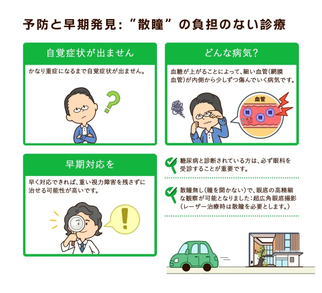
◯ You do not notice symptoms until the disease is quite advanced.
○ It is a disease in which high blood sugar gradually damages the thin blood vessels of the retina (retinal vessels) from the inside.
◯ If it is dealt with early, there is a good chance it can be treated without leaving serious visual impairment.
✔ If you have been diagnosed with diabetes, it is essential to see an eye doctor.
✔ High-definition observation of the fundus is now possible without pupil dilation (without widening the pupil):Ultra-widefield fundus imaging
(Pupil dilation is needed during laser treatment.)
» Advances in retinal imaging: ultra-widefield fundus imaging
[Diabetes]
Glucose in the blood (blood sugar) is known to act harmfully on blood vessels.
When blood sugar rises after a meal, the pancreas, located behind the stomach, immediately secretes insulin to bring it down.
When this function weakens and blood sugar stays high, blood vessels are damaged from the inside.
If this continues for a long time, diabetic complications appear first in the tissues with the thinnest blood vessels in the body (the kidneys, peripheral nerves, and retina).
» Diabetes and the Eyes
Short videos from the American Academy of Ophthalmology (AAO) and the National Eye Institute (NEI), NIH:
Each video is followed by a summary of its explanation.
What Is the Retina?
How we see with the retina [46-second video]
・Light passes through the cornea, pupil, lens, and vitreous and reaches the retina.
・In the center of the retina, the “macula,” and the surrounding retina, light is converted into signals, which travel through the optic nerve to convey information to the brain.
Diagnosis of Diabetic Retinopathy
Non-proliferative diabetic retinopathy [27-second video]
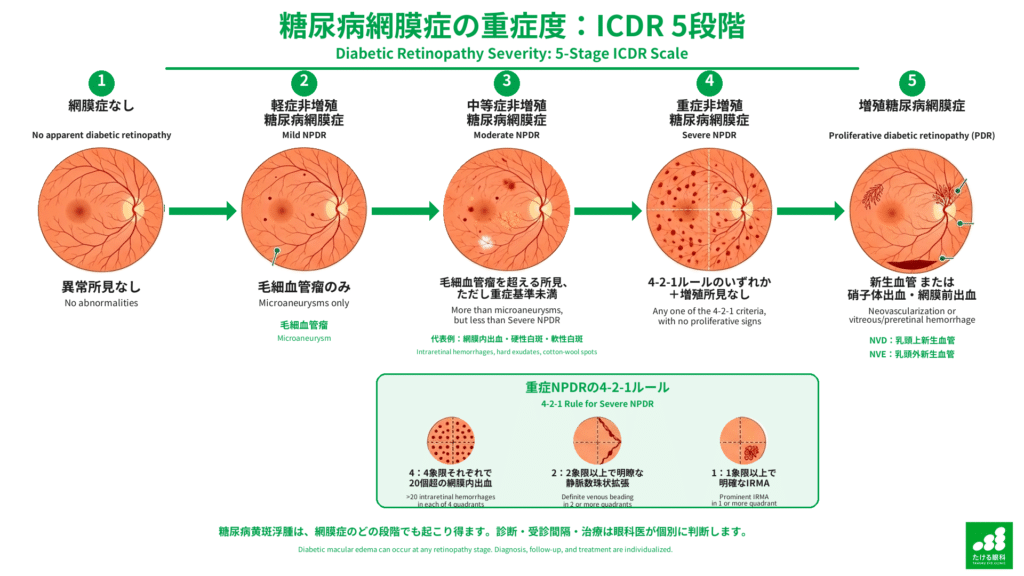
・Non-proliferative diabetic retinopathy: water and fat leak from damaged retinal vessels and collect inside the retina.
These deposits are called “hard exudates” (the areas shown as white dots).
[Examination and Diagnosis]
We examine the fundus to determine whether retinopathy is present and how far ischemia has progressed, and we tell you when to come back next.
As needed, we use devices such as fundus photography, optical coherence tomography, OCT angiography, and static visual field testing, and we explain all results by showing you the images.
(Addendum)
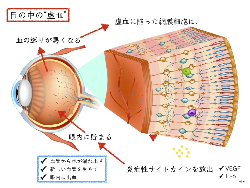
Inflammatory cytokines such as VEGF (vascular endothelial growth factor) accumulate inside the eye.
Proliferative diabetic retinopathy [42-second video]
・Proliferative diabetic retinopathy: blood flow in many retinal vessels stops, and the retina no longer receives the blood flow it should.
(The areas that look pale)
・The retina tries to grow new blood vessels.
These new vessels (abnormal vessels) not only fail to deliver proper blood flow but can also bleed or form proliferative tissue on the retinal surface, causing retinal detachment.
・New vessels can also appear in the angle, where fluid drains from the eye, raising eye pressure and leading to visual impairment from glaucoma.
The ischemic retina is the root of the trouble.
Retinal cells become inflamed, and various proteins (cytokines) accumulate in the eye and act even more harmfully on the retinal vessels.
・Increased permeability of retinal vessels: the vessel walls weaken and water leaks out
→ It collects in the center of the retina (the macula) and reduces vision: diabetic macular edema
・Retinal neovascularization: new, fragile vessels form in response to the retina’s lack of oxygen
→ Bleeding inside the eye (vitreous hemorrhage)
・Others: retinal detachment (tractional retinal detachment), secondary glaucoma, and so on
Diabetic retinopathy detected by a dilated exam [1 minute 53 second video]
・“Comprehensive dilated eye exam: the first step in preventing blindness”
・By widening the pupil and observing the whole fundus, the doctor can detect signs of diabetic retinopathy.
・Through the pupil widened with eye drops, the doctor can see the back of the eye clearly: the retina, optic nerve, and macula.
・In early diabetic retinopathy, small spots called microaneurysms are seen. Water can leak from the vessels of microaneurysms and collect in the retina, reducing vision.
・If new vessels grow and bleed, serious vision loss can result.
・People with diabetes are at risk of diabetic retinopathy.
・A dilated exam lets the doctor detect subtle changes in the fundus.
・It is important for people aged 60 and over to have a fundus exam once a year.
・It is also important to consult an eye doctor early if you notice any change in your vision.
・People thought to be at higher risk, for example because of race, age, or family history (relatives with eye disease), are recommended to have a fundus exam at least once a year.
Fluorescein angiography [25-second video]
・A yellow fluorescent dye called fluorescein is given through a drip in the arm and travels through the heart to the blood vessels throughout the body.
・Images are taken continuously as the dye reaches the retina, and we observe dye leaking from vessels and areas with poor blood flow.
EyeSmart — American Academy of Ophthalmology
* At our clinic, we evaluate the blood vessels of the retina and choroid with OCT angiography.
Below: OCT angiography images

✔ Blood vessels can be examined without the risk of an allergy to a contrast agent.
Treatment of Diabetic Retinopathy
Panretinal photocoagulation [41 seconds]
・The standard treatment for proliferative diabetic retinopathy (PDR) uses a laser beam.
・Photocoagulation of the retina as a whole is called “panretinal photocoagulation.”
・After surface anesthesia with eye drops, a laser beam is applied to the retina, avoiding the macula, the center used for seeing.
・Photocoagulation shrinks abnormal vessels and reduces the risk of bleeding into the eye and, in more severe cases, of retinal detachment.
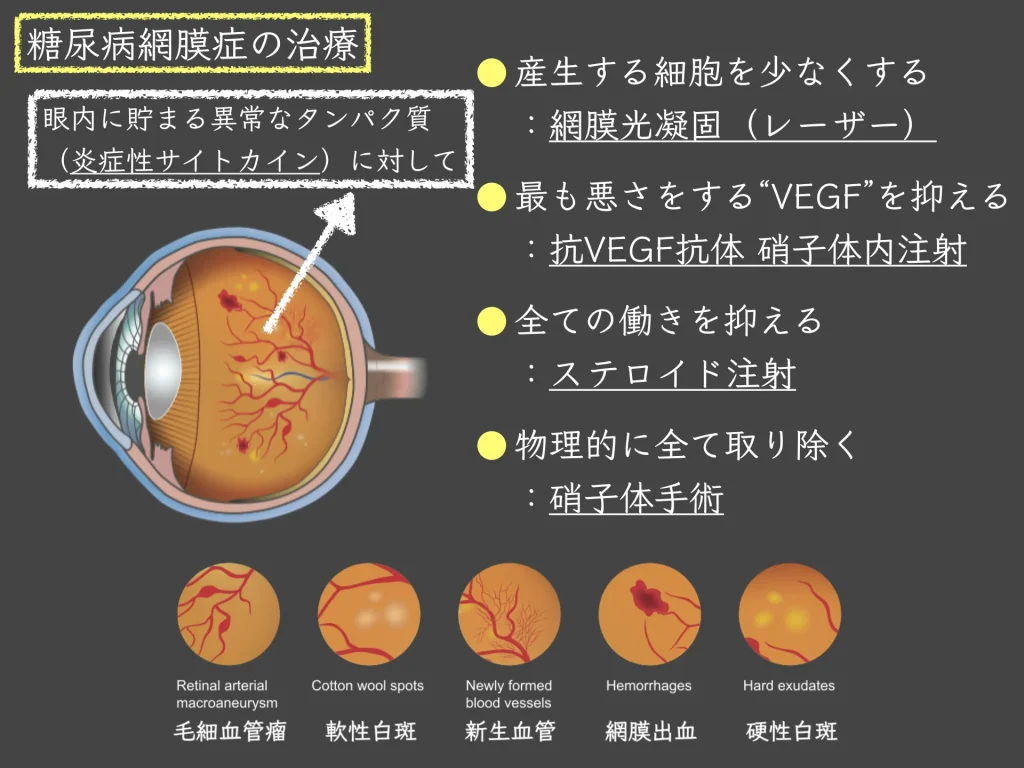
Retinal photocoagulation (laser treatment)
: Slows progression of the disease by shutting down the function of the ischemic retina.
Posterior sub-Tenon steroid injection
: A steroid suspension (triamcinolone acetonide) is injected outside the eyeball to calm inflammation inside the eye.
Anti-VEGF drugs: suppressing the growth of abnormal vessels [48 seconds]
・Anti-VEGF treatment targets a protein that increases inside the eye, VEGF (vascular endothelial growth factor).
・Anti-VEGF drugs
- suppress the growth of abnormal vessels in the eye
- prevent bleeding from abnormal vessels
- prevent proliferative tissue from spreading inside the eye
and so on.
・After anesthesia, the drug is injected directly into the eye with a fine needle.
・Treatment usually requires multiple injections over several months or more.
Intravitreal anti-VEGF injection
: Slows progression of the disease by suppressing the action of VEGF (vascular endothelial growth factor), one of the cytokines inside the eye.
Vitrectomy for diabetic retinopathy [59 seconds]
・Surgery is performed under a microscope.
・Instruments are inserted into the eye to remove blood and proliferative tissue.
・Endolaser photocoagulation: slows progression of the disease by shutting down the function of the ischemic retina.
・Gas or silicone oil may be placed in the eye for a detached retina.
Vitrectomy
: The main purpose is to remove the cytokines that accumulate in the eye.
In addition, it is a treatment for quite advanced diabetic retinopathy, such as removal of proliferative membranes, endophotocoagulation, and repair of retinal detachment.

