![Takeru Eye Clinic | Takatori Shopping Street, Sawara-ku, Fukuoka City [Nishijin Station / Fujisaki Station]](https://takeru-eye.com/wp-content/uploads/2022/10/takeru_logo_for-WP-header.png)
○ We have assembled a range of instruments to assess the condition of your eyes comprehensively.
○ We also offer various ophthalmic laser treatments (for glaucoma, retinal disease, and posterior capsule opacification).
○ Each test is performed by certified orthoptists (CO/ORT).

Auto ref-keratometer / tonometer / pachymeter
TONOREF III (NIDEK)
- After measuring the refractive error (myopia, hyperopia, and astigmatism), its axis, and the corneal curvature (autorefractor function), the device measures intraocular pressure (tonometer function).
- It also measures corneal thickness (pachymeter function) and automatically calculates a corrected intraocular pressure value.
- These data are used to determine the prescription for glasses and contact lenses and as a reference for the visual acuity test.
- The puff of air against the eye during pressure measurement is gentler than with older models.

Visual acuity test
SSC-370 type D (NIDEK)
- Visual acuity is measured based on the data from the auto ref-keratometer.
- The test is performed at a distance of 1 m.
Because the test can be done at a short distance, you can concentrate on it. - It is less affected by the brightness of the room and does not require vertical eye movements.
- The conventional 5 m visual acuity test is also available for those who need it.

Slit-lamp microscopy
BQ900/LED (Haag-Streit)
- Made by Haag-Streit of Switzerland, which has had a long-standing reputation.
- We examine the surface of the eye (cornea and conjunctiva), the anterior chamber, the lens, and the eyelids in detail under the microscope.
- Using a special lens, we also examine the fundus (vitreous and retina).
- Precise intraocular pressure measurement is also performed.
- Dry eye testing (tear film evaluation) is also performed.

Handheld slit lamp
XL-1 (Rexxam)
- Used for people who find it difficult to rest their chin and forehead on the Haag-Streit slit lamp (children under 2 years old and people who cannot sit upright, such as those in wheelchairs).
- A handheld slit-lamp microscope with LED illumination.

Monitor in the examination room
U2715H (Dell)
- Images from the slit-lamp microscope can be captured.
- All other examination images are displayed on a vertical monitor at the patient’s eye level while we explain them.
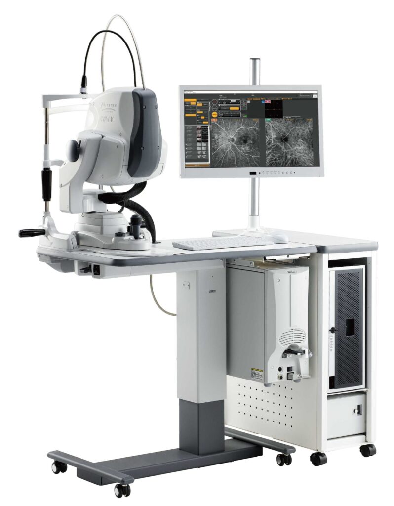
Confocal scanning diode laser ophthalmoscope
Optical coherence tomography (OCT) / OCT angiography
Mirante (NIDEK)
- Ultra-widefield fundus camera: 163° field of view, with panoramic images that capture the fundus out to its far periphery
- No mydriasis required: a “fundus camera that is not dazzling”
- The “MRI of the eye”: it shows cross-sections of the retina and vitreous, helping with early detection, diagnosis, and treatment of retinal disease and glaucoma.
- OCT angiography (OCTA): shows the state of the retinal and choroidal blood vessels without using contrast dye.
- Glaucoma and retinal diseases in general.
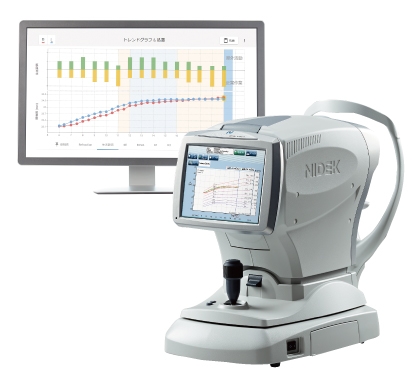
Optical biometer for axial length measurement / Myopia Viewer
AL-Scan M (NIDEK)
- An optical device that measures the axial length (the length from the cornea to the retina) and the corneal radius of curvature (the degree of corneal curvature), both needed for myopia management.
- It measures axial length quickly and without contact, and changes in the measurements over time can be shown on a graph.
- Myopia is a condition in which light comes to focus in front of the retina. In most cases it is “axial myopia,” in which the axial length grows excessively during the school-age years. The device is useful for
- managing the progression of myopia.
MV-1 (NIDEK)
- Software that, in addition to graphs of axial length and refractive power over time, lets you enter lifestyle and treatment history and visualize their correlation with axial length.
- It is used to explain changes in axial length and future treatment plans.
Other conditions that require axial length measurement
- High hyperopia and microphthalmos
- High myopia
- Glaucoma
- Preoperative cataract evaluation
- Vitreoretinal diseases, etc.

Non-mydriatic fundus camera
AFC-330 (NIDEK)
- Fundus photographs can be taken without using eye drops to dilate the pupil (mydriatics).
- Imaging is possible at low light levels, reducing the glare of conventional cameras.
- Captures detailed images of the macula and optic disc.
- Photographs are taken in the same chair as the Mirante above, without moving.
- Glaucoma and retinal diseases in general.

Corneal topography / total eye refraction analysis system
OPD (Optical Path Difference) scan III (NIDEK)
- Corneal shape analysis and refractive power analysis of the entire eye
- Vision simulation: shows in images the cause of “blurry vision even though visual acuity is good”
- We explain whether the cause of the blur lies in the cornea or deeper inside the eye.
- Wavefront analysis: identifies higher-order aberrations and irregular astigmatism
- Measurement of pupil diameter in bright and dark conditions
- Progression analysis of corneal diseases such as keratoconus, judging the indication for cataract surgery, whole-eye refraction analysis before and after surgery, support when glasses or contact lenses do not give good vision, and assistance with contact lens prescription, among others

Retinal photocoagulator
Green laser GYC-500 (NIDEK)
- Retinal diseases are treated by applying laser to the retina at the back of the eye.
- Main target diseases: diabetic retinopathy, retinal vein occlusion, retinal tears, retinal breaks associated with retinal detachment, central serous chorioretinopathy, etc.

Laser device for glaucoma and posterior capsule opacification
Tango Ophthalmic Laser (Ellex)
- Laser treatment for glaucoma (SLT; selective laser trabeculoplasty)
- For posterior capsule opacification, an incision is made with a YAG laser.
- Main target diseases: open-angle glaucoma, ocular hypertension, exfoliation glaucoma, pigmentary glaucoma, steroid-induced glaucoma, and after cataract surgery

Static perimeter
HFA III 840 (Carl Zeiss)
- We use the Humphrey Field Analyzer, the world standard for visual field testing.
- With the HFA III 840 that we have adopted, a measurement can now take as little as about 2 minutes per eye.
- You look at a single point without moving your eyes, and we examine the range you can see. Lights are shown from various positions, and you press a button when you see one. We examine the central 30° and 10° of the visual field in detail.
- Glaucoma progression analysis (Guided Progression Analysis (GPA™)):
Analysis of visual field changes over time (Visual Field IndexTM (VFITM )), and we explain the prediction of the remaining visual field. - Shorter test time / better insight into glaucoma
https://www.zeiss.co.jp/meditec/product-portfolio/perimetry/humphrey-field-analyzer-3.html
About ventilation during Humphrey visual field testing (from Carl Zeiss):
The Humphrey Field Analyzer (700 and 800 series) has a function that ventilates the inside of the dome. It also automatically adjusts the background luminance to small changes in brightness, even if the room is not completely dark. As long as the examination room is reasonably dark, the test can be performed with the curtains or doors open, without completely partitioning the room with curtains or the like. If too much light comes in to be adjusted for, an error message is displayed.

Kinetic perimeter (Goldmann-type perimeter)
L-1560 (Inami)
- It is the most widely used perimeter in the world and also serves as the reference standard for automated perimeters.
- An orthoptist examines visual field constriction and defects over a wider range than with the Humphrey, while observing the patient’s responses.
- It is also used to diagnose advanced glaucoma and brain diseases.

Accommodation analyzer
Accommo-Ref 2 (Right Manufacturing Co.)
- Accommodation analysis: objectively measures the accommodative response when the eye tries to focus at several distances (33 cm, 50 cm, 1 m, ∞) and the degree of ciliary muscle tension needed to maintain that accommodation. The results are displayed visually as an Fk-map (Frequency of Kinetic reaction), which shows accommodative disorders.
- Accommodative tension, accommodative spasm, accommodative panic, presbyopia, presbyopia with accommodative tension, IT eye syndrome (VDT syndrome / techno-stress eye syndrome), eye strain, etc.
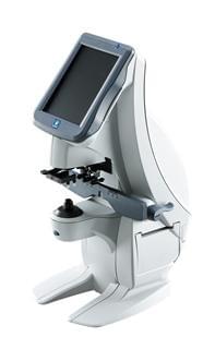
Auto lensmeter
LM-7 (NIDEK)
- It can accurately measure the prescription of glasses and hard contact lenses.
- It can also measure progressive lenses (multifocal glasses).
- Equipped with a UV transmittance measurement function
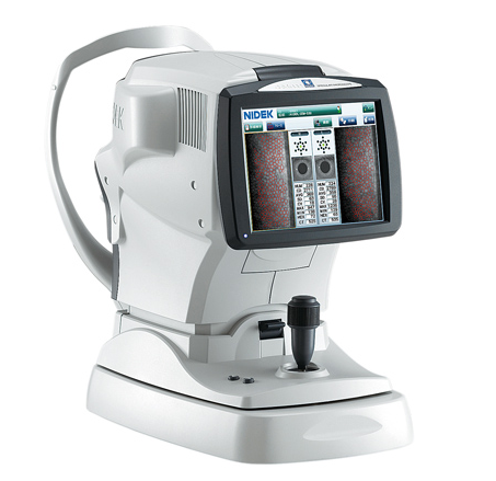
Corneal endothelial cell imaging device (specular microscope)
CEM-530 (NIDEK)
- It examines the state of the “corneal endothelial cells,” which are important for keeping the cornea transparent.
- The cornea (the black part of the eye) has a five-layer structure. The hexagonal endothelial cells in the innermost layer decrease with age, intraocular surgery, lack of oxygen, and so on, and once damaged they are known not to regenerate. If they become extremely few, the cornea can become cloudy and white (bullous keratopathy). For this reason, regular observation is needed for the following people.
- Contact lens wearers, people before and after cataract, glaucoma, or vitrectomy surgery, and people with corneal disease, etc.

Hess chart projector (Hess red-green test)
HE-183 (Handaya)
- A device that evaluates whether the movements of both eyes are normal.
- It can detect abnormalities of the nerves and muscles around the eyeball.
- Head trauma, eye injury, orbital blowout fracture, extraocular myositis (diabetic and other), intracranial diseases such as brain tumor and cerebral hemorrhage, strabismus, thyroid eye disease, etc.
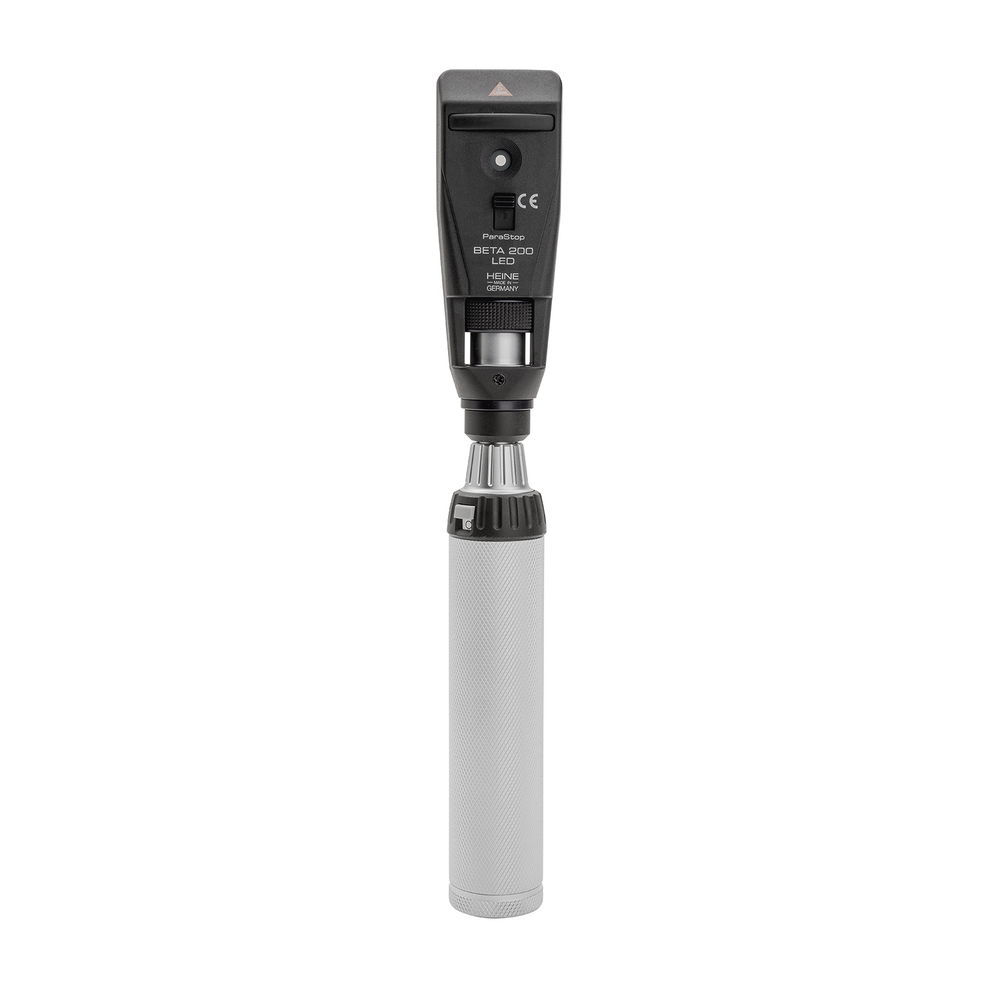
Retinoscope
HEINE BETA 200 LED Retinoscope (HEINE)
- Retinal reflex test for babies (early detection of congenital cataract, retinoblastoma, and similar conditions)
- Refractive errors such as myopia, hyperopia, and astigmatism
- Screening for strabismus and amblyopia
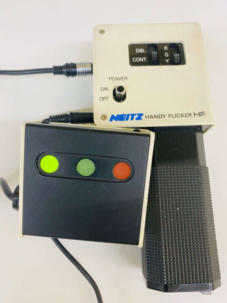
Central flicker test
Handy Flicker HF (Neitz)
- It measures the frequency at which a flickering light can be distinguished.
- It can detect abnormalities of the optic nerve.
- Optic neuritis, optic neuropathy

Biological microscope
ECLIPSE E200 (Nikon)
- Gram staining is performed to estimate the bacteria causing eye infections (mainly of the conjunctiva and cornea).
- Depending on the type of inflammatory cells, allergic and infectious inflammation can be distinguished.
- Based on these results, appropriate eye drops are selected.
- (A bacterial culture or other tests may be needed for an accurate diagnosis.)
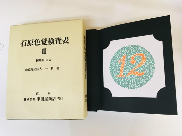
Color vision test plates
Ishihara Color Vision Test Plates II (Handaya)
- It is regarded as the most accurate test for distinguishing normal color vision from congenital color vision deficiency, and is used worldwide.
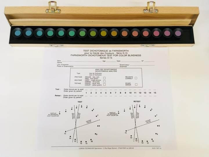
Color vision tester (Panel D-15)
15D Farnsworth test (Leneau Technology)
- It determines the “degree” of color vision deficiency.
- In this test, you arrange 15 color caps in order of similarity.

Prism bars
Berens prism bar K-1291 (Inami)
- Eye alignment test: when there is a deviation in eye movement, its angle is measured.
- Strabismus and amblyopia

Stereo fly test (Titmus stereo test)
Stereo Fly Test (Stereo Optical)
- Binocular vision test: stereopsis is measured using polarized glasses.
- Strabismus and amblyopia
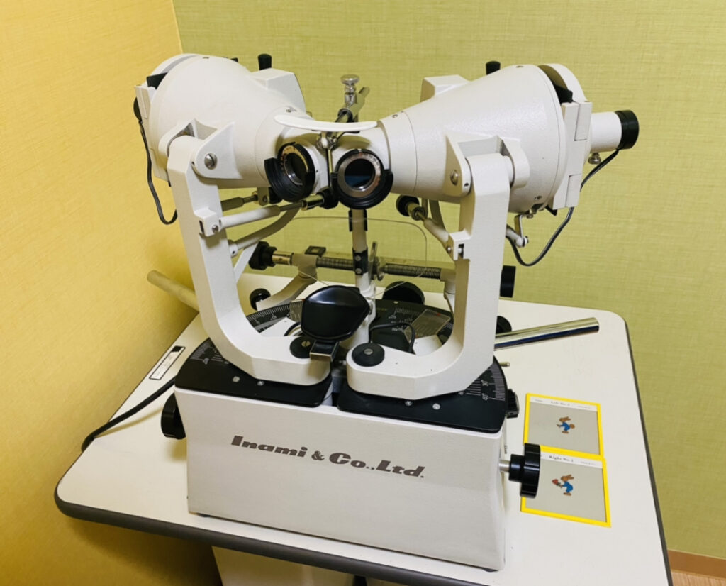
Synoptophore (major amblyoscope)
Synoptiscope (Inami)
- A device that measures “how correctly the two eyes are being used together.”
- Binocular vision test: measurement and training of simultaneous perception, fusion, and stereopsis
- Measurement of the angle of strabismus
- Strabismus and amblyopia

Spot Vision Screener(photoscreener)
Spot Vision Screener (Welch Allyn Japan)
- From 6 months of age: screening for refractive errors and eye alignment
- Used mainly for early detection of strabismus and amblyopia; useful for the 3-year-old checkup
- Myopia, hyperopia, astigmatism, anisometropia, and anisocoria.
- Tested from 1 meter away (non-contact), so children are not frightened.
- If an abnormality is found, a detailed eye examination by our full-time orthoptist can also be performed on the same day.
- http://welchallyn.jp/visionscreener/
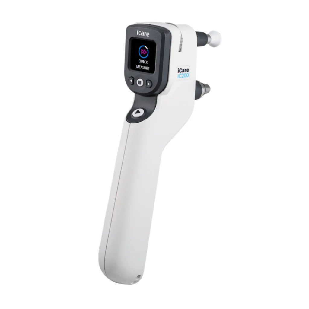
Handheld tonometer(iCare handheld tonometer)
iCare IC200 (ME Technica)
- A medical device that uses an extremely small, lightweight probe that touches the cornea momentarily and measures intraocular pressure from the speed at which it rebounds.
- Features:
- No anesthetic eye drops are needed, and the test is painless
- Accurate readings are shown on a digital display
- Can be measured in any position: sitting, supine, or lying on the side
- Intended for:
- Diagnosis and follow-up of glaucoma
- Measuring intraocular pressure in children and bedridden patients
- Managing intraocular pressure after surgery
- https://www.metechnica.co.jp/products/product228.html

