![Takeru Eye Clinic | Takatori Shopping Street, Sawara-ku, Fukuoka City [Nishijin Station / Fujisaki Station]](https://takeru-eye.com/wp-content/uploads/2022/10/takeru_logo_for-WP-header.png)
From September 2021: we introduced the ultra-widefield fundus camera “Mirante.”
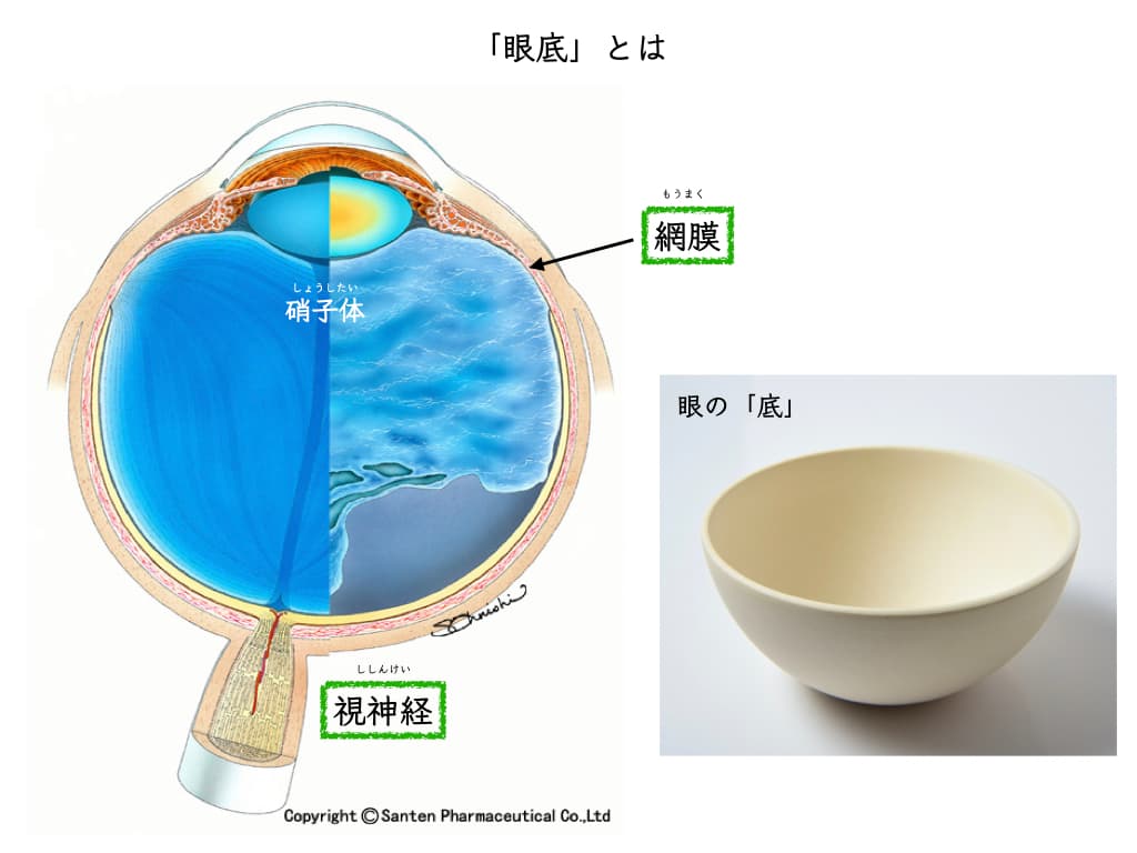
A fundus examination without dilating the pupils (no dilating drops) lets us see the state of the “retina and vitreous” inside the eye.
The images combine the three RGB colors (red, green, blue) and are very beautiful, making this a precise fundus examination.
Mirante is a multifunction device that can obtain images of the inside of the eye from many angles: multimodal imaging
It can also tell “whether there is any concern about glaucoma.”
(If a more detailed examination is needed, we may also perform a fundus examination with dilating drops.)
Ultra-widefield fundus photography makes it possible to perform a thorough fundus examination even if you come by car.
It is also a safe fundus examination for people who are pregnant (or may be pregnant).
With OCT angiography, we evaluate the FAZ in order to catch early changes of retinal diseases such as diabetic retinopathy.
Advances in Retinal Imaging: Ultra-Widefield Fundus Photography
Confocal scanning diode laser ophthalmoscope NIDEK Mirante(NIDEK Mirante)

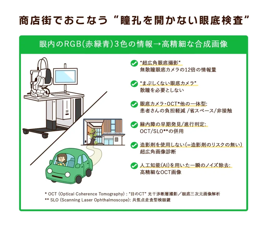
[A “fundus examination without dilating the pupils” offered on a local shopping street]
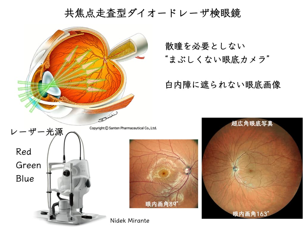
○ “Ultra-widefield fundus photography”: 12 times the information of a non-mydriatic fundus camera
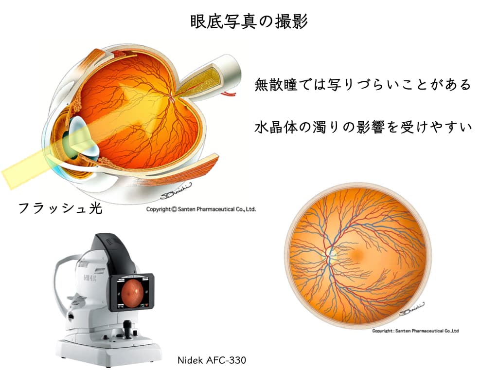
○ No pupil dilation needed (no dilating drops): a “fundus camera that isn’t dazzling”
○ Fundus camera, OCT*, and more in one unit: less burden on patients / space saving / non-contact
○ Early detection and progression assessment of glaucoma: OCT/ SLO** used together
○ Ultra-widefield imaging without contrast dye (= no risk from contrast dye)
○ Instant noise removal using artificial intelligence: high-definition OCT images
* OCT (Optical Coherence Tomography) = “CT of the eye”: optical coherence tomography / three-dimensional analysis of the fundus
** SLO (Scanning Laser Ophthalmoscope): confocal scanning ophthalmoscope
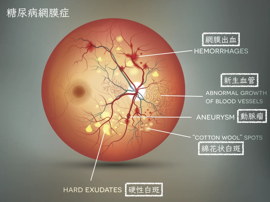
Used in fundus diseases in general, such as diabetic retinopathy, retinal vein occlusion, retinal and vitreous diseases in children, and uveitis1。
Floaters: detailed examination, and so on.
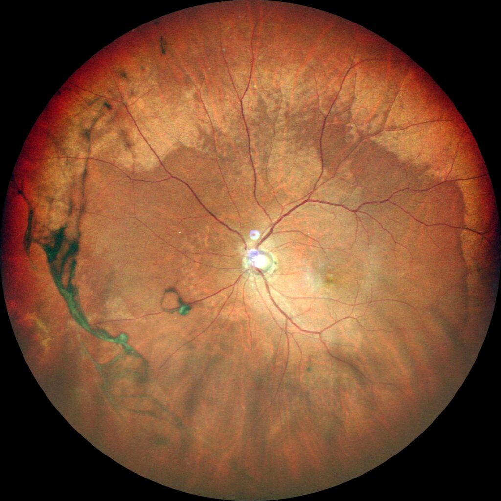
Ultra-widefield fundus photography: 163° internal field of viewto a panoramic photograph of the far periphery of the fundus
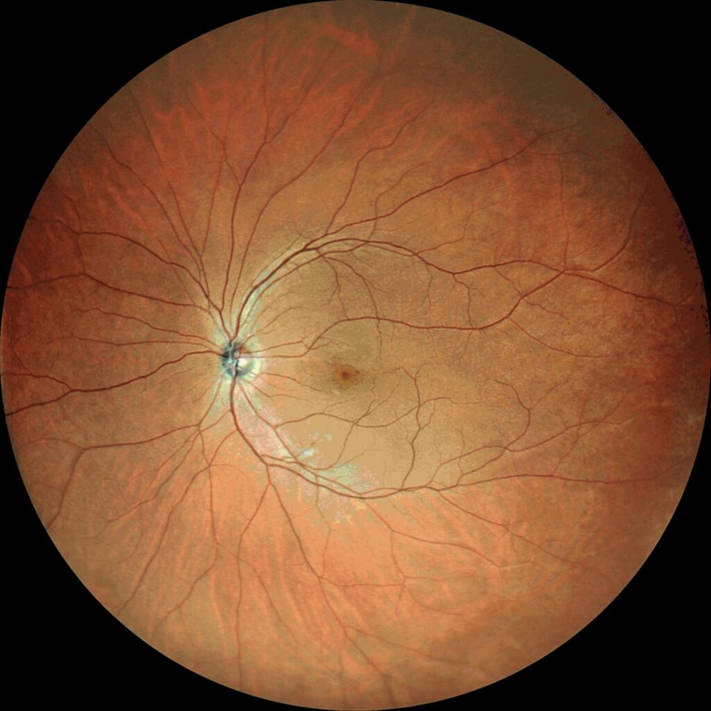
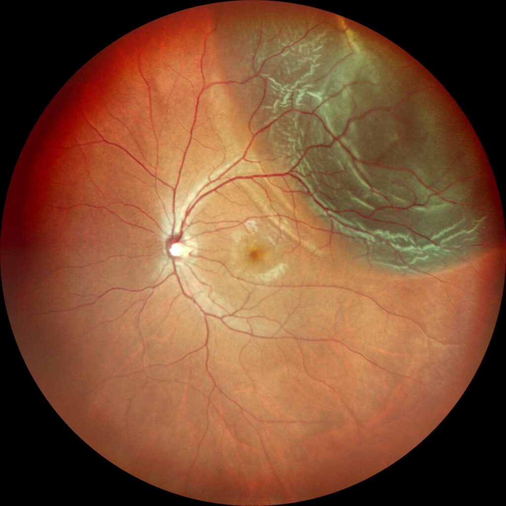
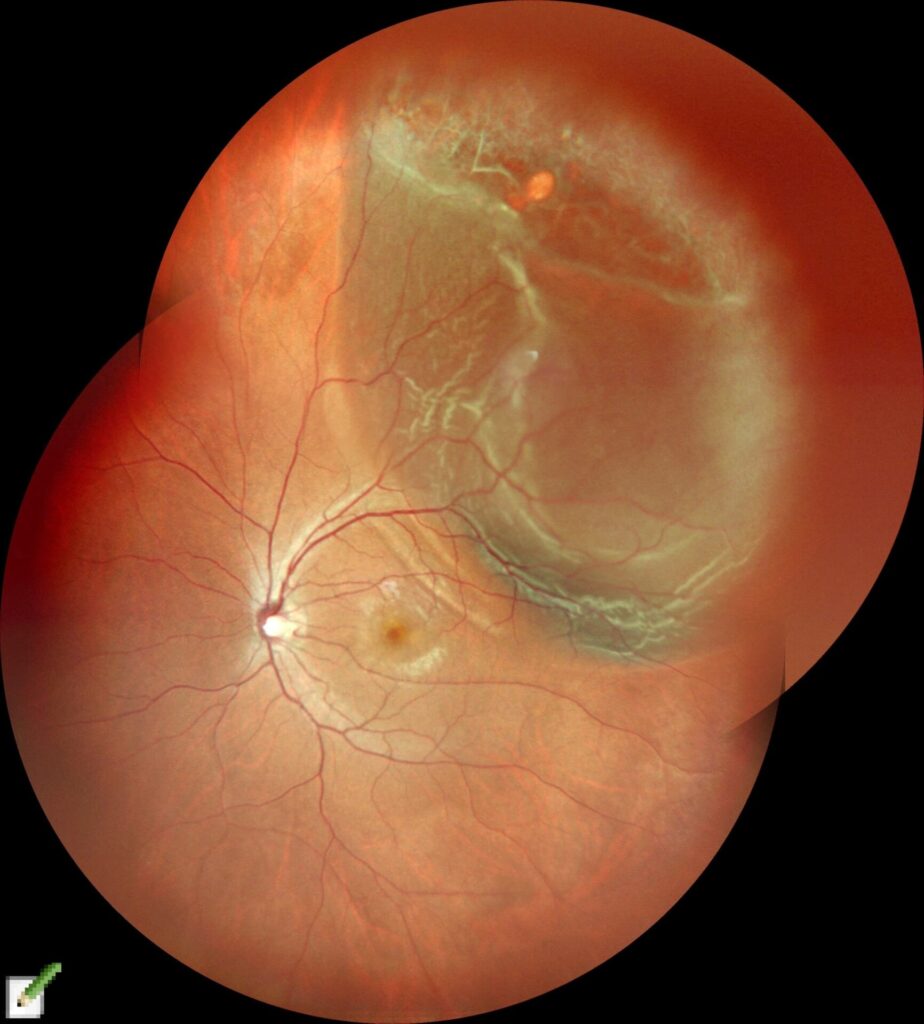
A dramatic increase in the amount of information obtained: it meets the definition of ultra-widefield2.
Imaging of the far periphery of the fundus: fundus panoramic photographs
High definition (HD): a “4K image of the fundus”
“Ultra-widefield fundus photography: 12 times the information of a non-mydriatic fundus camera”
No pupil dilation needed: confocal scanning laser ophthalmoscope (SLO)
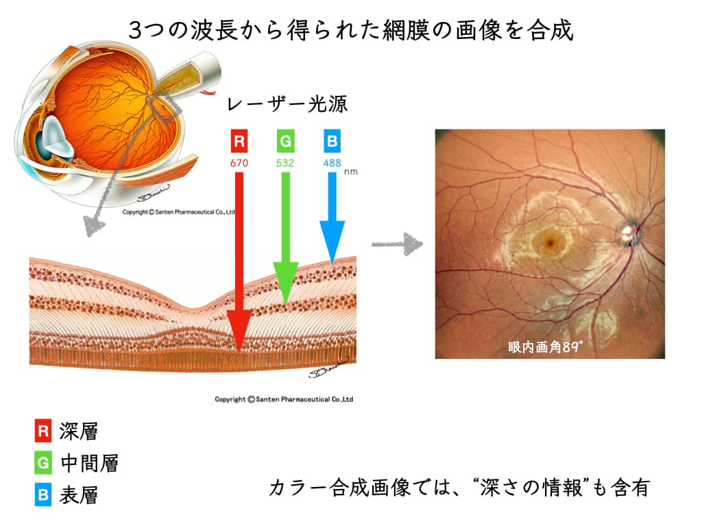
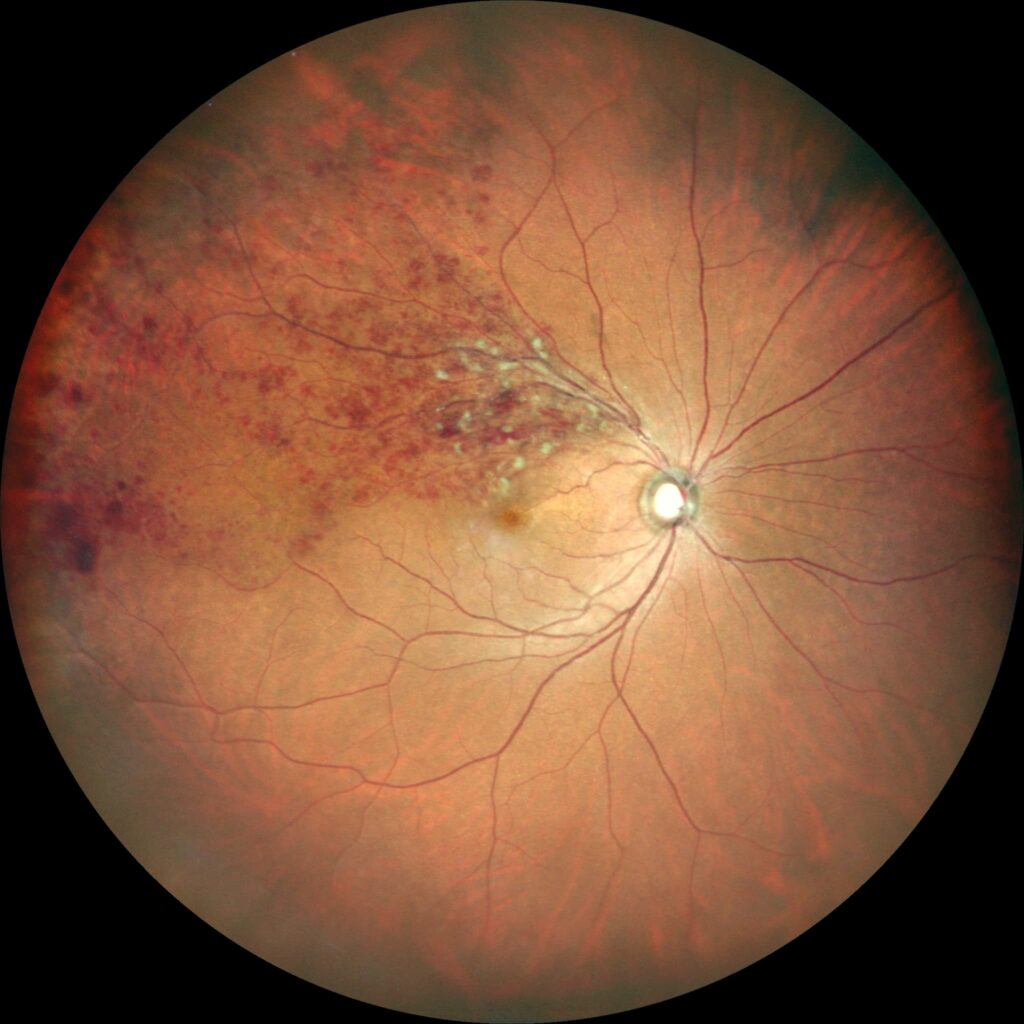
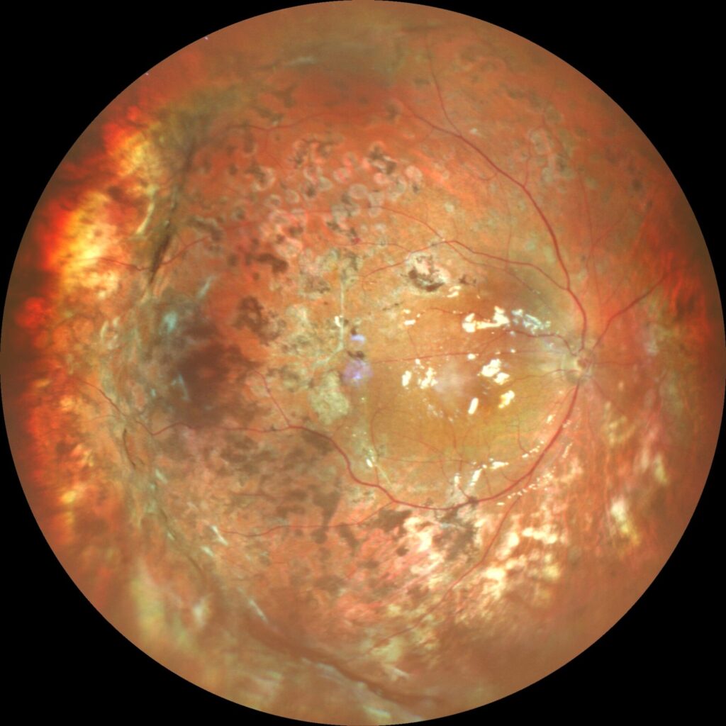
Red / Green / Blue linear light sources, each with its own wavelength
→ Light enters through a small pupil, and the instrument receives the reflection from the layers of the retina
→ The RGB images are combined and presented as one image
It is not affected by clouding of the lens (cataract).
“A fundus camera that isn’t dazzling”
* Vitreous opacities are visualized, so it is useful for evaluatinguveitis.
No need to move: an integrated imaging device
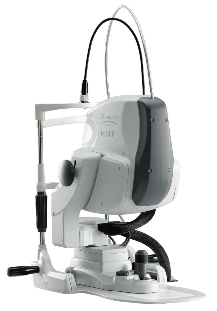
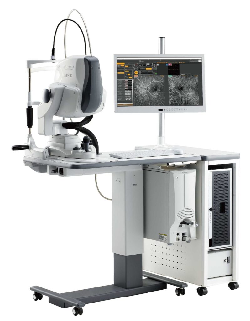
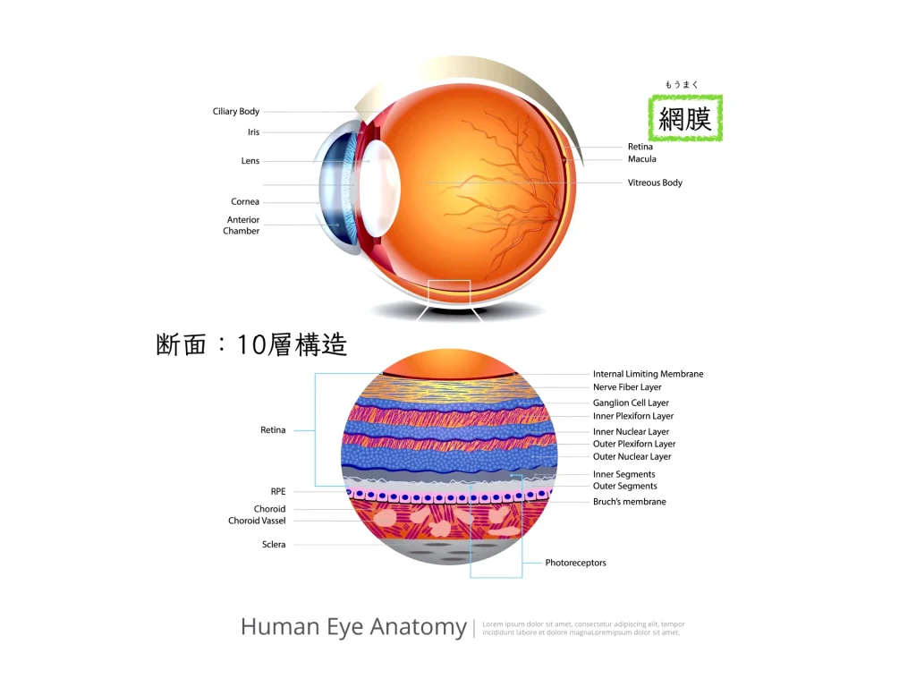
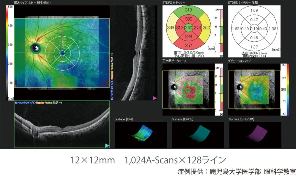
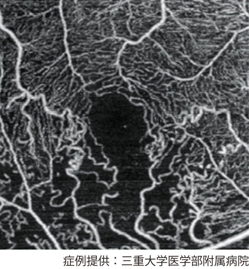
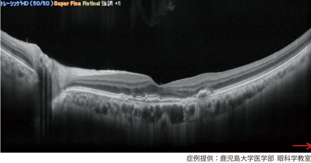
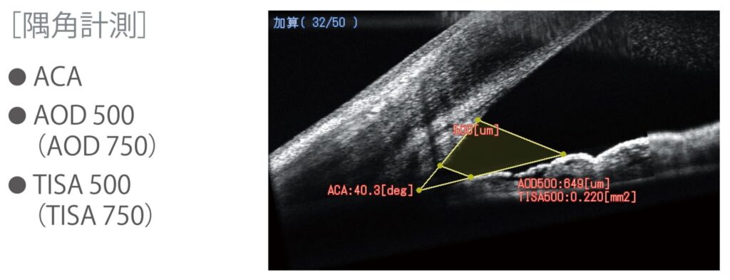
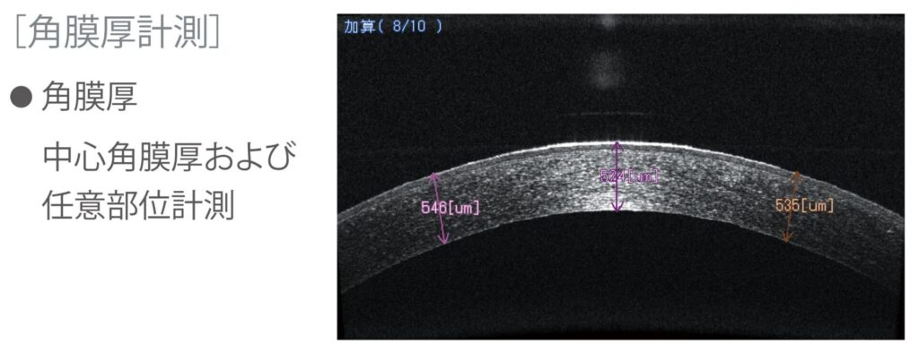
✅ OCT (3D fundus imaging): expanded imaging range
✅ OCTA (OCT angiography): expanded imaging range
✅ Anterior segment OCT: used for glaucoma, corneal diseases, and others
With added functions,
✅ Ultra-widefield fundus photography (163° internal field of view to panoramic photographs of the far periphery)
✅ Fundus autofluorescence
* You do not need to move to obtain all of these images.
Imaging of the fundus is finished while you stay seated.
The conventional non-mydriatic camera is also kept in a position where it can be used from the same chair.
In every case, the distance between the eyeball (cornea) and the device is about 9–15 mm.
“Less burden on patients,” “space saving,” “non-contact”
Identifying “retinal nerve fiber layer defects”: used together with confocal scanning laser imaging (SLO)
Imaging is also useful for diseases without symptoms.
Early detection and progression assessment of glaucoma:
OCT images give detailed information about the retinal nerve fibers.
SLO also helps with early detection of glaucoma and assessment of disease progression.
A lot of information can be obtained from the Blue/Green wavelengths of light acquired instantly3,4。
○ Three-dimensional fundus imaging (OCT)
○ Confocal scanning laser imaging (SLO)
“Early detection / progression assessment of glaucoma: OCT and SLO used together”OCT/SLOの併用」
Identifying “retinal ischemic areas”:using confocal scanning laser imaging (SLO)5
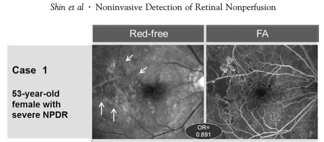
→ “Retinal ischemic areas” that match the Red-free (Blue) portion
An evaluation essential for judging disease activity in diabetic retinopathy, retinal vein occlusion, and others
The “poorly perfused areas (retinal ischemic areas)” that have been identified with fluorescein angiography (FAG)
without the risk of contrast dye allergy or shock,
may be identified by an “alternative” means
“Ultra-widefield imaging without contrast dye (= no risk from contrast dye)”
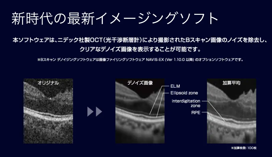
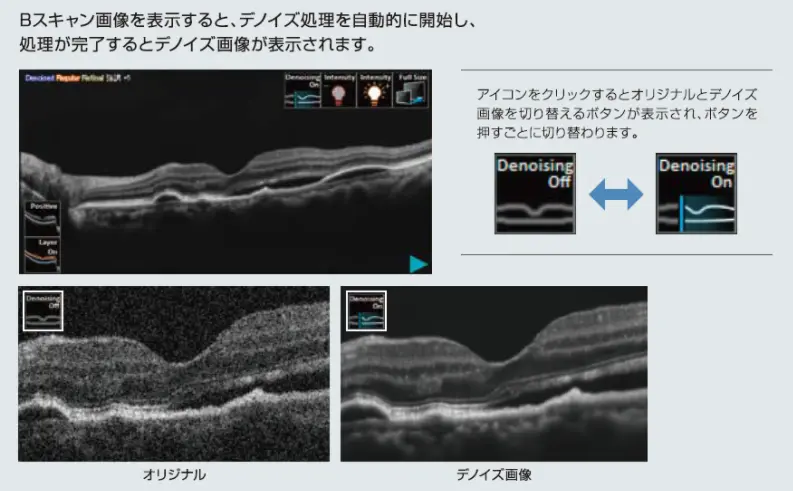
High-definition images: noise removal using AI (artificial intelligence) technology
“Instant noise removal using artificial intelligence: clear OCT images”
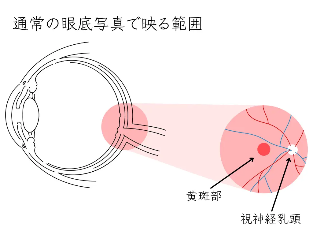
Fundus Examinations Used Until Now: Comparing Advantages and Disadvantages
Advantages and disadvantages of non-mydriatic fundus examination
Advantages
- Because the pupil is not dilated, the exam can be done without eye drops.
- You can return to daily life right after the exam, and driving is not a problem.
- It can be done with peace of mind even during pregnancy.
Disadvantages
- Some fine abnormalities may be missed.
Advantages and disadvantages of fundus examination with dilation
Advantages
- A detailed exam is possible: eye drops (Midrin®-P or Neosynesin® ophthalmic solution) dilate the pupil so that fine details of the fundus can be seen.
An exam by an ophthalmology specialist using a slit-lamp microscope combined with a condensing lens. - Early detection of abnormalities: dilation allows minute lesions and abnormalities to be found early, so appropriate treatment is possible.
Disadvantages
- Waiting time before the exam: you need to wait 20–30 minutes after the drops are instilled.
- Glare and blurred vision: because light is shone during the exam, you feel glare during the exam. This is because more light enters the eye.
In addition, symptoms such as “hard to see,” “glare,” “blurred,” and “difficulty focusing” last 4–5 hours.
(This varies between individuals.) - The pupil does not return quickly: it takes time for the pupil enlarged by the dilating drops to return (constrict), so vision is temporarily blurred.
- Risk during pregnancy: using dilating drops during pregnancy carries risks.
- Driving restriction: vision is blurred after the exam, so you need to refrain from driving.
- Sunglasses?: sunglasses may be needed to reduce glare after the exam.
- Difficult to come by car: after dilation vision is blurred, so it is difficult to come by car or motorcycle.
Eye Imaging Diagnosis: A New Era
As a result, in the imaging analysis of retinal diseases in general,
in a short time,
with less burden on the patient,
we are now in an era where the amount of information obtained from the fundus has increased dramatically.
Medical professionals strive to keep their knowledge up to date.
(References)
1. Patel, S.N., Shi, A., Wibbelsman, T.D., Klufas, M.A., 2020. Ultra-widefield retinal imaging: an update on recent advances. Ophthalmol Eye Dis 12, 2515841419899495. https://doi.org/10.1177/2515841419899495
2. Choudhry, N., Duker, J.S., Freund, K.B., Kiss, S., Querques, G., Rosen, R., Sarraf, D., Souied, E.H., Stanga, P.E., Staurenghi, G., Sadda, S.R., 2019. Classification and Guidelines for Widefield Imaging: Recommendations from the International Widefield Imaging Study Group. Oph Retina 3, 843–849. https://doi.org/10.1016/j.oret.2019.05.007
3. Tassel, S.H.V., Asrani, S.G., 2019. Possible Undiagnosed Glaucoma Detected by MultiColor Imaging of Retinal Diseases. Ophthalmology Glaucoma 2, 22–27. https://doi.org/10.1016/j.ogla.2018.11.003
4. Kim, Y.H., Ahn, J., Kim, K.E., 2021. Multicolor imaging for detection of retinal nerve fiber layer defect in myopic eyes with glaucoma. American Journal of Ophthalmology. https://doi.org/10.1016/j.ajo.2021.07.022
5. Shin, Y.U., Lee, B.R., Kim, S., Lee, W.J., 2012. A Novel Noninvasive Detection Method for Retinal Nonperfusion Using Confocal Red-free Imaging. Ophthalmology 119, 1447–1454. https://doi.org/10.1016/j.ophtha.2012.01.036