![Takeru Eye Clinic | Takatori Shopping Street, Sawara-ku, Fukuoka City [Nishijin Station / Fujisaki Station]](https://takeru-eye.com/wp-content/uploads/2022/10/takeru_logo_for-WP-header.png)

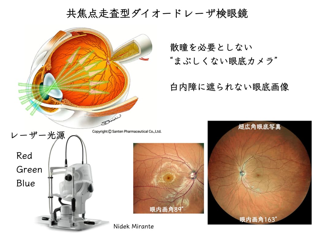
Retinal imaging without dilating the pupils turned out to give higher-definition images than I had expected.
“I’m dazzled for a while after a fundus examination.” “I can’t drive home after my pupils are dilated.”
: inconveniences like these for patients have disappeared.
It has been a little over two weeks since we began using the Mirante® (NIDEK).
These are days of astonishment at the transformation of ophthalmic imaging 😳.
We obtain high-definition images for uveitis, retinal disease and glaucoma.
* SLO (Scanning Laser Ophthalmoscope): confocal scanning laser ophthalmoscope
Retinal disease and glaucoma: high-quality diagnostic imaging and convenience for patients

The evolution of retinal imaging, starting from the “posterior pole”
The important parts for “being able to see” are the “macula” and the “optic nerve.”
Posterior pole: the central part of the fundus.
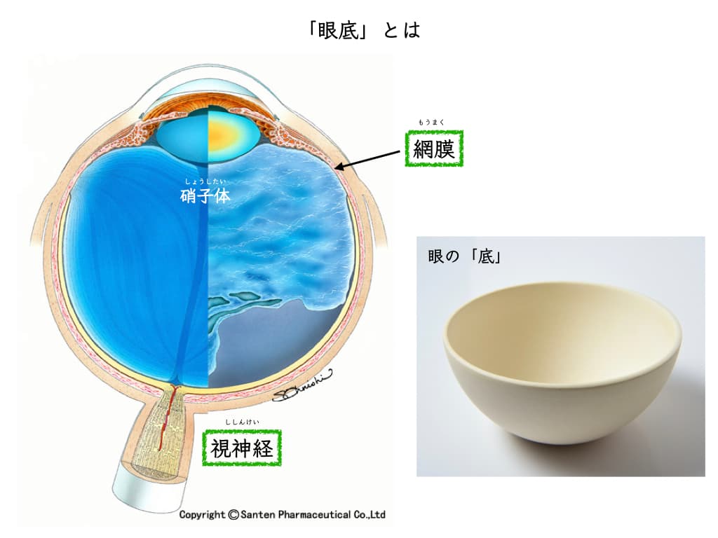
When I was a resident in 2001, OCT did not yet exist.
Days spent working hard to examine the “macula” after dilation with a slit-lamp microscope and a preset lens..
The patients were dazzled.. (I’m sorry)
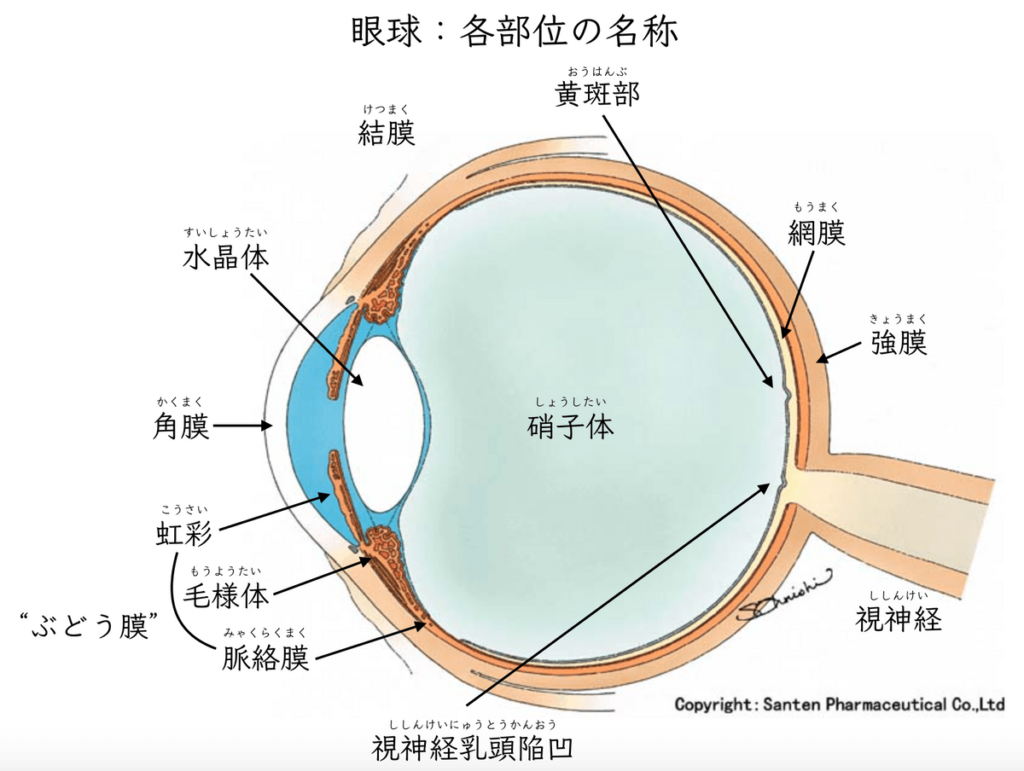
Using the same method, I can now see the state of the “optic nerve (optic disc cupping)” directly in an instant.
Direct screening can be done for almost all patients.
For an ophthalmologist toview the “optic nerve” directly, a dilated (pupil-widening) examination is not needed.
: early detection of glaucoma
* OCT (Optical Coherence Tomography) : “CT of the eye” — optical coherence tomography / three-dimensional fundus image analysis
OCT has now evolved so that images of the macula and optic nerve that anyone can understand at a glance are obtained instantly.
It is also a machine that plays a major role when we need detailed information on the state of “optic disc cupping.”
Having evolved further, now
we are in an era in which beautiful images can be obtained from the posterior pole all the way to the periphery.
“Color information” expressed in multiple colors (RGB)
It is a composite image from three laser light sources (red, blue and green).
RGB is called the “three primary colors of light.”
The way smartphone and computer displays and TVs produce color is the same, and it can express almost all colors.
Expressing color with codes
Hexadecimal color codes and RGB values
Hex code
#000000
Hexadecimal numbers are lined up in the order red, green, blue.
(A comes after 9)
00–09-0A–0F-10–19-1A–1F-20—FF (maximum value)
Converted to decimal,
0A is 10 and FF is 255
#000000: black
#FF0000: red
#00FF00: green
#0000FF: blue
#FFFFFF: white
Conversion tool (hex–rgb):
https://www.webfx.com/web-design/hex-to-rgb/
Example: color codes of SNS icons (corporate colors)


#06c755


(For reference)
Printed materials use the CMYK format
Cyan / Magenta / Yellow / Key Plate (black)
: This is why printing an RGB screen as it is often makes the colors look a little darker.
The retina’s “color information” and “depth information”
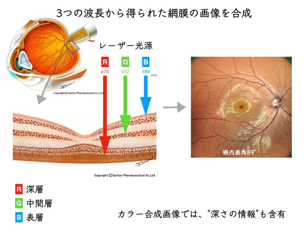
○ Color information
→ Composite of RGB
○ Depth information
→ Uses the retinal layers that absorb each RGB wavelength
The information and state of the retina that the ophthalmologist sees
• without dilating the pupil,
• in high definition,
appear as images that are obvious to anyone.
Most retinal and uveal diseases can now be examined without dilation.
“You can drive home after a fundus examination.”
It has become a gentler situation for patients, too.
(Even so, when necessary, we may ask for a fundus examination after dilation.)
Retinal conditions captured without dilation
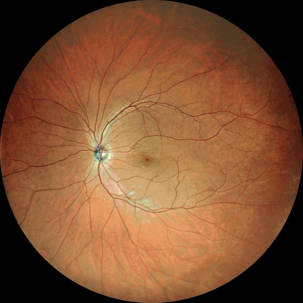
Soon after we began using the SLO, we were able to capture the following changes clearly.
• Nerve fiber layer defects (NFLD) in glaucoma
NFLD that is not clear even on OCT seems to be depicted
• Peripheral retinal vasculitis in sarcoidosis uveitis
• Retinal ischemia associated with anemia
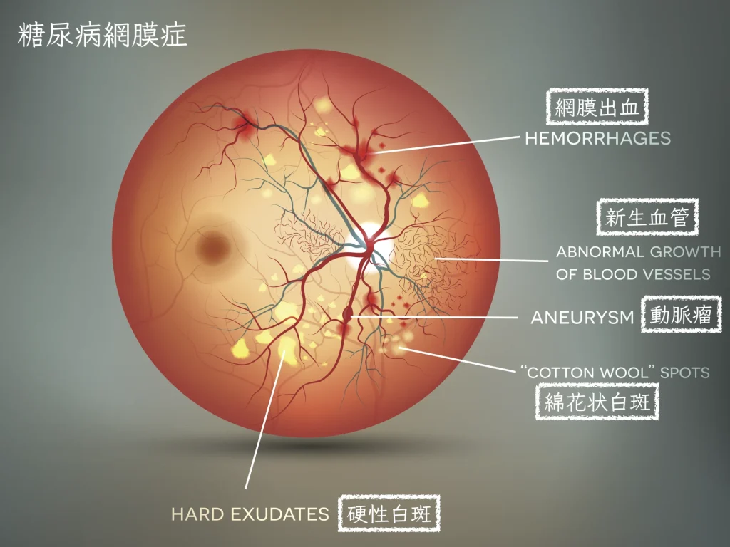
• Changes of diabetic retinopathy: hemorrhages and white spots
• Hemorrhage and retinal edema associated with branch retinal vein occlusion
• Retinal tears and retinal detachment
• Shallow retinal detachment
• Epiretinal membrane (macular pucker)
• Imaging through the small pupils of people in their 90s and older
(Pupil size becomes smaller in inverse proportion to age)
• Macular diseases
Age-related macular degeneration, central serous chorioretinopathy, macular degeneration and choroidal neovascularization associated with high myopia
The intraocular field of view is 163°
: Images of the farther periphery can be obtained using panoramic composite photographs.
Compositing is also completed in a few seconds..
All without dilation (= patient convenience remains high)
We obtain RGB images that are clear to anyone’s eyes.
With the arrival and evolution of OCT, explanation and understanding of diseases of the posterior pole of the fundus,
the “macula” and “optic nerve,” have advanced.
The SLO expresses fine detail from the center out to the surrounding areas.
Obtaining and recording findings of disease from the ophthalmologist’s eye and experience:
it is a machine that changes the very foundation of the care we have given until now.
What is “intuition”?
Is it a judgment guided by past experience and knowledge?
While valuing the “ophthalmologist’s intuition” gained with the slit-lamp microscope and preset lens,
I would like to continue practicing for a long time together with the new retinal imaging.