![Takeru Eye Clinic | Takatori Shopping Street, Sawara-ku, Fukuoka City [Nishijin Station / Fujisaki Station]](https://takeru-eye.com/wp-content/uploads/2022/10/takeru_logo_for-WP-header.png)

- Diabetes can cause complications throughout the body, including retinopathy, nephropathy and neuropathy.
- The most feared eye complication is diabetic retinopathy.
- Blood circulation in the retina worsens, and the retina no longer receives enough nourishment.
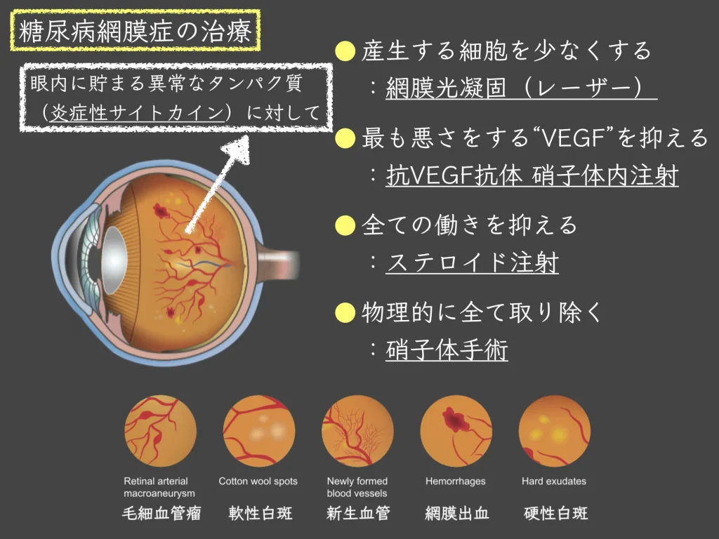
- Proteins called inflammatory cytokines are released and accumulate inside the eyeball, damaging blood vessels.
- This eventually causes “macular edema,” in which fluid collects in the center of the retina and vision declines.
A state of high blood sugar damages blood vessels from the inside.
(This is the result of complex immune reactions inside and outside the vessels.)
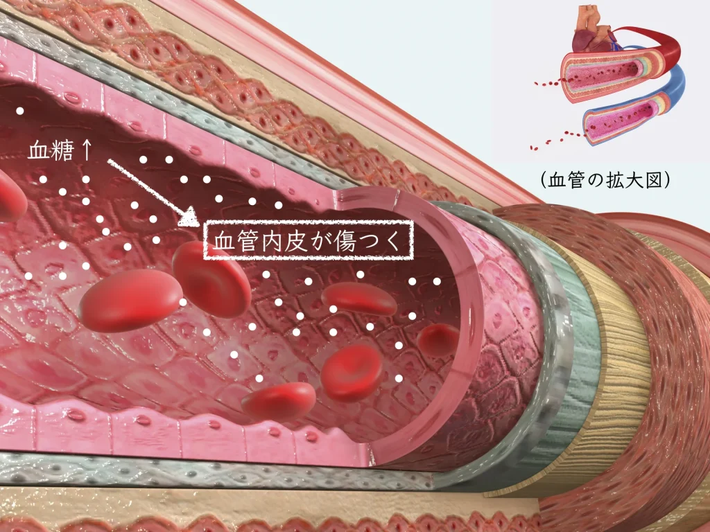
Starting with the thinnest vessels:
– the blood vessels of the retina
– the blood vessels of the kidney
– the blood vessels that supply the nerves
Besides the three major complications (retinopathy, nephropathy and neuropathy), diabetes causes complications throughout the body1
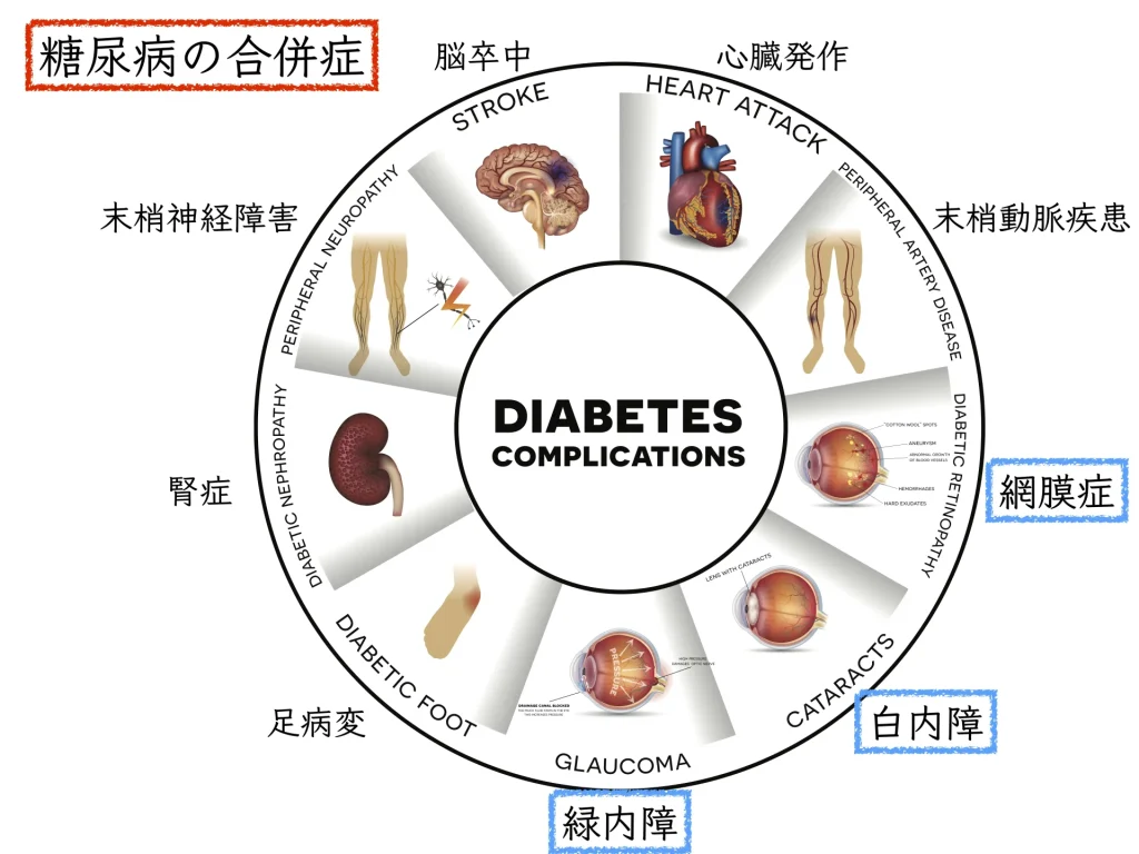

Diabetic retinopathy is the most feared complication of the eye.
Blood flow in the retina worsens: “ischemia”2
-1024x768.webp)
Depending on which part of the retina is damaged, some areas become hard to see.
(The image above is only an example.)
A retina that is not receiving enough nourishment becomes distressed and releases proteins called inflammatory cytokines into the eye.
The inside of the eye is a closed space: harmful proteins keep accumulating.
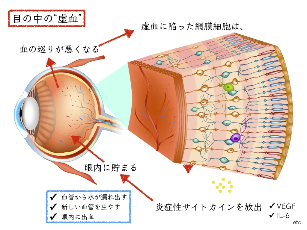
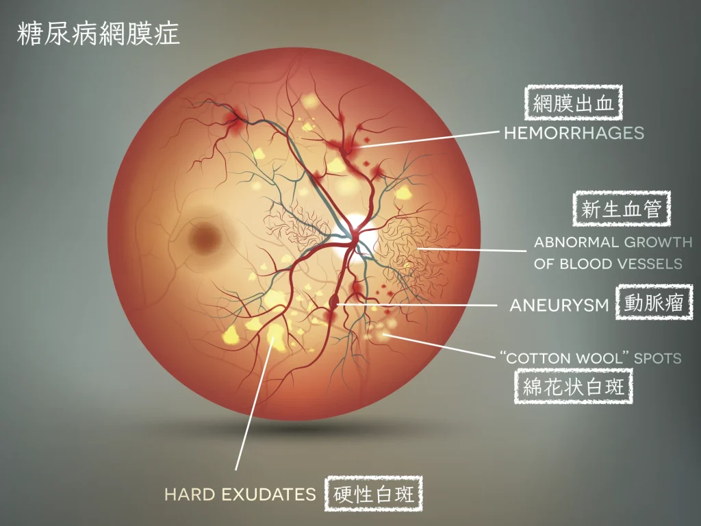
The walls of damaged vessels become fragile, and the watery part of the blood leaks out of them.
Fluid collecting in the macula, “macular edema”: a cause of reduced vision.
Causes and treatment of diabetic retinopathy: “High blood sugar causes inflammation inside the eye”

The role of inflammatory cytokines: two key factors in diabetic retinopathy
We found that, inside the eyeball (the vitreous cavity) in diabetic retinopathy, a number of inflammatory cytokines commonly accumulate.
This is a research result from my time in the Department of Ophthalmology at Kyushu University3.
VEGF (vascular endothelial growth factor)4
– Increases the permeability of blood vessels
A state in which fluid collects in the macula, the most important part for seeing: edema
– Makes new blood vessels grow
New vessels are fragile and cause bleeding inside the eye (vitreous hemorrhage).
IL-6 (interleukin-6)
IL-6is also an important factor linked to worsening of diabetic retinopathy.5,6
What is standard treatment? The Diabetic Retinopathy Practice Guideline
Standard treatment for diabetes can also help control the “cytokines” that cause inflammation.
* Standard treatment: treatment backed by large-scale scientific evidence
In ophthalmology, we provide care in line with theDiabetic Retinopathy Practice Guideline.7
The diagnosis of diabetic retinopathy must be made comprehensively, combining various tests in addition to fundus findings.
Diabetic Retinopathy Practice Guideline (1st edition). Nippon Ganka Gakkai Zasshi (Journal of the Japanese Ophthalmological Society) 124(12): 955-981, 2020
A standard eye examination with a slit-lamp microscope and a handheld condensing (preset) lens, and
imaging: care using OCT, OCTA and SLO.
Fundus examination without dilating the pupilhas become possible, and times have changed.
Keep inflammatory cytokines from being produced as much as possible: retinal photocoagulation (laser)
A treatment that burns the retina, the source of the inflammatory cytokines:
To preserve the macula, which is vital for seeing, the surrounding retina is “thinned out.”
Retina that has been physically burned no longer releases cytokines.
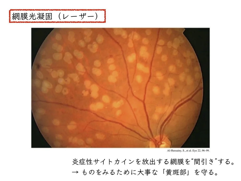
Inflammation also occurs when the laser is applied to the retina.
An inflamed eye can again develop macular edema.
This can therefore be a reason why vision drops during treatment.
Laser treatment is given a little at a time, with about a week between sessions for each eye.
Suppress all the actions of inflammatory cytokines: steroid treatment
Steroid injection: STTA (sub-Tenon triamcinolone acetonide injection)
A needle slipped behind the eyeball deposits a steroid suspension behind the eye.
The steroid soaks (sustained release) through the sclera, the wall of the eyeball, into the eye and suppresses all inflammation.
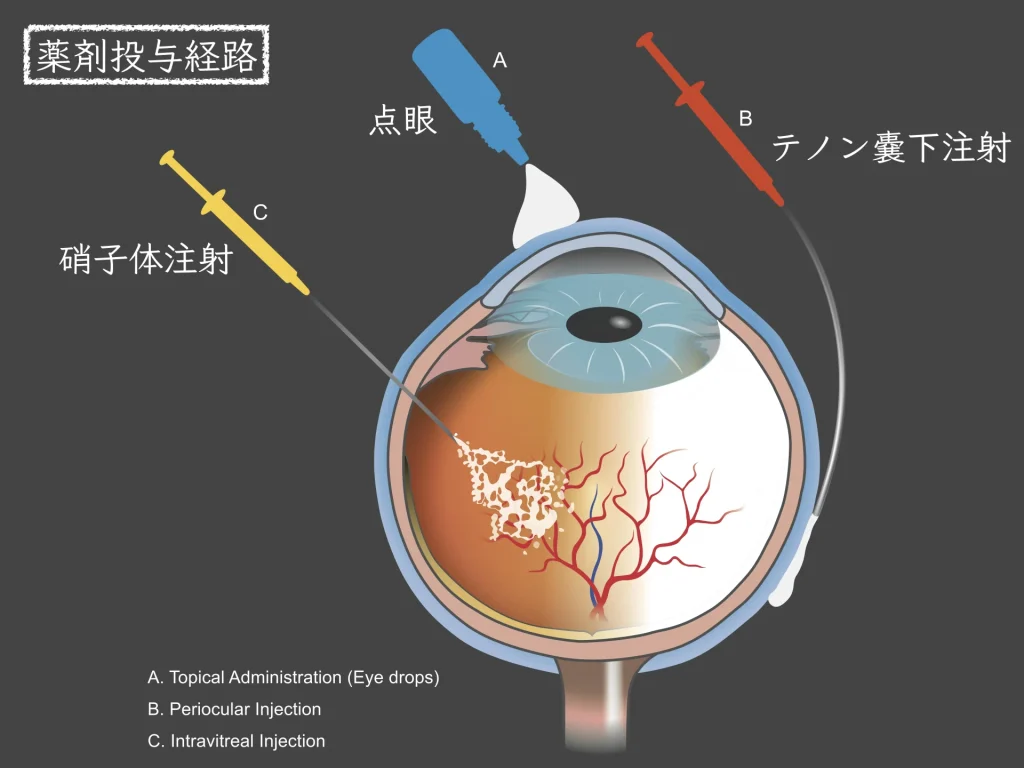
The production of inflammatory cytokines is suppressed.
After about three months, the effect wears off and the inflammation returns.
We watch for the eye complications of steroids: cataract and a rise in eye pressure (steroid-induced glaucoma).
There is also a steroid injection given inside the eye (intravitreal steroid injection).
Suppress the cytokine doing the most harm: anti-VEGF therapy
It is known that VEGF is the factor doing the most harm in the pathology of diabetic retinopathy.8
An antibody drug that neutralizes VEGF is injected directly into the vitreous cavity.
Here too, the inflammation returns within a few months.
The dramatic effect of anti-VEGF intravitreal injections surprised the world.
Repeated injections are needed,
and the complications of suppressing VEGF can no longer be overlooked.9
Physically remove all of the inflammatory cytokines and the “scaffold” they sit on: vitrectomy
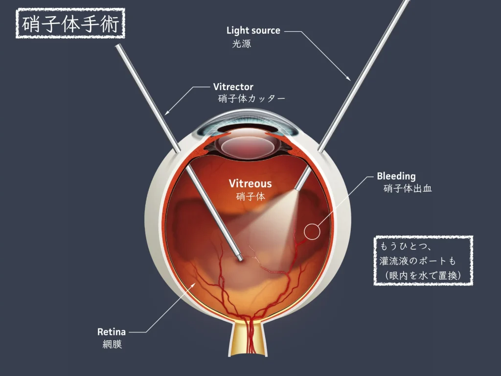
The gel inside the eye, the “vitreous,” is thoroughly removed.
Laser is also applied from inside the eye at that time.
Compared with laser applied from outside the eye, it can be delivered more effectively.
References
- Harding, J.L., Pavkov, M.E., Magliano, D.J., Shaw, J.E., Gregg, E.W., 2019. Global trends in diabetes complications: a review of current evidence. Diabetologia 62, 3–16. https://doi.org/10.1007/s00125-018-4711-2
- Bresnick, G.H., De Venecia, G., Myers, F.L., Harris, J.A., Davis, M.D., 1975. Retinal Ischemia in Diabetic Retinopathy. Archives of Ophthalmology 93, 1300–1310. https://doi.org/10.1001/archopht.1975.01010020934002
- Yoshimura, T., Sonoda, K.-H., Sugahara, M., Mochizuki, Y., Enaida, H., Oshima, Y., Ueno, A., Hata, Y., Yoshida, H., Ishibashi, T., 2009. Comprehensive Analysis of Inflammatory Immune Mediators in Vitreoretinal Diseases. PLoS ONE 4, e8158. https://doi.org/10.1371/journal.pone.0008158
- Antonetti, D.A., Silva, P.S., Stitt, A.W., 2021. Current understanding of the molecular and cellular pathology of diabetic retinopathy. Nat Rev Endocrinol 17, 195–206. https://doi.org/10.1038/s41574-020-00451-4
- Mason, R.H., Minaker, S.A., Lahaie Luna, G., Bapat, P., Farahvash, A., Garg, A., Bhambra, N., Muni, R.H., 2022. Changes in aqueous and vitreous inflammatory cytokine levels in proliferative diabetic retinopathy: a systematic review and meta-analysis. Eye 1–51. https://doi.org/10.1038/s41433-022-02127-x
- Yao, Y., Li, R., Du, J., Long, L., Li, X., Luo, N., 2019. Interleukin-6 and Diabetic Retinopathy: A Systematic Review and Meta-Analysis. Current Eye Research 44, 564–574. https://doi.org/10.1080/02713683.2019.1570274
- Diabetic Retinopathy Practice Guideline (1st edition). Nippon Ganka Gakkai Zasshi (Journal of the Japanese Ophthalmological Society) 124(12): 955-981, 2020
- Aiello, L.P., Avery, R.L., Arrigg, P.G., Keyt, B.A., Jampel, H.D., Shah, S.T., Pasquale, L.R., Thieme, H., Iwamoto, M.A., Park, J.E., 1994. Vascular endothelial growth factor in ocular fluid of patients with diabetic retinopathy and other retinal disorders. N Engl J Med 331, 1480–1487. https://doi.org/10.1056/NEJM199412013312203
- Chatziralli, I., Touhami, S., Cicinelli, M.V., Agapitou, C., Dimitriou, E., Theodossiadis, G., Theodossiadis, P., 2021. Disentangling the association between retinal non-perfusion and anti-VEGF agents in diabetic retinopathy. Eye 36. https://doi.org/10.1038/s41433-021-01750-4