![Takeru Eye Clinic | Takatori Shopping Street, Sawara-ku, Fukuoka City [Nishijin Station / Fujisaki Station]](https://takeru-eye.com/wp-content/uploads/2022/10/takeru_logo_for-WP-header.png)

“I was told I might have glaucoma (glaucoma suspect)”
“I was told I am pre-glaucoma or borderline for glaucoma”
“I was told I have a touch of glaucoma”
“I was told I have an enlarged optic disc cup, and when I searched online, it seemed to mean glaucoma.”
Depending on the condition, there is often nothing to worry about.
From a state with almost no glaucoma
to mild glaucoma
to advanced glaucoma
The word “glaucoma” spans a gradient.
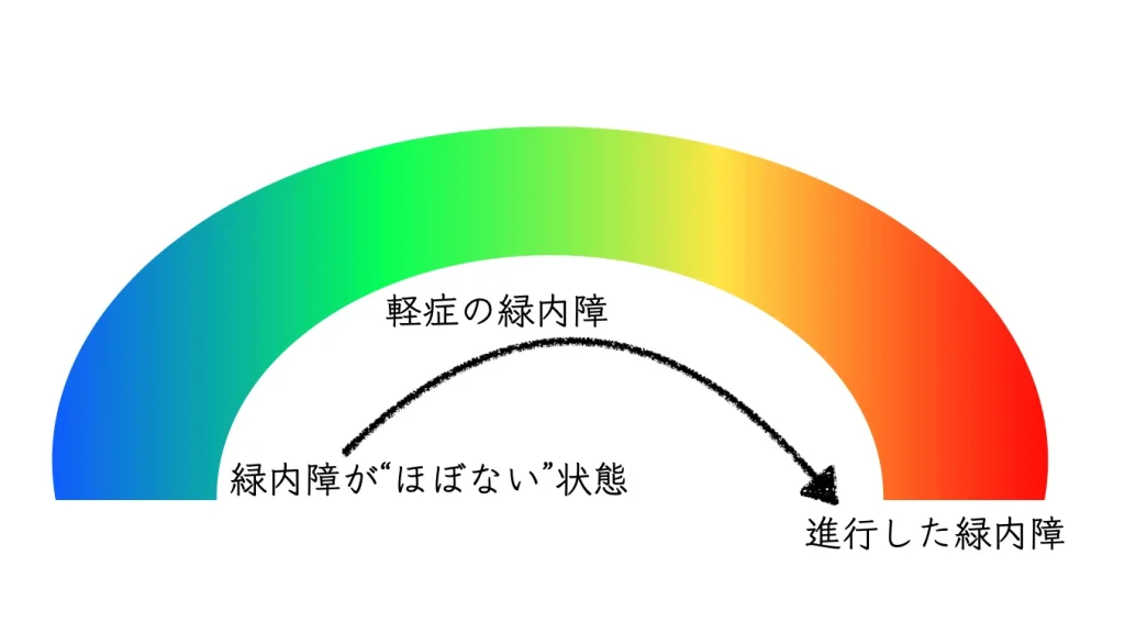
Whether there is truly nothing to worry about depends on two things:
- your current condition
- and your outlook going forward
In the clinic, I will talk with you about both.

The Broad Meaning of “Glaucoma”: A Gradient in the Word

Degeneration of the Retina and Optic Nerve: Changes in Form
• Image analysis to look at the shape of the nerves in the eye
: OCT, SLO, fundus photography, etc.
The retina is the part of the eye that corresponds to the film in a camera. We examine the state of the retina and optic nerve in detail.

This is a screening to check whether there are any abnormalities in the form of the retina and optic nerve.
Degeneration of the Retina and Optic Nerve: Changes in Function
When a structural abnormality is confirmed, a visual field test is performed to evaluate abnormalities in the function of the retina and optic nerve.
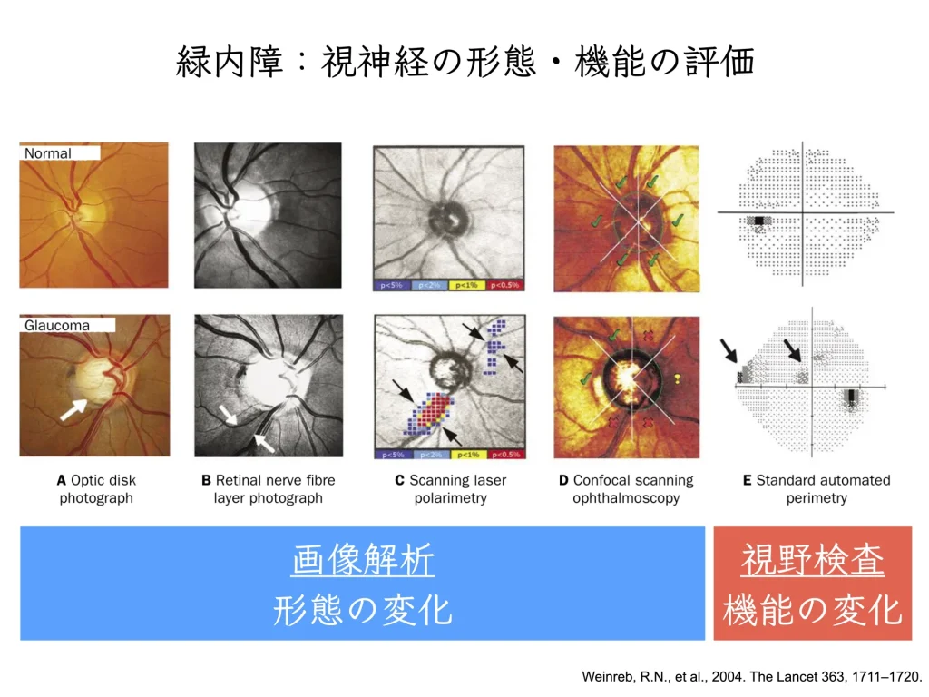
The figure above is reproduced from a figure in The Lancet (2004)1.
The visual field test is important for determining how well you can see various parts of your visual field.
This test helps confirm whether blind spots or reduced sensitivity (decreased sensitivity) are present.
It is also useful for evaluating nerve damage and investigating the cause of reduced vision.
With advances in imaging, we are now in an era in which even smaller changes can be detected.
Changes in form and changes in function can be shown side by side.
Glaucoma continuum
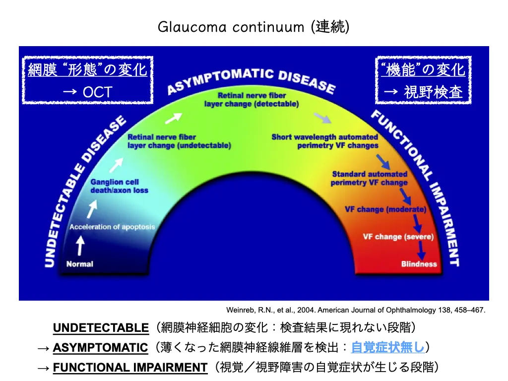
At the point when changes in form are captured by image analysis:
it is thought that neurodegeneration had already been progressing in the invisible world (the microscopic world)1.
If left alone, neurodegeneration progresses further.

After some time has passed, enlargement of the abnormality on OCT images (progression of the structural change) appears in the test results.

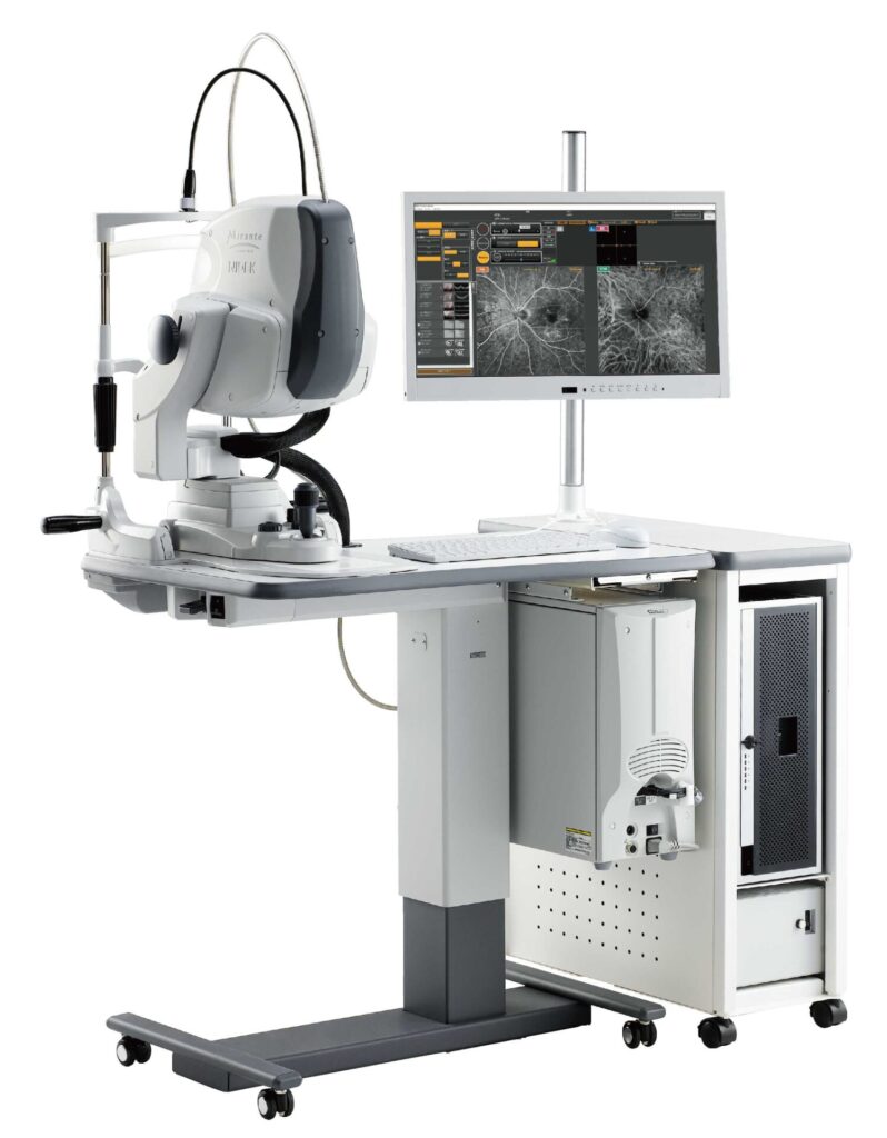
Multimodal imaging
Multiple images are used to evaluate the form of the retina and optic nerve from multiple angles.
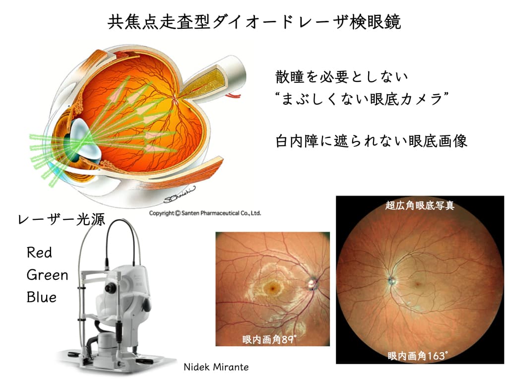
Confocal scanning laser ophthalmoscope (Mirante®)
: findings are displayed as a combination of various images.
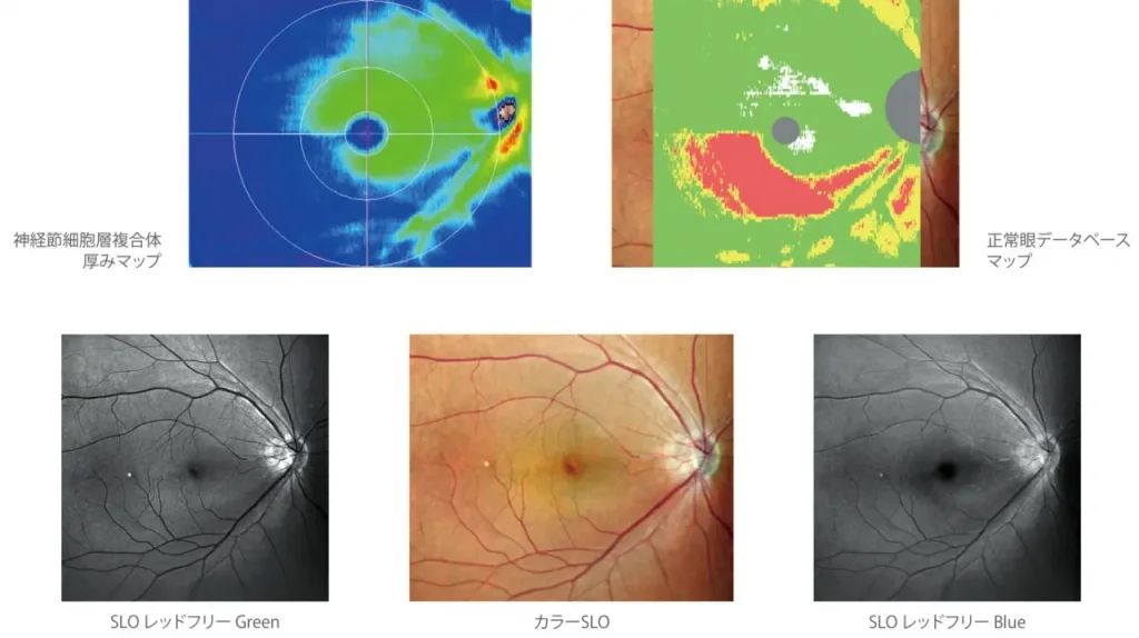
Detecting abnormalities of the retina and optic nerve at an early stage
Since many patients present in the early stages of the disease, the goal of treatment is to arrest, delay, or limit progression of predisposing ocular hypertension or early optic nerve damage to significant visual impairment.1
Weinreb, R.N., et al., 2004. American Journal of Ophthalmology 138, 458–467.
For Each Person: Grasp and Fully Understand Your Current Glaucoma Status
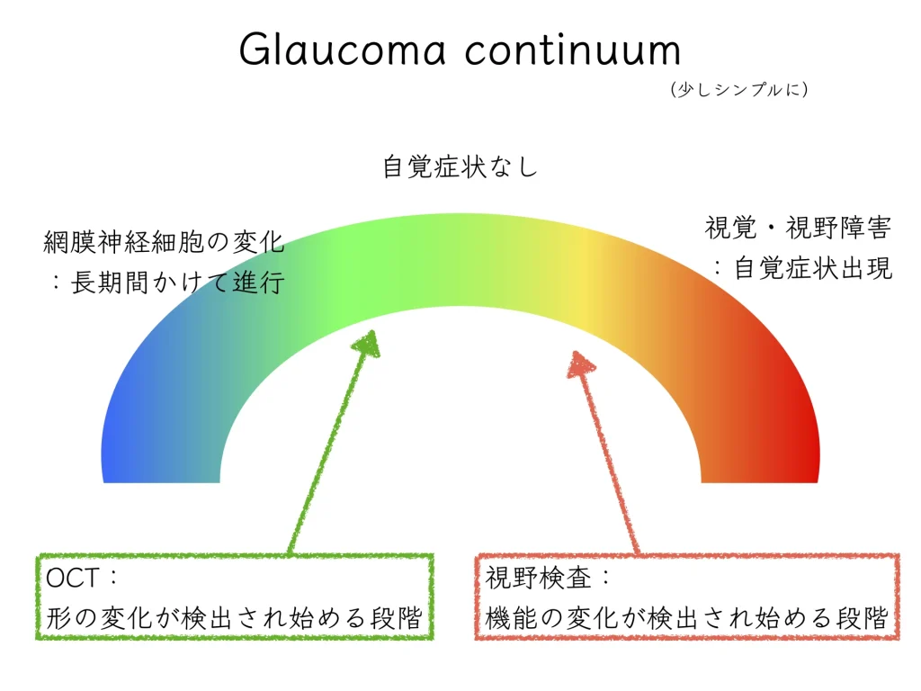
“Glaucoma” is a single word, but it is a word with a gradient.
When searching for medical information, it is difficult to arrive at information suited to the individual.
If you come across information about the worse end, it can lead to excessive worry and anxiety.
Medical devices have continued to advance compared with the past.
• Your current condition
• Your outlook going forward
I place importance on explaining these and on helping you understand them.
References
- Weinreb, R.N., Khaw, P.T., 2004. Primary open-angle glaucoma. The Lancet 363, 1711–1720. https://doi.org/10.1016/S0140-6736(04)16257-0
- Weinreb, R.N., Friedman, D.S., Fechtner, R.D., Cioffi, G.A., Coleman, A.L., Girkin, C.A., Liebmann, J.M., Singh, K., Wilson, M.R., Wilson, R., Kannel, W.B., 2004. Risk assessment in the management of patients with ocular hypertension. American Journal of Ophthalmology 138, 458–467. https://doi.org/10.1016/j.ajo.2004.04.054