![Takeru Eye Clinic | Takatori Shopping Street, Sawara-ku, Fukuoka City [Nishijin Station / Fujisaki Station]](https://takeru-eye.com/wp-content/uploads/2022/10/takeru_logo_for-WP-header.png)

“My blood tests say my liver values are fine. So drinking is okay, isn’t it?”
Alcohol may affect the optic nerve through pathways that liver function tests cannot evaluate.
It has become clear that alcohol can affect the optic nerve, an extension of the brain, in ways that eye pressure alone cannot explain1,2.
A large Korean observational study reported an association in which people who stopped drinking after being diagnosed with glaucoma had a lower risk of severe visual impairment and blindness than those who kept drinking3.
Not only the total amount consumed but also the pattern of “how one drinks” may be involved in the fate of the optic nerve.
This article explains this from the mechanisms to regional differences.
The More Alcohol, the More Association with Eye Pressure, Glaucoma and Thinning of Retinal Nerves
Current research reports a tendency for greater alcohol intake to be associated with higher eye pressure, glaucoma, and thinning of the retinal nerve1,2.
In a large Korean observational study of people diagnosed with glaucoma, those who stopped drinking after diagnosis had a lower risk of severe visual impairment and blindness than those who kept drinking3. However, because this is an observational study, it has not been proven that stopping alcohol prevents progression.
At present, it is not known what amount of drinking is “safe for the optic nerve.” For people with glaucoma, especially those whose OCT or visual field is trending worse, it is worth considering reducing how much and how often they drink.
Glaucoma Is Also a “Brain Disease Inside the Eye”
Glaucoma is known as a disease in which the optic nerve is damaged by factors such as eye pressure.
However, the picture of glaucoma that recent research has revealed is far more complex than this.
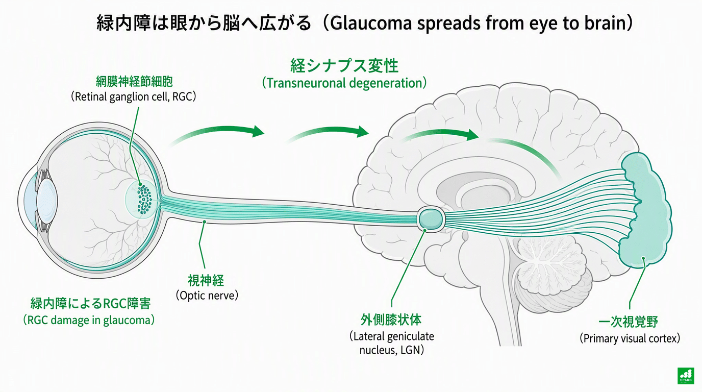
Retinal ganglion cells (RGCs) on the inner side of the retina are the cells whose axons (nerve fibers) bundle together to form the optic nerve. They are genuine central nervous system cells, derived during development from the brain (diencephalon)4.
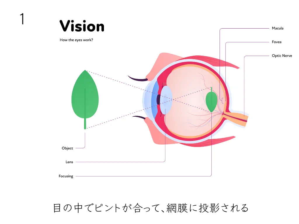
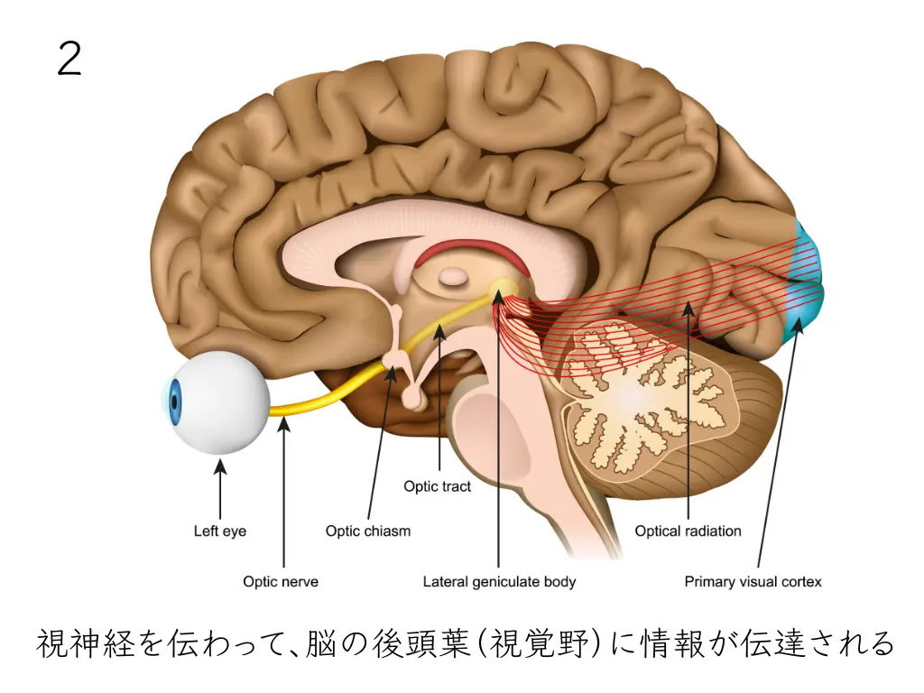
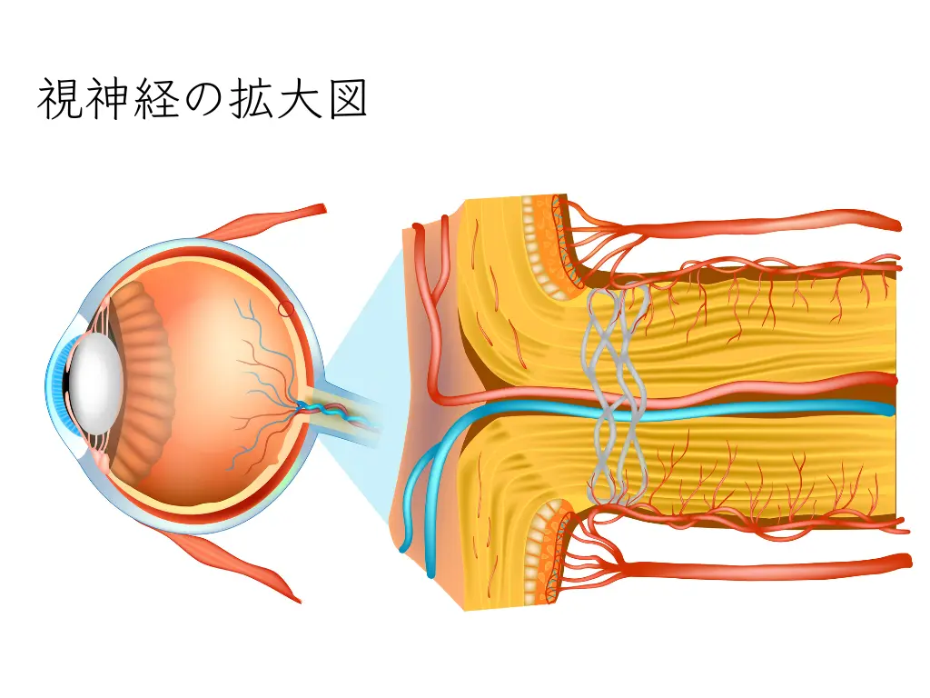
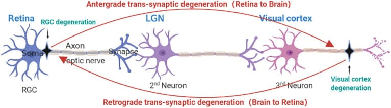
When RGCs are lost, the relay nucleus in the brain that received their signals, the lateral geniculate nucleus (LGN), also gradually atrophies. A chain called “transneuronal degeneration,” in which degeneration spreads further to the primary visual cortex (occipital lobe), has been reported in animal models and in human imaging and autopsy studies4.
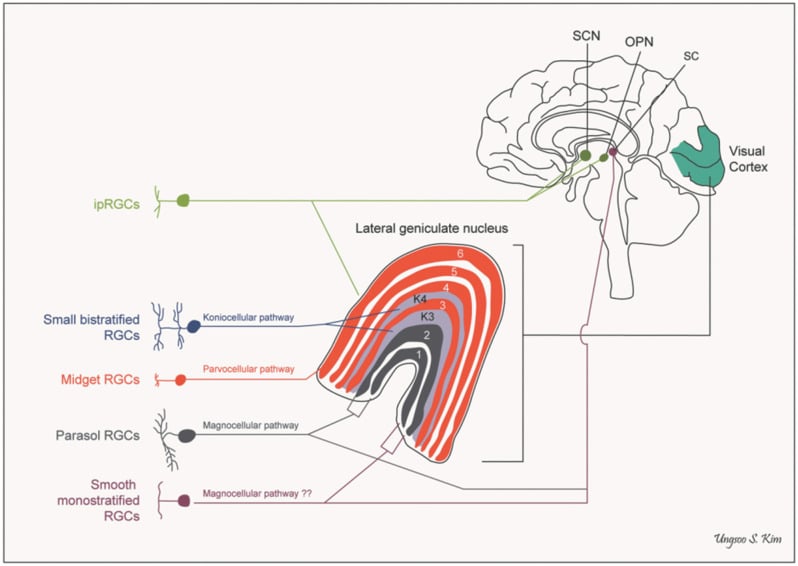
In other words, glaucoma is now thought to have aspects of a neurodegenerative disease, in which changes that start in the eye can spread to the brain.
How Alcohol Damages Nerves
The mechanisms described from here on are leading hypotheses based mainly on cell and animal experiments. Associations in humans are shown later with large-scale data.
It is well known that the brains of heavy drinkers tend to atrophy. It was once explained as “shrinking from dehydration,” but it is now understood as loss and degeneration of nerve cells caused by multifaceted cytotoxicity5.
First, there isdirect toxicity from acetaldehyde and reactive oxygen species (ROS).
Alcohol is metabolized not only in the liver but also in the brain. In the brain the enzyme catalase mainly handles its breakdown, but with chronic heavy drinking the enzyme CYP2E1 increases and generates large amounts of reactive oxygen species during breakdown. These reactive oxygen species cause oxidative damage to lipids, proteins and DNA of nerve cells5.
Next ismitochondrial dysfunction.
Mitochondria are the organelles that act as the cell’s power plants, and when they are damaged, production of energy (ATP) falls. Nerve cells, which consume a great deal of energy, are more vulnerable to this effect5.
Third ischronic neuroinflammation. Microglia (the immune cells of the brain) are activated and continue to release inflammatory cytokines, so neurodegeneration advances little by little5.
Fourth issuppression of the mechanisms that protect cells. It has been reported that chronic drinking suppresses the NRF2 defense system, which protects cells from oxidative stress, and autophagy, which clears away damaged components5.
In addition, the effect on brain-derived neurotrophic factor (BDNF), which supports nerve cell survival, is also being studied, mainly in animal experiments. There are reports that BDNF signaling decreases with chronic alcohol exposure6.
These Overlap with the Mechanisms of RGC Death in Glaucoma
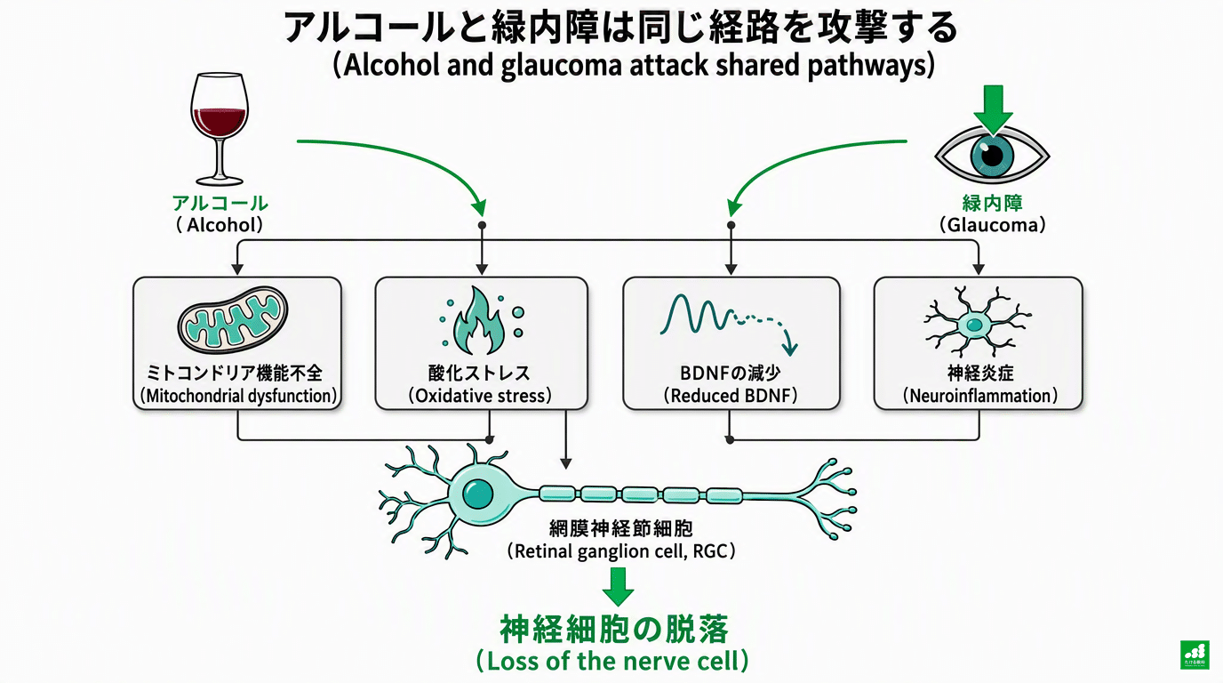
The mechanisms listed above largely overlap with the process by which RGCs die in glaucoma.
In other words, alcohol and glaucoma attack the same “weak points” in similar ways.
| Shared pathway of damage | Effect of alcohol | Effect in glaucoma |
|---|---|---|
| Mitochondrial dysfunction | Decreased ATP production | Energy depletion and apoptosis of RGCs |
| Oxidative stress | Production of reactive oxygen species via CYP2E1 | Excess reactive oxygen species due to eye pressure and ischemia |
| Neuroinflammation | Microglial activation | Microglial activation and inflammation of the optic nerve |
| Reduced BDNF signaling | Reported to decrease with chronic exposure | Shortage of trophic signals due to impaired axonal transport |
| Glutamate excitotoxicity | Rebound excitation after heavy drinking | RGC death from excitotoxicity within the retina |
* This table is a schematic summary based on research reports, including cell and animal experiments4,5,6. The degree to which the evidence is established differs between items.
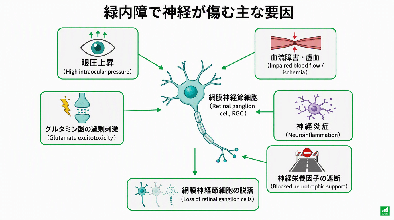
Looking only at eye pressure means overlooking the burden on the optic nerve from drinking that is separate from eye pressure.
Even now, the only target whose effectiveness in glaucoma treatment is established is “eye pressure.”
Eye drops, laser and surgery are all treatments to lower eye pressure. Treatments that act directly on neuroinflammation or oxidative stress such as those listed above have not yet been put into practice.
As one of the few factors besides eye pressure that patients can change themselves, there is value in reviewing how you drink.
Loss of Nerve Cells Seen on OCT: “The Result After Drinking”
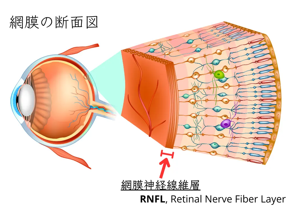
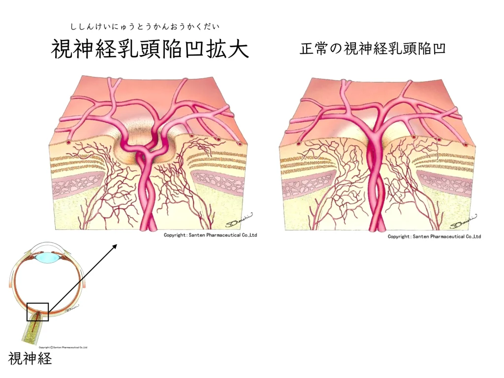
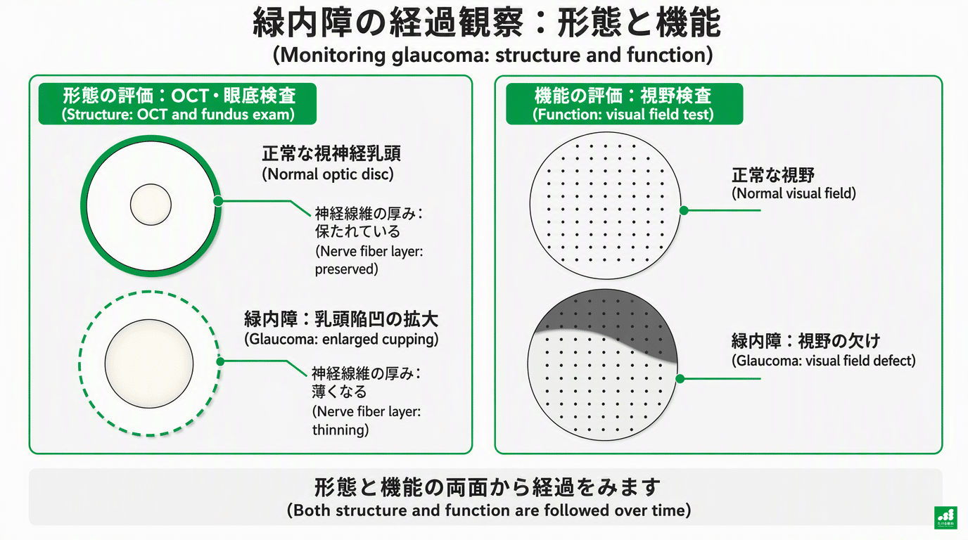
Optical coherence tomography (OCT), used for follow-up of glaucoma, is a test that can measure the thickness of the retinal nerve fiber layer (RNFL) and the ganglion cell–inner plexiform layer (GCIPL) in micrometers.
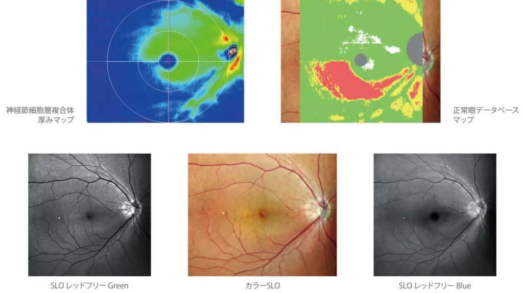
In a study of 445 Korean glaucoma patients, the peripapillary RNFL of the heavy-drinking group was significantly thinner than that of the abstaining group (65.0 μm vs 70.9 μm).
The macular GCIPL was also significantly thinner in both the mild-drinking group (65.8 μm) and the heavy-drinking group (63.8 μm) than in the abstaining group (68.1 μm)7.
A large study using the UK Biobank in the United Kingdom (OCT analysis of about 36,000 people) reported that an association with thinning of the macular GCIPL and RNFL appears once drinking exceeds about 50 g of pure alcohol per week. Below that amount no clear association was seen, and the picture changes beyond a certain amount. The study calls this a “threshold effect”1.
Regular brain MRI is not realistic in terms of cost and effort, but OCT can be done in a few minutes in the clinic with little burden.
For glaucoma patients with drinking habits, OCT provides a way to objectively track changes in the retina and optic nerve.
How Much Is Risky? Guidelines from Large-Scale Data
How much drinking raises the risk?
The association of drinking with eye pressure and open-angle glaucoma has also been confirmed in a systematic review and meta-analysis that pooled multiple studies8.
In the same UK Biobank study (Stuart et al. 2023), the group that drank the most had 1.36 times higher odds of glaucoma than the group that drank the least1. Fifty grams of pure alcohol per week corresponds to a little over 2 gō of sake (1 gō = 180 mL) or about 2.5 medium bottles (500 mL) of beer.
In a study using the U.S. NIH’s diverse population database (All of Us) (Meller et al. 2025), people who drank four or more drinks per week had 1.22 times the odds (a 22% increase) of glaucoma (primary open-angle glaucoma), and this dose–response relationship was more pronounced in women2.
Drinking Patterns Change How Disease Appears

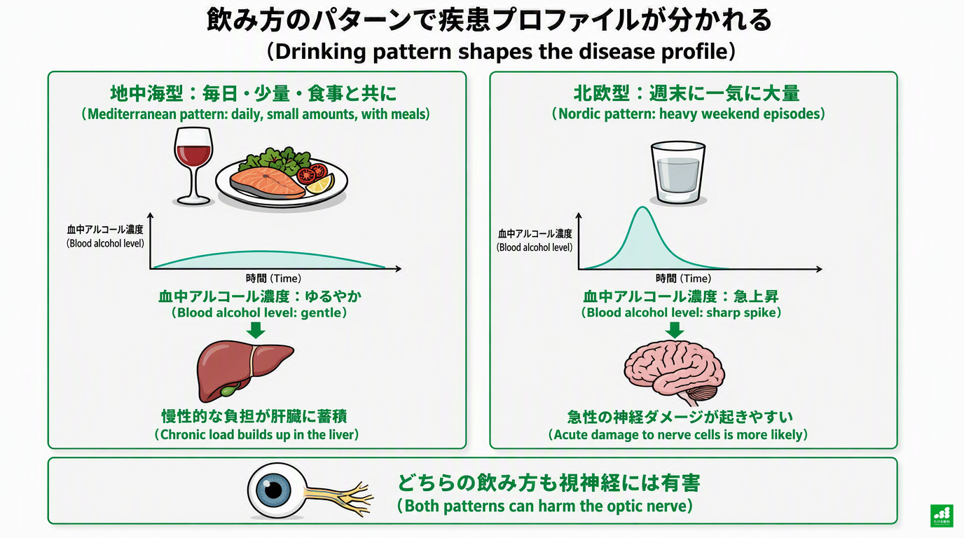
The health effects of alcohol are thought to depend not only on the total amount but also on “how one drinks”9.
In the Mediterranean region (Spain, Italy and elsewhere), the traditional style is to drink a small amount of wine with meals every day.
In epidemiological reviews, the Mediterranean pattern of drinking a small amount slowly with food and avoiding binge drinking is supported as a relatively less harmful option for people who currently drink9. Drinking with food slows gastric emptying and suppresses a sharp rise in blood alcohol concentration.
In contrast, it has been pointed out that in Northern and Eastern Europe, a style of not drinking on weekdays and drinking large amounts of spirits at once on weekends is relatively common.
A comparative study of 17 European countries showed that alcohol-related mortality, including alcoholic psychosis, dependence and cirrhosis, is at a high level in Northern and Eastern Europe10. Heavy drinking in a short time rapidly raises blood alcohol concentration and places a large acute burden on the nervous system.
Japan Also Has a “North–South Difference”
This pattern is reproduced to some extent within Japan as well.
A nationwide survey of the causes of liver cirrhosis in Japan (Enomoto et al. 2024) named three regions, Hokkaido, Tohoku and Kyushu, as having a particularly high proportion of alcoholic cirrhosis11. In a study using the national claims (receipt) database, the standardized claim ratio (SCR) for liver cancer was also the highest in Kyushu nationwide, with a median of 13412(Regional differences in liver cancer also involve factors other than drinking, such as viral hepatitis).
Kyushu has a traditional shochu culture, and a habit of drinking a small amount slowly every day is also seen. Drinking every day in this way is also a style in which the weekly total easily adds up.
The Ohsaki Study, which followed about 23,000 Japanese men, reported an association in which suicide risk rose linearly with daily alcohol intake (hazard ratio 2.4 at 45.6 g or more per day), showing how great the effect of heavy drinking on the nervous system and mental state can be13.
With any style of drinking, the burden grows as the total amount and frequency increase. There may be differences in how the burden arises depending on drinking pattern, but they cannot be asserted as regional differences within Japan9,10.
People Who “Quit” After Diagnosis Had Lower Risk
A study using Korean national health insurance data (Jeong et al. 2023) followed the drinking patterns after new diagnosis of glaucoma (open-angle glaucoma) in people with drinking habits, and their subsequent visual outcomes. The subjects were 13,643 people3.
| Drinking pattern after diagnosis | Risk of blindness or severe visual impairment (abstainers as reference) |
|---|---|
| Quit drinking (21%) | Reference (37% lower risk than those who kept drinking: AHR 0.63) |
| Continued: 3 days a week or fewer | AHR 1.42 (95% CI 0.97-2.08, not significant but a rising trend) |
| Continued: 4 or more days a week | AHR 2.56 (95% CI 1.52-4.33, significantly higher) |
* Source: reference 3. AHR = adjusted hazard ratio.
The lowest risk was in people who quit drinking at the time of diagnosis. Even among those who continued, no statistically significant increase in risk was confirmed for those drinking 3 days a week or fewer.
However, a tendency toward higher risk remained. In the analysis based on amount, a significant increase in risk was seen even with small amounts (less than 105 g per week)3.
There seems to be no need to be pessimistic and think that “you must not drink a single drop once diagnosed.”
Still, in this data, the lowest risk was in those who abstained.
If quitting is difficult, a realistic first step is to stop the “almost every day” habit and lower the frequency and amount3.
This study is observational, and drinking amounts are self-reported. Other lifestyle habits of those who quit may also have influenced the results, so it has not been proven that “quitting prevents progression”3.
A Point Specific to Japanese People: ALDH2 Gene Variant
About 40% of Japanese people carry a gene variant (rs671) that weakens the activity of aldehyde dehydrogenase 2 (ALDH2), the enzyme that processes the breakdown product of alcohol.
These are the so-called “people whose faces turn red when they drink”14,15.
In people with this constitution, highly toxic acetaldehyde tends to stay in the blood at high concentrations for longer.
On the other hand, in the Korean study of 445 people mentioned earlier, the ALDH2 genotype itself did not show a significant association with RNFL or GCIPL thickness.
Regardless of genotype, heavy drinking was associated with GCIPL thinning7. Rather than “whether you are the type whose face turns red,” what matters is managing the amount and frequency of drinking itself.
You may also hear people say, “I don’t turn red, so I’m fine,” or “I turn red, but I’ve trained myself and become stronger.”
When a flushing reaction occurs, it is a sign that acetaldehyde is accumulating in the body.
It is not good to force yourself to keep drinking14,15.
Practical Guidelines for Drinking in Kyushu

In Fukuoka and elsewhere in Kyushu, there is a shochu tradition of “drinking with food, slowly, and diluted.” In the sense of avoiding a rapid rise in blood alcohol concentration, this way of drinking is itself reasonable.
On top of that, here are guidelines for amount and frequency as numbers.
In the UK Biobank study, an association with thinning of the retinal nerve became noticeable beyond about 50 g of pure alcohol per week1. However, this is a boundary suggested by observational research, and it is not a standard meaning that “50 g or less is safe” or that “the optic nerve is protected.”
Here is only a conversion to help you grasp the amounts. For 25% shochu, 1 gō (180 mL) is about 36 g of pure alcohol, and one glass of shochu with hot water or water (0.5 gō) is about 18 g.
Please use this as reference information when thinking about a target for cutting down.
A habit of drinking “every day without fail” tends to add up the weekly total even if each serving is small, and in terms of frequency it matches the pattern with the highest risk in research (4 or more days a week)3. Start by increasing the number of days you do not drink.
Summary: What You Can Do Starting Today
With some thought, enjoying alcohol and protecting your optic nerve can be reconciled.
In the Korean study, the lowest risk was in those who quit drinking3. If quitting is difficult, reducing both the amount and the frequency and increasing the number of days you do not drink is a realistic step. Even at 3 days a week or fewer, safety has not been confirmed.
At present, it is not known what amount of drinking is “safe for the optic nerve.” The 50 g per week at which associations began to stand out in research (a little over 2 gō of sake, or about 2.5 medium bottles of beer) is reference information for thinking about a target for cutting down1.
When OCT results are trending worse, it may be an opportunity to look back not only at eye pressure but also at drinking habits.
I hope you can use your regular tests as an opportunity to review your lifestyle as a whole.
The statement “my liver numbers are normal, so drinking is fine” does not guarantee the safety of the optic nerve. The brain and the eye are made of nerve of the same origin, and depending on how you drink, that nerve may be under continuous strain.
Once the optic nerve is lost, it does not regenerate.
To keep good vision and visual field for a long time, reviewing how you drink a little is, though quiet, an important option.

References
- Stuart KV, Madjedi K, Luben RN, et al. The Association of Alcohol Consumption With Glaucoma and Related Traits: Findings From the UK Biobank. Ophthalmol Glaucoma. 2023;6(4):366-379. https://doi.org/10.1016/j.ogla.2022.11.008
- Meller LLT, Saseendrakumar BR, Mahmoudinezhad G, et al. Association Between Alcohol Use and Primary Open Angle Glaucoma. J Glaucoma. 2025;34(2):69-76. https://doi.org/10.1097/IJG.0000000000002529
- Jeong Y, Kim SH, Kang G, Yoon HJ, Kim YK, Ha A. Visual Impairment Risk After Alcohol Abstinence in Patients With Newly Diagnosed Open-Angle Glaucoma. JAMA Netw Open. 2023;6(10):e2338600. https://doi.org/10.1001/jamanetworkopen.2023.38600
- You M, Rong R, Zeng Z, Xia X, Ji D. Transneuronal Degeneration in the Brain During Glaucoma. Front Aging Neurosci. 2021;13:643685. https://doi.org/10.3389/fnagi.2021.643685
- Ruiter-Lopez L, Khan MAS, Wang X, Song BJ. Roles of Oxidative Stress and Autophagy in Alcohol-Mediated Brain Damage. Antioxidants (Basel). 2025;14(3):302. https://doi.org/10.3390/antiox14030302
- Davis MI. Ethanol-BDNF interactions: still more questions than answers. Pharmacol Ther. 2008;118(1):36-57. https://doi.org/10.1016/j.pharmthera.2008.01.003
- Han YS, Kim YW, Kim YJ, Park KH, Jeoung JW. Alcohol consumption is associated with glaucoma severity regardless of ALDH2 polymorphism. Sci Rep. 2020;10(1):17422. https://doi.org/10.1038/s41598-020-74470-6
- Stuart KV, Madjedi K, Luben RN, et al. Alcohol, Intraocular Pressure, and Open-Angle Glaucoma: A Systematic Review and Meta-analysis. Ophthalmology. 2022;129(6):637-652. https://doi.org/10.1016/j.ophtha.2022.01.023
- Barbería-Latasa M, Gea A, Martínez-González MA. Alcohol, Drinking Pattern, and Chronic Disease. Nutrients. 2022;14(9):1954. https://doi.org/10.3390/nu14091954
- Mackenbach JP, Kulhánová I, Bopp M, et al. Inequalities in Alcohol-Related Mortality in 17 European Countries: A Retrospective Analysis of Mortality Registers. PLoS Med. 2015;12(12):e1001909. https://doi.org/10.1371/journal.pmed.1001909
- Enomoto H, Akuta N, Hikita H, et al. Etiological changes of liver cirrhosis and hepatocellular carcinoma-complicated liver cirrhosis in Japan: Updated nationwide survey from 2018 to 2021. Hepatol Res. 2024;54(8):763-772. https://doi.org/10.1111/hepr.14047
- Nakano M, Kawaguchi M, Nakano D, et al. Regional Difference of Liver Cancer in Japan: A Real-world Evidence Using the National Database of Health Insurance Claims. Intern Med. 2025;64(15):2263-2269. https://doi.org/10.2169/internalmedicine.4820-24
- Nakaya N, Kikuchi N, Shimazu T, et al. Alcohol consumption and suicide mortality among Japanese men: the Ohsaki Study. Alcohol. 2007;41(7):503-510. https://doi.org/10.1016/j.alcohol.2007.08.001
- Chen CH, Kraemer BR, Mochly-Rosen D. ALDH2 variance in disease and populations. Dis Model Mech. 2022;15(6):dmm049601. https://doi.org/10.1242/dmm.049601
- Chen CH, Ferreira JCB, Gross ER, Mochly-Rosen D. Targeting aldehyde dehydrogenase 2: new therapeutic opportunities. Physiol Rev. 2014;94(1):1-34. https://doi.org/10.1152/physrev.00017.2013