![Takeru Eye Clinic | Takatori Shopping Street, Sawara-ku, Fukuoka City [Nishijin Station / Fujisaki Station]](https://takeru-eye.com/wp-content/uploads/2022/10/takeru_logo_for-WP-header.png)

Many parents must feel anxious as their child’s myopia progresses year after year.
Others may be worried about the future health of their own eyes because their own myopia is strong.
It is easy to think, “It’s fine because I can see with glasses or contact lenses,” but in fact the progression of myopia is not just a matter of visual acuity.
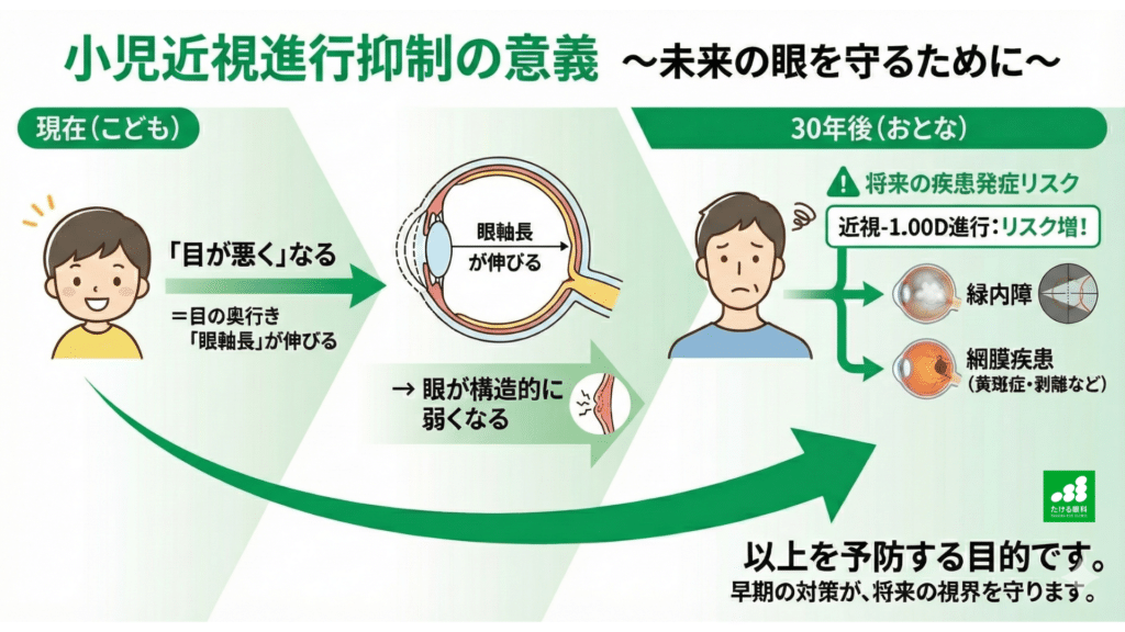
Each time myopia progresses and the glasses prescription increases by 1 diopter (D),
it has been shown that the risk of eye diseases that can cause serious visual impairment later in life rises by a statistically significant amount.
“Eyesight getting worse” = the axial lengthincreases = the eyeball becomes structurally weaker
→ Complications of myopia later in life: what prevents them is “myopia control in children』
In this article, I explain in detail, based on scientific data, just how much those risks rise.
Over many years as an ophthalmologist, I have seen many patients with diseases that can seriously affect visual function, such as glaucoma, retinal detachment, and myopic maculopathy.
And through daily practice I strongly feel that the progression of myopia since childhood is deeply involved in the background of these diseases.
When I see a child’s myopia progressing now, as an ophthalmologist I cannot help predicting the path that child may follow in the future.
The current refractive error and the length of the eye (axial length) are important clues for judging what risks a person may face later on.
Common sense in ophthalmology is changing, and an era has arrived in which myopia in children can be controlled.
I believe it is important to convey, as early as possible, that myopia is not just a problem of visual acuity but an important issue that also affects future eye health.
We have introduced a new device (July 17, 2026)
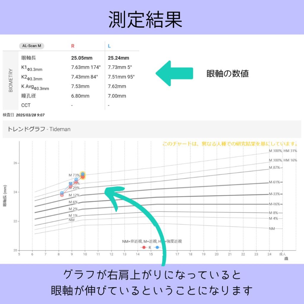
We have switched to the new measuring device “OA-1,” which shows how many millimeters the axial length grows per year (the speed of progression) as a slope and lets us compare it with the percentile curves of Japanese children (ages 4 to 15).
Why does the risk of eye disease rise as myopia progresses?
First, it is important to understand how myopia progresses.
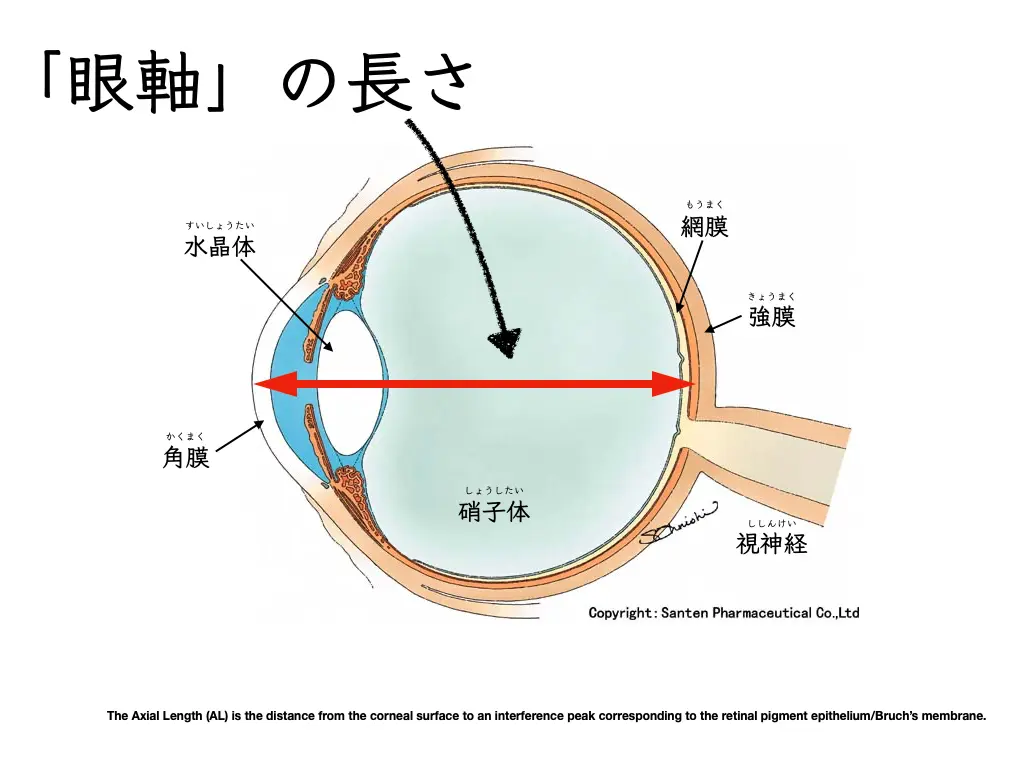
In most cases, myopia progresses as the depth of the eyeball, called the “axial length,” increases.
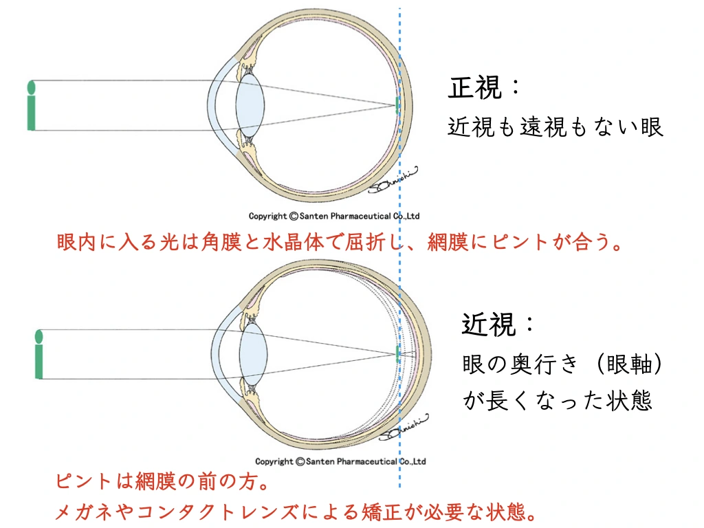
Think of the eyeball as a balloon.
When you put too much air into a balloon and inflate it greatly, the rubber becomes thin and weak.
In the same way,
as the eyeball stretches, the delicate tissues at the back of the eye, such as the retina and the optic nerve, are also stretched, and the mechanical strain makes them structurally fragile.
This “stretched and weakened” state is the root cause of the various diseases that can develop later.
So what diseases are at increased risk, and by how much?
Degree of myopia and the risk of major eye diseases
Multiple reliable studies have shown a clear association between the degree of myopia and the risk of various eye diseases.
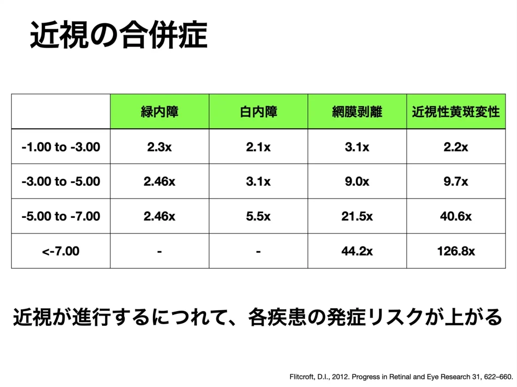
The table below summarizes the main diseases for which the progression of myopia may affect future vision.
| Disease | Increase in risk per −1.00 D of progression (approximate) | Approximate risk by degree of myopia (compared with emmetropia or mild myopia) |
|---|---|---|
| Myopic maculopathy (MMD) | About 1.67 -fold¹ | High myopia (over −6.00 D): about 40 -fold² |
| Retinal detachment (RRD) | — | Moderate or higher (over −3.00 D): about 10 -fold² |
| Glaucoma (POAG) | — | Low myopia: about 1.6-fold / moderate to high: about 2.9 -fold⁴ |
Here are the three diseases that deserve particular attention.
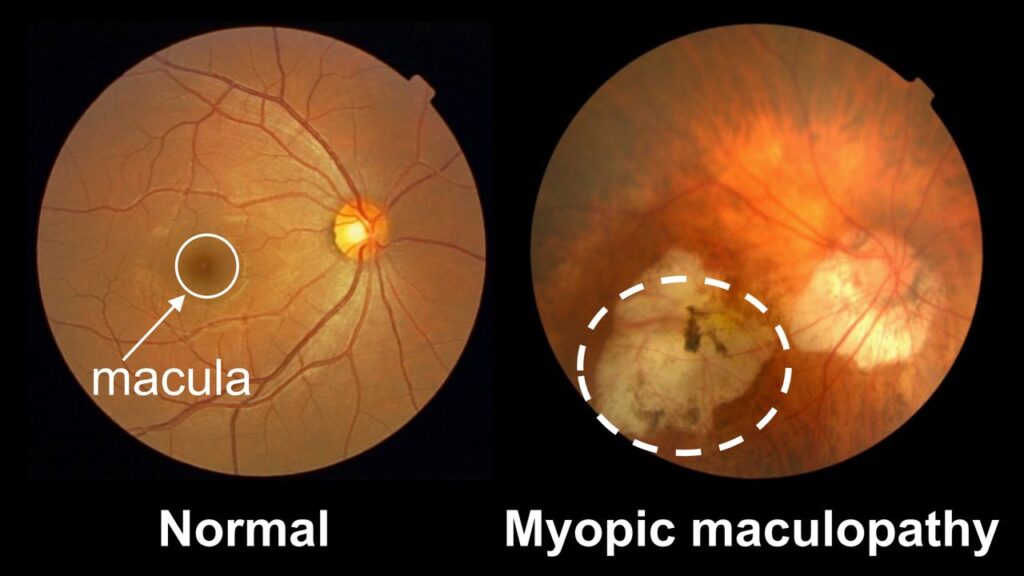
Myopic maculopathy
A disease in which the tissue called the “macula,” which is responsible for central vision, is damaged as the eyeball stretches.
Once it develops, the center of vision becomes distorted or dark, leading to vision loss that is difficult to recover.
One study reported that each −1.00 D of myopia progression raises the risk (odds ratio) of developing myopic maculopathy about 1.67-fold¹. In addition, the risk does not simply increase in proportion to the degree of myopia.
For example, compared with mild myopia of less than −3.00 D, the risk is about 10 times higher in moderate myopia of −3.00 D to −6.00 D and rises sharply to about 40 times in high myopia beyond −6.00 D².
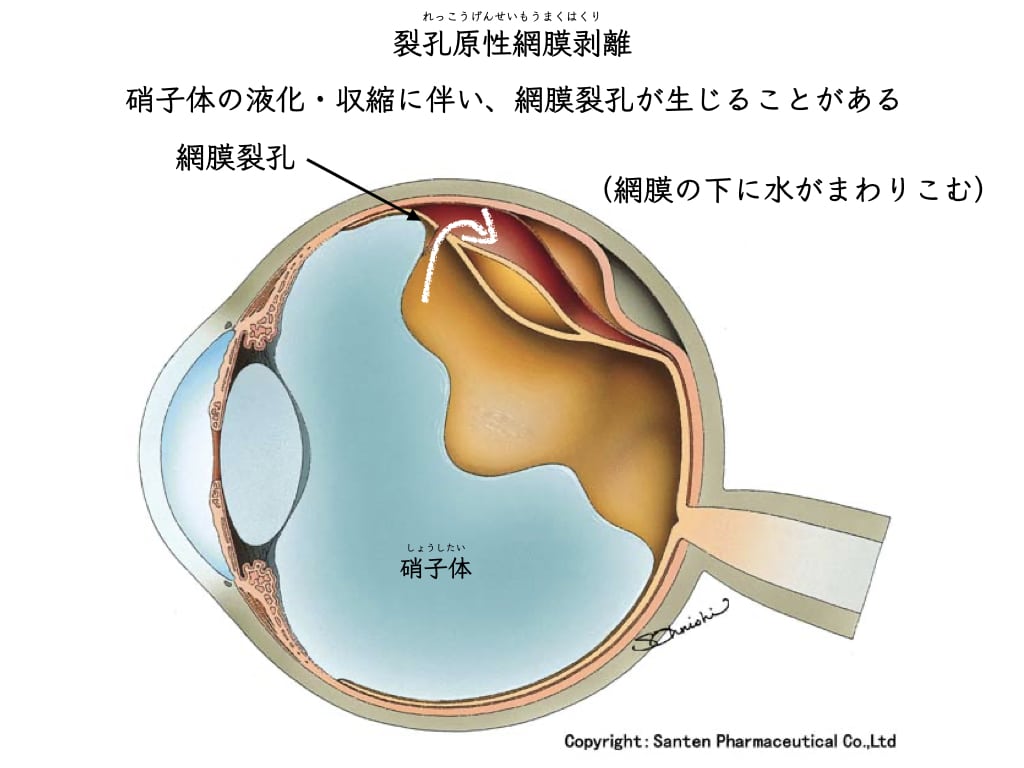
Rhegmatogenous Retinal Detachment (RRD)
A disease in which a hole (retinal tear) opens in the stretched, thinned retina and the retina detaches from there.
It starts with a sudden increase infloatersor a visual field defect, requires emergency surgery, and can have a permanent effect on visual function.

By degree of myopia, it has been reported that the risk is about 3 to 4 times higher in mild myopia (−1.00 D to −3.00 D) and about 10 times higher in myopia beyond −3.00 D².
Surveys in Japan show that retinal detachment peaks not only in the elderly but also in young people in their twenties³.
This suggests that the progression of myopia since childhood can lead to serious consequences at a relatively young age.
Primary Open-Angle Glaucoma (POAG)
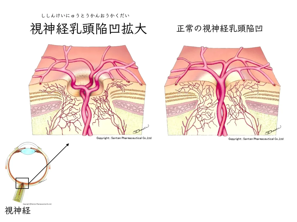
Glaucoma is a disease in which the optic nerve is damaged and the visual field gradually narrows, and it is one of the leading causes of visual impairment in Japan.
Intraocular pressure (the firmness of the eye) is the main risk factor, but myopia is also an important independent risk factor.
As the eyeball stretches, structural strain is thought to be placed on the base of the optic nerve, making it more easily damaged.
An analysis combining multiple studies has shown that the stronger the myopia, the higher the risk of glaucoma⁴.
It is also reported that, compared with emmetropia (no myopia), people with mild myopia have about 2.3 times the risk, and the risk rises to about 2.9 times in moderate to high myopia⁴.
In Japan in particular, the great majority of glaucoma is “normal-tension glaucoma,” in which glaucoma progresses even though intraocular pressure is within the normal range.
A large survey in Japan found glaucoma in about 5.8% of people aged 40 and over⁵, and given this high prevalence, the added risk from myopia cannot be ignored.
Axial length: another important measure
So far I have explained things in terms of the glasses prescription (diopters), but what reflects the risk more directly is the length of the eyeball itself, the “axial length.”
Recent studies report that for every 1 mm increase in axial length, the risk of glaucoma rises about 1.4-fold⁵ and the risk of myopic maculopathy about 1.9-fold⁶, showing how important structural changes of the eye are.
In particular, it is known that when the axial length exceeds 26 mm, the risk of many diseases rises sharply. For this reason, in eye care we have come to place importance on measuring a child’s axial length and on how to slow its growth.
Summary: slowing myopia progression is an important preparation for future eye health
“Myopia progressing by −1.00 D” does not simply mean that the lenses of the glasses become a little thicker.
It means thatthe eyes you will use for the rest of your life are moving in a direction in which they become more prone to structural weakness.The risks of these diseases:
- myopic maculopathy
- retinal detachment
- glaucoma
increase steadily with every diopter of myopia progression.
It is also known that the risk rises sharply once the degree of myopia passes a certain level.
Slowing the progression of myopia from childhood has a long-term meaning as “preventive medicine”: not only keeping good vision, but protecting the eyes from serious eye diseases in the future.
If you have worries or questions about myopia progression, it is important to see an eye doctor regularly and receive examinations and advice from a specialist.
References
- Bullimore MA, Brennan NA. Myopia Control: Why Each Diopter Matters. Optom Vis Sci. 2019 Jun;96(6):463-465. doi: 10.1097/OPX.0000000000001367. PMID: 31116165.(https://pubmed.ncbi.nlm.nih.gov/31116165/)
- Flitcroft DI. The complex interactions of retinal, optical and environmental factors in myopia aetiology. Prog Retin Eye Res. 2012 Nov;31(6):622-60. doi: 10.1016/j.preteyeres.2012.06.004. Epub 2012 Jul 4. PMID: 22772022.(https://pubmed.ncbi.nlm.nih.gov/22772022/)
- Haga A, et al. The Incidence of Rhegmatogenous Retinal Detachment in Kumamoto, Japan, Between 2009 and 2011. J Clin Exp Ophthalmol. 2015;6(3):428. (https://www.longdom.org/open-access/the-incidence-of-rhegmatogenous-retinal-detachment-in-kumamoto-japan-between-2009-and-2011-51639.html)
- Haarman AEG, Enthoven CA, Tideman JWL, Tedja MS, Verhoeven VJM, Klaver CCW. The Complications of Myopia: A Review and Meta-Analysis. Invest Ophthalmol Vis Sci. 2020 Apr 9;61(4):49. doi: 10.1167/iovs.61.4.49. PMID: 32347918; PMCID: PMC7401976. (https://iovs.arvojournals.org/article.aspx?articleid=2765517)
- Fujiwara K, Yasuda M, Hata J, Nakano S, Hashimoto S, Ueda E, Nakamura S, Murakami Y, Nakamuro T, Iwase A, Araie M, Tawara A, Kubota T, Yoshitomi T, Ninomiya T, Sonoda KH. Prevalence of Glaucoma and Its Systemic Risk Factors in a General Japanese Population: The Hisayama Study. Transl Vis Sci Technol. 2022 Nov 1;11(11):11. doi: 10.1167/tvst.11.11.11. PMID: 36378141; PMCID: PMC9672895.(https://pmc.ncbi.nlm.nih.gov/articles/PMC9672895/)
- Mu T, He HL, Chen XY, Fang YX, Xu J, Jin ZB. Axial length as a predictor of myopic macular degeneration: a meta-analysis and clinical study. Eye (Lond). 2025 Jul;39(10):2073-2082. doi: 10.1038/s41433-025-03782-6. Epub 2025 Apr 25. PMID: 40281120; PMCID: PMC12209455. (https://pubmed.ncbi.nlm.nih.gov/40281120/)
- Iwase A, Suzuki Y, Araie M, Yamamoto T, Abe H, Shirato S, Kuwayama Y, Mishima HK, Shimizu H, Tomita G, Inoue Y, Kitazawa Y; Tajimi Study Group, Japan Glaucoma Society. The prevalence of primary open-angle glaucoma in Japanese: the Tajimi Study. Ophthalmology. 2004 Sep;111(9):1641-8. doi: 10.1016/j.ophtha.2004.03.029. PMID: 15350316. (https://pubmed.ncbi.nlm.nih.gov/15350316/)
- Tideman JW, Snabel MC, Tedja MS, van Rijn GA, Wong KT, Kuijpers RW, Vingerling JR, Hofman A, Buitendijk GH, Keunen JE, Boon CJ, Geerards AJ, Luyten GP, Verhoeven VJ, Klaver CC. Association of Axial Length With Risk of Uncorrectable Visual Impairment for Europeans With Myopia. JAMA Ophthalmol. 2016 Dec 1;134(12):1355-1363. doi: 10.1001/jamaophthalmol.2016.4009. PMID: 27768171.(https://pubmed.ncbi.nlm.nih.gov/27768171/)
- Ueda E, Yasuda M, Fujiwara K, Hashimoto S, Ohno-Matsui K, Hata J, Ishibashi T, Ninomiya T, Sonoda KH. Trends in the Prevalence of Myopia and Myopic Maculopathy in a Japanese Population: The Hisayama Study. Invest Ophthalmol Vis Sci. 2019 Jul 1;60(8):2781-2786. doi: 10.1167/iovs.19-26580. PMID: 31260519.(https://iovs.arvojournals.org/article.aspx?articleid=2737593)
- Marcus MW, de Vries MM, Junoy Montolio FG, Jansonius NM. Myopia as a risk factor for open-angle glaucoma: a systematic review and meta-analysis. Ophthalmology. 2011 Oct;118(10):1989-1994.e2. doi: 10.1016/j.ophtha.2011.03.012. PMID: 21684603.(https://pubmed.ncbi.nlm.nih.gov/21684603/)