![Takeru Eye Clinic | Takatori Shopping Street, Sawara-ku, Fukuoka City [Nishijin Station / Fujisaki Station]](https://takeru-eye.com/wp-content/uploads/2022/10/takeru_logo_for-WP-header.png)

“I’ve been going to the eye doctor for years, but every time all I hear is ‘nothing has changed.’ I don’t really understand what hasn’t changed, and it worries me.”
I sometimes hear this from patients who have been told they have superior segmental optic hypoplasia (SSOH).
SSOH looks very much like glaucoma, but it arises differently: it is an inborn shape of the optic nerve.
Often there is not enough time or material to explain SSOH fully, and I feel it is a diagnosis that easily leaves people thinking, “I felt like I understood, but once I got home, I still didn’t.”
In this article, I explain as simply as I can, starting from how the optic nerve is built, what SSOH is and why people keep hearing “nothing has changed.”
What This Article Covers (Summary)
SSOH is a condition in which part of the optic nerve is present in smaller numbers from birth. It generally does not progress and is thought to be closer to an individual trait of body shape, like height, finger length or hair texture.
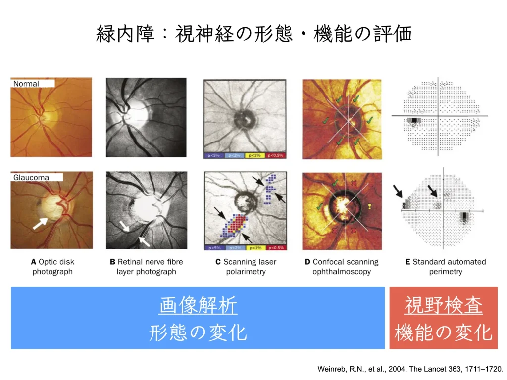
It is confusing with glaucoma because fundus photographs and OCT show the same finding of “a thin nerve.” However, the location of the thinning, and whether it changes over time, differ.
For people with SSOH, “nothing has changed” is good news. It means that no clear worsening has been found in the shape of the optic nerve or the visual field compared with earlier tests. However, because absence of change alone cannot rule out glaucoma, I judge it together with other findings.
What the Optic Nerve Is For, and What Shape It Has

The retina at the back of the eye corresponds to the film in a camera. The retinal cells that receive light convert the information into electrical signals and send them to the brain.
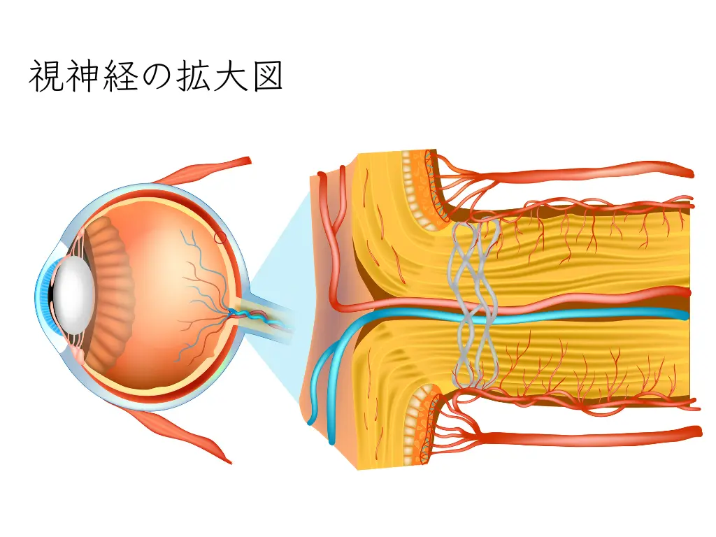
The “cable” that carries those signals is the optic nerve.
The optic nerve is not one thick wire but a bundle of thin nerve fibers (axons). A study that counted fibers under the microscope in human optic nerves reported an average of about 1.16 million, about 820,000 in people with fewer and about 1.5 million in people with more1.
Here I will share an analogy I often use in the exam room.
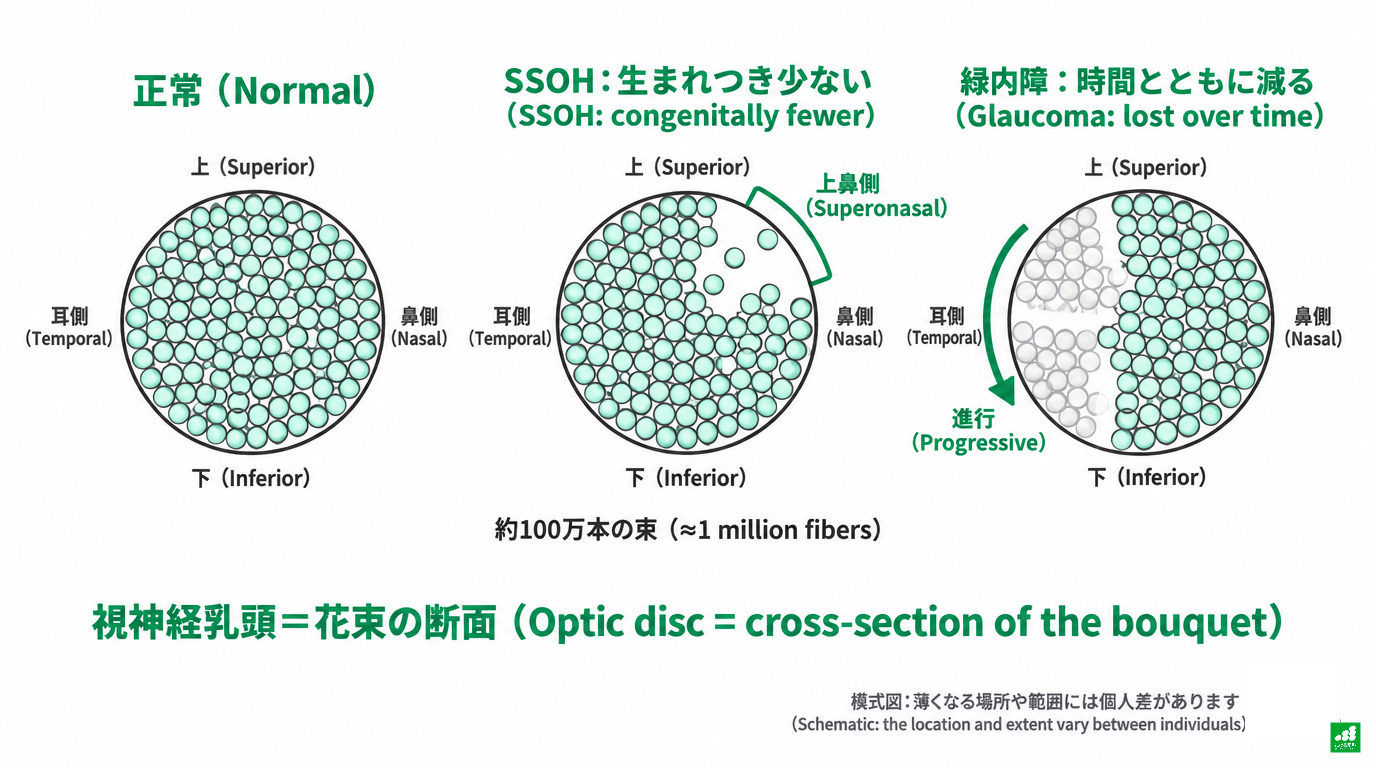
The optic nerve is like a bouquet of about one million roses.
Fibers from the upper half of the retina are bundled neatly on the upper side of the bouquet, and fibers from the lower half on the lower side. The “optic disc” you see when you look into the back of the eye is the cross-section of this bouquet.
It is known that the number of optic nerve fibers varies greatly between individuals from the start1,2. Eyes with larger optic discs tend to have more fibers2. In studies comparing people of different ages, the age-related loss is estimated at roughly 4,000 to 5,400 fibers per year1,2. The figure “one million” is only a rough guide.
What SSOH Is: The “Upper Corner” of the Bouquet Has Fewer Stems from Birth
SSOH is a condition in which the number of fibers in the upper part of this bouquet (mostly the section called superonasal) is smaller from birth.
“Hypoplasia” means a condition related to development from birth, rather than a later change like that of glaucoma. It is easy to picture it as having been formed with fewer fibers from the start.
At the back of the eye, the upper half of the retina handles the lower half of the visual field. The relationship is upside down.
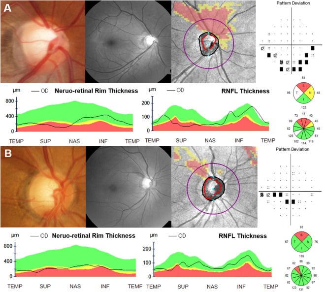
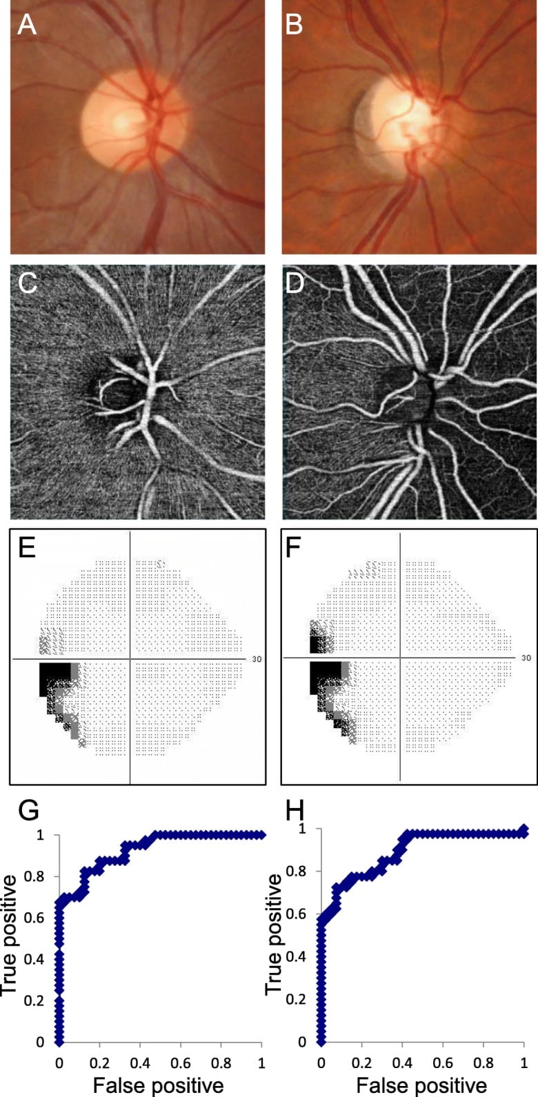
Therefore, in SSOH, where the upper side of the bouquet has fewer fibers, a “hard-to-see area” may appear in the lower part of the visual field test. A wedge-shaped defect connected to the blind spot (Mariotte blind spot) is typical3,4.
On the other hand, fibers leading to the center of the retina that sees fine detail (the macula) are often preserved, and visual acuity is almost always normal4,5. Few people notice any difficulty seeing in daily life, and it is often found by chance at health checkups or visits for other reasons.3.
The History of SSOH: Children of Mothers with Diabetes
The name SSOH has a bit of history.
In 1977, Petersen and Walton in the United States reported a common feature in 17 children born to mothers with diabetes: good vision, but hypoplasia of part of the optic disc with a corresponding visual field defect6. This was the first report to link optic nerve hypoplasia to a possible cause.
In 1989, Kim et al. examined 10 people (20 eyes) with diabetic mothers in detail and gave the condition the name SSOH7. They also summarized four features of the disc (the central retinal artery enters from a higher position, the upper disc is pale, there is a pale halo along the upper edge, and the upper nerve fiber layer is thin).
In 1998, Landau et al. in Switzerland prospectively examined 34 children born to mothers with type 1 diabetes and found SSOH in 3 (8.8%)8. In this report, because the upper part of the disc looks missing, it was also called a “topless disc.”
In Japan, SSOH That Looks “Just Like Glaucoma” Is Common
In Japan, the frequency of SSOH was reported in 2004 based on a large-scale eye examination conducted in Tajimi City, Gifu Prefecture, in 2000–20013,4.
Among 14,431 people aged 40 and older with evaluable fundus photographs, SSOH was found in 37 (about 0.3%)3. In this survey, that is about 1 in 400 people. It is by no means rare.

Dr. Tetsuya Yamamoto, who led this survey, made an important point about SSOH in Japanese people. In the Japanese SSOH he encountered in practice, there was neither the “pale upper disc” feature seen in Western reports nor a history of maternal diabetes, and some cases showed findings resembling glaucoma4.
In other words, in Japan SSOH first became known as a “condition easily mistaken for glaucoma.”
Most glaucoma in Japanese people is normal-tension glaucoma (NTG), in which the optic nerve is damaged even though eye pressure is within the normal range. In SSOH as well, eye pressure is often normal, part of the disc is thin, and visual field defects may be seen. Because they share so much, it is a diagnosis that must always be distinguished in glaucoma care4.

How Is It Different from Glaucoma? “Location” and “Time”
Even though both mean “the nerve is thin,” SSOH and glaucoma differ in two ways.

The first is the location of thinning.
In glaucoma, damage often starts from the inferotemporal (lower outer) or superotemporal (upper outer) part of the disc, and visual field defects spread from the upper and lower nasal areas.
In SSOH, at the superonasal part of the disc (upper inner, closer to the nose), the rim (the edge of the disc where nerve fibers gather) is thin, the nerve fiber layer at the same location is thin, and the visual field shows a wedge-shaped defect in the lower field connected to the blind spot4.
When the thickness of the nerve fiber layer around the disc is measured with OCT (optical coherence tomography), it has been reported that in SSOH it is thin mainly from the upper to the superonasal side, while the temporal fibers leading to the macula are relatively preserved5,9,10.

In addition, in a Japanese study that examined disc cross-sections in detail, in all 20 SSOH eyes examined, the retinal pigment epithelium was seen extending over the edge of the disc11.
In a study using the HRT, a device that examines disc shape, 6 of 13 SSOH eyes were classified as “outside normal limits” by one of the criteria used to detect glaucoma. Shape features centered on the superonasal area were also seen, so it is important not to decide on glaucoma from automated classification alone12.
The second is whether it changes over time.
Glaucoma is, in most cases, a progressive disease in which, without treatment, optic nerve fibers gradually decrease and visual field defects widen.
Because SSOH is an inborn shape, it is thought not to progress in principle. In a report from Niigata University that followed young patients for an average of a little over 8 years, there was no change in the visual field defects13.
There are also reports of long-term follow-up from Japan14. Furthermore, in the study by Abe et al., “a non-progressive visual field defect” is included in the diagnostic criteria for SSOH9.
| Item | SSOH | Glaucoma (especially NTG) |
|---|---|---|
| Origin | Inborn shape (congenital) | Disease that develops later (acquired) |
| Location of thinning | Often most noticeable in the upper to superonasal area | Often most noticeable in the inferotemporal and superotemporal areas |
| Visual field defect | Wedge-shaped in the lower field, often connected to the blind spot | Nasal step, arcuate defects, etc. |
| Change over time | Basically non-progressive for changes due to SSOH | May progress; speed varies between individuals |
| Visual acuity | Often preserved | Tends to be preserved early on, but may decline once the center is affected |
| Treatment | SSOH alone usually does not need pressure-lowering treatment; follow-up | Pressure-lowering treatment, etc., considered according to the condition |
* Sources: Yamamoto 2019, Unoki 2002, Takagi 20084,5,13. There are exceptions in individual patients.
Why Do People Keep Hearing “Nothing Has Changed”?
Having read this far, I think the meaning of “nothing has changed” becomes clear.
What people with SSOH are checking at the eye clinic is “whether this time’s bouquet is the same as last time’s.”
Eye pressure, disc photographs, OCT nerve fiber layer thickness, the visual field. If these have not clearly changed compared with last time, the inborn shape is being maintained as it is, and no clear progression is found in this round of tests.
So for SSOH, “nothing has changed” is good news.
Even so, there is a reason for continued visits. Cases in which people with SSOH also developed glaucoma have been reported15. It cannot be said that “because you have SSOH, you will never get glaucoma.” It has also been discussed that individual differences in the number of optic nerve fibers may relate to the “reserve” when disease occurs. However, this has not been directly confirmed in people with SSOH2.
That is why taking photographs and OCT about once a year and confirming “it’s the same shape this year, too” is meaningful.

At our clinic, I explain while showing a graph of OCT-measured nerve fiber layer thickness arranged over time. What matters is not that it is “thin” but that “the thinness has not changed,” and a graph often conveys this better than words alone. If you would like to compare with your previous data, please let me know at your visit.
Relationship with Maternal Diabetes: What Is Known and What Is Not
Explanations of SSOH always bring up “the mother’s diabetes.” I write about this carefully so that readers do not needlessly blame themselves or their families.
First, what is known.
The early Western reports found SSOH while examining children born to mothers with diabetes6,7,8. In the Korea National Health and Nutrition Examination Survey (5,612 people), 6 of 14 people with SSOH had a maternal history of diabetes, compared with 428 of 5,598 people without SSOH. Comparing the two, a statistical association between SSOH and maternal diabetes was confirmed16.
Next, what is not known.
In Japan, SSOH has also been reported in people whose mothers had no history of diabetes4,13. Studies that showed an association were also cross-sectional surveys or case series, and the mechanism of “how blood sugar during pregnancy affects optic nerve development” remains at the hypothesis stage8,16.
In the study by Landau et al., even with maternal diabetes, 31 of the 34 children born had no SSOH8. It does not always occur when diabetes is present, nor does it never occur when it is absent.
A 2022 review also positions maternal diabetes as a “major risk factor,” while concluding that the exact pathophysiology of SSOH is “still not elucidated”17.
In summary, maternal diabetes is “one factor reported to be associated with SSOH” and is not at the stage where it can be called the cause. Whether or not your mother had diabetes, I believe there is no need to think that something you or your family did is the cause.
There is still much we do not know about how SSOH comes about. No prevention specific to SSOH, and no standard treatment that increases the optic nerve fibers one has from birth, has been established. I believe there is no need to connect the cause to your own or your family’s responsibility.
What I Look at in an Actual Exam
In examining SSOH, I combine the following tests to confirm the “shape” and the “absence of change.”
| Test | What is looked at |
|---|---|
| Fundus photograph | Thinness of the rim in the upper to superonasal part of the disc, how the vessels enter, dark bands of the nerve fiber layer |
| OCT | Nerve fiber layer thickness around the disc (a pattern in which the upper to superonasal side is thin and the temporal side is relatively preserved), and comparison with the previous exam |
| Visual field test | Presence of a wedge-shaped defect in the lower field connected to the blind spot, and comparison with the previous exam |
| Eye pressure | The eye pressure value and its changes so far (glaucoma cannot be ruled out even when pressure is in the normal range) |
When I first judge that it is SSOH, I may repeat the same tests after a little interval to confirm whether there is any change, in order to make sure it is not glaucoma. It takes time until the diagnosis is confirmed because I am being careful.
Once it is judged to be SSOH only, eye drops or other treatment is in principle not necessary. For many people, a check about once a year becomes the form of follow-up. The interval between visits is decided in discussion, according to test results and whether glaucoma is also present.
Our Approach at Takeru Eye Clinic (Takatori Shopping Street, Sawara-ku, Fukuoka)
At our clinic, I try to tell people with SSOH the following.
The optic nerve is a bouquet of about one million roses, and the upper corner has slightly fewer from birth. This is closer to an individual trait of body shape than to a disease, and SSOH itself is thought not to progress in principle. And “nothing has changed” is good news that the shape is being maintained.
At our clinic, for people whose condition is stable, about once a year is a guideline for checking the course with fundus photographs, OCT and, when needed, visual field tests. The content and interval of tests are decided according to the possibility of coexisting glaucoma and previous results.
What I Want to Tell Patients
For people with SSOH, being told “nothing has changed” again and again is good news: regular tests have confirmed that there is no change.
The shape of the bouquet you were born with is part of your own body. Checking it every year and being able to say “it’s the same this year, too” is, I believe, how to live with this diagnosis.
If anything in the explanation is hard to understand, please feel free to ask me anything.
References
- Jonas JB, Müller-Bergh JA, Schlötzer-Schrehardt UM, Naumann GO. Histomorphometry of the human optic nerve. Invest Ophthalmol Vis Sci. 1990;31(4):736-744. https://pubmed.ncbi.nlm.nih.gov/2335441/
- Jonas JB, Schmidt AM, Müller-Bergh JA, Schlötzer-Schrehardt UM, Naumann GO. Human optic nerve fiber count and optic disc size. Invest Ophthalmol Vis Sci. 1992;33(6):2012-2018. https://pubmed.ncbi.nlm.nih.gov/1582806/
- Yamamoto T, Sato M, Iwase A. Superior segmental optic hypoplasia found in Tajimi Eye Health Care Project participants. Jpn J Ophthalmol. 2004;48(6):578-583. https://pubmed.ncbi.nlm.nih.gov/15592784/
- Yamamoto T. Superior segmental optic hypoplasia as a differential diagnosis of glaucoma. Taiwan J Ophthalmol. 2019;9(2):63-66. https://pmc.ncbi.nlm.nih.gov/articles/PMC6557073/
- Unoki K, Ohba N, Hoyt WF. Optical coherence tomography of superior segmental optic hypoplasia. Br J Ophthalmol. 2002;86(8):910-914. https://pubmed.ncbi.nlm.nih.gov/12140214/
- Petersen RA, Walton DS. Optic nerve hypoplasia with good visual acuity and visual field defects: a study of children of diabetic mothers. Arch Ophthalmol. 1977;95(2):254-258. https://pubmed.ncbi.nlm.nih.gov/836211/
- Kim RY, Hoyt WF, Lessell S, Narahara MH. Superior segmental optic hypoplasia. A sign of maternal diabetes. Arch Ophthalmol. 1989;107(9):1312-1315. https://pubmed.ncbi.nlm.nih.gov/2783063/
- Landau K, Bajka JD, Kirchschläger BM. Topless optic disks in children of mothers with type I diabetes mellitus. Am J Ophthalmol. 1998;125(5):605-611. https://pubmed.ncbi.nlm.nih.gov/9625543/
- Abe M, Omodaka K, Kikawa T, Nakazawa T. Radial peripapillary capillary density in superior segmental optic hypoplasia measured with OCT angiography. BMC Ophthalmol. 2020;20(1):199. https://pubmed.ncbi.nlm.nih.gov/32448227/
- Yagasaki A, Sawada A, Manabe Y, Yamamoto T. Clinical features of superior segmental optic hypoplasia: hospital-based study. Jpn J Ophthalmol. 2019;63(1):34-39. https://pubmed.ncbi.nlm.nih.gov/30367298/
- Hayashi K, Tomidokoro A, Konno S, Mayama C, Aihara M, Araie M. Evaluation of optic nerve head configurations of superior segmental optic hypoplasia by spectral-domain optical coherence tomography. Br J Ophthalmol. 2010;94(6):768-772. https://pubmed.ncbi.nlm.nih.gov/20508053/
- Miki A, Shirakashi M, Yaoeda K, Fukushima A, Takagi M, Abe H. Optic nerve head analysis of superior segmental optic hypoplasia using Heidelberg retina tomography. Clin Ophthalmol. 2010;4:1193-1199. https://pubmed.ncbi.nlm.nih.gov/21060671/
- Takagi M, Abe H, Hatase T, Yaoeda K, Miki A, Shirakashi M. Superior segmental optic nerve hypoplasia in youth. Jpn J Ophthalmol. 2008;52(6):468-474. https://pubmed.ncbi.nlm.nih.gov/19089568/
- Hayashi K, Tomidokoro A, Aihara M, Tsuji H, Shirato S, Araie M. Long-term follow-up of superior segmental optic hypoplasia. Jpn J Ophthalmol. 2008;52(5):412-414. https://pubmed.ncbi.nlm.nih.gov/18991047/
- Ohguro I, Ohguro H. A case of superior segmental optic hypoplasia accompanied by a glaucomatous optic neuropathy. Clin Ophthalmol. 2008;2(2):475-478. https://pubmed.ncbi.nlm.nih.gov/19668741/
- Seo S, Lee CE, Kim DW, et al. Prevalence and risk factors of superior segmental optic hypoplasia in a Korean population: the Korea National Health and Nutrition Examination Survey. BMC Ophthalmol. 2014;14:157. https://doi.org/10.1186/1471-2415-14-157
- Wu JH, Lin CW, Liu CH, Weinreb RN, Welsbie DS. Superior segmental optic nerve hypoplasia: A review. Surv Ophthalmol. 2022;67(5):1467-1475. https://pubmed.ncbi.nlm.nih.gov/35189184/