![Takeru Eye Clinic | Takatori Shopping Street, Sawara-ku, Fukuoka City [Nishijin Station / Fujisaki Station]](https://takeru-eye.com/wp-content/uploads/2022/10/takeru_logo_for-WP-header.png)

In summer 2021, I had the opportunity to introduce an ultra-widefield fundus imaging device, and it has changed our eye care dramatically.
Almost everything about it has been good.
The points are listed below.
New Eye Care (Retina): Four Years After Introducing the Mirante® Ultra-Widefield Fundus Camera

Burdensome Pupil Dilation Is No Longer Needed
With Mirante®, fundus examination is possible without eye drops to widen the pupil.
The burden on patients is reduced, and you can return to daily life right after the exam.
In conventional fundus examination, we used pupil-dilating eye drops (Midrin®-P and Neosynesin® ophthalmic solution), which required a waiting time of 20 to 30 minutes.
Symptoms such as “hard to see,” “glare,” and “blurring” could last 4 to 5 hours.
Dilating drops also carry risks for women who are pregnant, and patients had to refrain from driving, which was inconvenient.
With Mirante®, however, a thorough fundus examination is possible even if you come by car.
We can now offer a safe fundus examination to women who are pregnant (or may be pregnant).
When the fundus needs to be observed in detail under a microscope, we perform the conventional fundus examination after pupil dilation.
“Vitreous Opacity” Inside the Eye Can Now Be Photographed Clearly
Mirante® can photograph vitreous opacity very clearly.
When inflammation occurs in uveitis, opacity of the vitreous can appear.
It is now possible to monitor the state of vitreous opacity.
It has also become easier to accurately diagnose conditions such as vitreous hemorrhage and floaters and to follow their course.
Patients can see their own symptoms visually, which helps them understand the meaning and effect of treatment.
Retinal Vascular Inflammation Is Easier to Observe
Posterior segment inflammation such as uveitis is easier to see, enabling early detection and appropriate treatment.
In uveitis, inflammation can arise from the peripheral blood vessels.
Even when you think the inflammation is limited to the front of the eye, it is important to look at the state of the vessels in the peripheral retina.
What is especially wonderful is that the state of the peripheral vessels can be captured clearly.
We record images of the peripheral retina over time to grasp the state of inflammation.
For inflammatory diseases of the posterior segment as well, it is important to proceed with diagnosis and treatment so that transparency is maintained.

Mirante®’s high-resolution images help us accurately grasp the extent and degree of inflammation, and they are also excellent for monitoring the effect of treatment.
Precious Time Can Be Used More Effectively
Even in a busy clinic, the posterior segment can be checked quickly, so serious conditions such as retinal tears and retinal detachment are not missed.
Efficiency of care has improved, and more patients can be seen in a short time.
Because results are available immediately, explanations to patients and decisions about the next steps can be made promptly.
Comparison With the Previous State (Follow-Up) Is Easier
Because high-quality images are obtained without pupil dilation, it is easy to compare with past images, and disease progression and treatment effect can be evaluated accurately.
Mirante® images are high-resolution and cover a wide area, so even subtle changes can be confirmed without being missed.
Treatment policy can be decided and changed appropriately.
Patient Comfort Has Improved, and Regular Checkups Are Easier
Because pupil dilation is unnecessary, patient comfort improves and reluctance to have regular eye exams decreases.
This promotes early detection and appropriate management.
In particular, the visual disturbance and glare that come with pupil dilation are reduced, so patients can undergo the exam with peace of mind.
An exam that takes only a short time is also a great advantage for patients with busy daily lives.
Diabetic Retinopathy: Regular Checkups Are Easier
The ultra-widefield fundus camera is particularly useful in the diagnosis and management of diabetic retinopathy (DR).
It can detect lesions in the peripheral retina that tend to be missed with a conventional fundus camera, making it possible to assess the risk of DR progression more accurately.
By providing appropriate treatment and preventive measures, this helps protect vision.
The burden on patients at regular checkups is also reduced, which makes long-term follow-up easier.
RGB Images: Beautiful Retinal Images Can Now Be Obtained
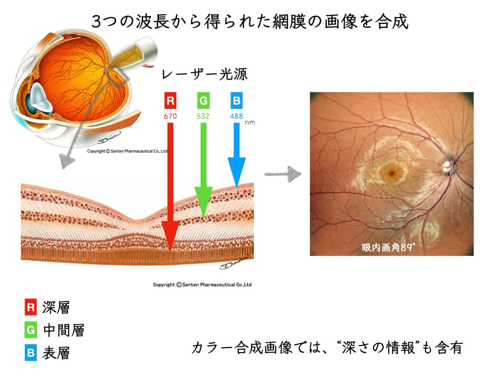
Mirante® provides composite images combining the three colors RGB (red, green, blue),
and yields very beautiful fundus images.
This allows detailed observation of the fundus and improves diagnostic accuracy.
High-quality images are also very effective when explaining the “current state of your eyes,” making it easier to gain understanding and agreement about treatment.
Whether “there is any concern about glaucoma” can also be understood from retinal images.
What the 163-Degree Field of View Shows
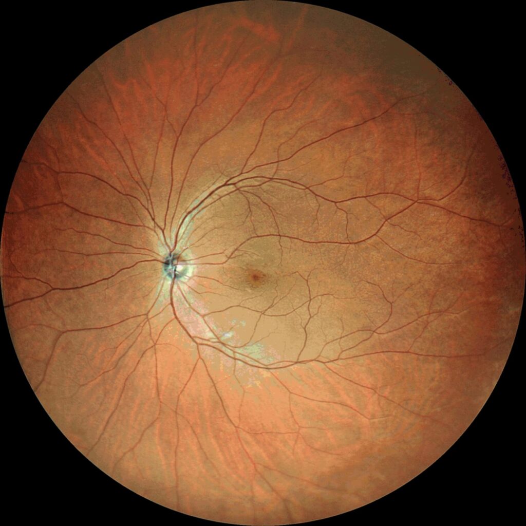
Mirante® has a wide 163-degree field of view and can photograph the retina in detail almost to its periphery.
This allows detection, without missing them, of lesions that are common in the periphery, such as retinal tears and uveitis inflammation.
Fundus images covering a wide area are particularly useful for detecting lesions at the pre-symptomatic stage.
What the 89-Degree Field of View Shows
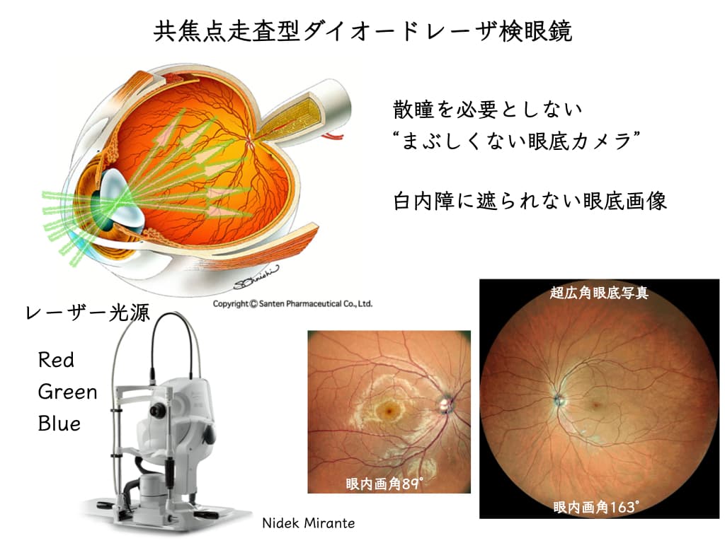
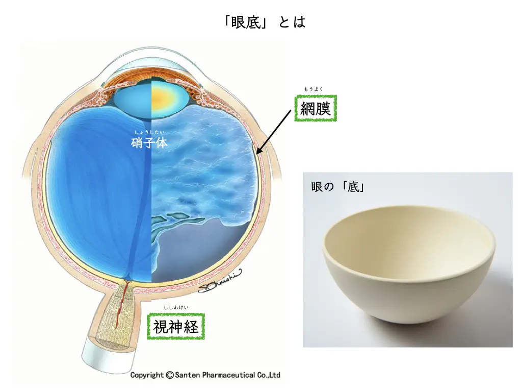
With its 89-degree field of view, Mirante® can photograph the macula clearly and allows detailed observation of macular lesions. Representative macular diseases include age-related macular degeneration (AMD), diabetic macular edema (DME), and macular hole.
Detailed observation of the macula is an important diagnostic item directly tied to preserving vision.
Panoramic Imaging
Panoramic imaging is a technique that stitches together multiple photographs to capture a wide area in a single image.
In photography, it can capture vast landscapes and large scenes at once.
Mirante® app update:
Multiple images can be stitched together automatically, which has greatly shortened the examination time.
Almost the entire fundus can be depicted.
This is useful for observing in detail lesions that are common in the periphery, such as retinal tears and uveitis inflammation.
Combining retinal images:
Integrating views from multiple angles makes a more comprehensive diagnosis possible.
What Autofluorescence Shows
Mirante® can also detect autofluorescence (FAF).
FAF is a non-invasive imaging technique that visualizes endogenous fluorescent substances such as lipofuscin in the retinal pigment epithelium (RPE).
This makes it possible to diagnose and manage diseases such as age-related macular degeneration (AMD), Stargardt disease, Best disease, and central serous chorioretinopathy (CSC). With FAF, early detection of disease and assessment of its progression become more accurate.
Overall, the introduction of the Mirante® ultra-widefield fundus camera has improved our diagnostic ability and greatly improved the patient experience.
All eye examinations are performed by certified orthoptists.
In this new era, technological development has been a major advance for both patients and medical staff.
Along with enjoying the wonders of advances in equipment,
we will put our expanding knowledge of retinal diseases and uveitis to use in patient care.
References
- Kumar, V., Surve, A., Kumawat, D., Takkar, B., Azad, S., Chawla, R., Shroff, D., Arora, A., Singh, R. and Venkatesh, P., 2021. Ultra-wide field retinal imaging: A wider clinical perspective. Indian Journal of Ophthalmology, 69(4), pp.824-835. https://www.ncbi.nlm.nih.gov/pmc/articles/PMC8012972/
- Tan, C.S., Sadda, S.R. and Hariprasad, S.M., 2014. Ultra-widefield retinal imaging in the management of diabetic eye diseases. Ophthalmic Surg Lasers Imaging Retina, 45(5), pp.363-366. https://eyesoneyecare.com/resources/how-ultra-widefield-imaging-can-boost-your-practice-and-benefit-your-patients/
- Wessel, M.M., Aaker, G.D., Parlitsis, G., Cho, M., D’Amico, D.J. and Kiss, S., 2012. Ultra-wide-field angiography improves the detection and classification of diabetic retinopathy. Retina, 32(4), pp.785-791. https://retinatoday.com/articles/2019-apr/when-ultra-widefield-retinal-imaging-is-most-useful
- Silva, P.S., El-Rami, H., Barham, R., Gupta, A. et al., 2017. Hemorrhage and/or Microaneurysm Severity and Count in Ultrawide Field Images and Early Treatment Diabetic Retinopathy Study Photography. Ophthalmology, 124(7), pp.970-976. https://pubmed.ncbi.nlm.nih.gov/28336057/
- Gross, J.G., Glassman, A.R., Liu, D. et al., 2018. Five-year outcomes of panretinal photocoagulation vs intravitreous ranibizumab for proliferative diabetic retinopathy: a randomized clinical trial. JAMA Ophthalmol, 136(10), pp.1138-1148. https://www.ophthalmologytimes.com/view/ultra-widefield-imaging-enhance-efficiency-for-practices
- Muqit, M.M., Marcellino, G.R., Henson, D.B. et al., 2013. Optos-guided pattern scan laser (Pascal)-targeted retinal photocoagulation in proliferative diabetic retinopathy. Acta Ophthalmol, 91(3), pp.251-258. https://pubmed.ncbi.nlm.nih.gov/22176513/
- Gupta, V., Al-Dhibi, H.A. and Arevalo, J.F., 2014. Retinal imaging in uveitis. Saudi J Ophthalmol, 28(2), pp.95-103. https://www.optos.com/blog/2016/Nov/two-reasons-your-diabetic-patients-need-ultra-widefield-retinal-imaging/
- Campbell, J.P., Leder, H.A., Sepah, Y.J. et al., 2012. Wide-field retinal imaging in the management of noninfectious posterior uveitis. Am J Ophthalmol, 154(5), pp.908-911. https://journals.sagepub.com/doi/10.1177/2515841419899495
- Lucente, A., Taloni, A., Scorcia, V. and Giannaccare, G., 2023. Widefield and Ultra-Widefield Retinal Imaging: A Geometrical Analysis. Life (Basel), 13(1), 202. https://pubmed.ncbi.nlm.nih.gov/36676151/
- Hussain, N., Edraki, M., Tahhan, R., Sanalkumar, N. et al., 2017. Telemedicine for diabetic retinopathy screening using an ultra-widefield fundus camera. Clin Ophthalmol, 11, pp.1477-1482. https://pubmed.ncbi.nlm.nih.gov/28860696/
- Nidek Mirante and Heidelberg Spectralis. 2021. Comparison between two multimodal imaging platforms: PubMed. https://pubmed.ncbi.nlm.nih.gov/33409677/
- EyeWiki. 2021. Fundus Autofluorescence. https://eyewiki.aao.org/Fundus_Autofluorescence