![Takeru Eye Clinic | Takatori Shopping Street, Sawara-ku, Fukuoka City [Nishijin Station / Fujisaki Station]](https://takeru-eye.com/wp-content/uploads/2022/10/takeru_logo_for-WP-header.png)
Blood vessels in the eye also reflect the condition of the whole body
◯ Retinal vein occlusion is a disease in which a vein in the retina becomes blocked, causing bleeding and swelling (macular edema).
◯ It is closely related to high blood pressure and arteriosclerosis, and becomes more common with age.
◯ Depending on where the blockage occurs, it is divided into branch retinal vein occlusion (BRVO) and central retinal vein occlusion (CRVO).
✔ If you have sudden blurring, reduced vision, or an area you cannot see, please see an eye doctor promptly.
✔ Along with treatment, managing your overall health, including blood pressure, is very important.
What Is Retinal Vein Occlusion (BRVO and CRVO)?
It is a disease in which the veins that carry blood from the retina back to the heart become blocked.
Blood upstream of the blockage has nowhere to go, causing bleeding and swelling in the retina.
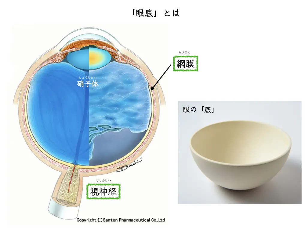
- Branch retinal vein occlusion (BRVO): A type in which a branch of the vein is blocked. It accounts for most cases of retinal vein occlusion. It tends to occur where an artery and a vein cross, and is strongly associated with high blood pressure.
- Central retinal vein occlusion (CRVO): A type in which the root of the vein (the central retinal vein) is blocked. Because it affects the whole retina, it tends to have a larger effect on vision and requires more careful follow-up.
Causes and Who Is at Risk
High blood pressure, arteriosclerosis, and aging are the main background factors.
It is also known to occur more often in people with diabetes, dyslipidemia, or glaucoma.
“Eye blood vessel disease” is also a mirror of the condition of the blood vessels throughout the body.
It is not uncommon for high blood pressure to be discovered when this disease develops.
Symptoms
• Sudden blurring or reduced vision in one eye
• Part of the visual field looks dark or missing
• Things look distorted (when swelling reaches the macula)
When bleeding does not reach the macula, there may be almost no symptoms.
It is a disease that is sometimes found by chance on fundus photographs at a health checkup.
Diagnosis
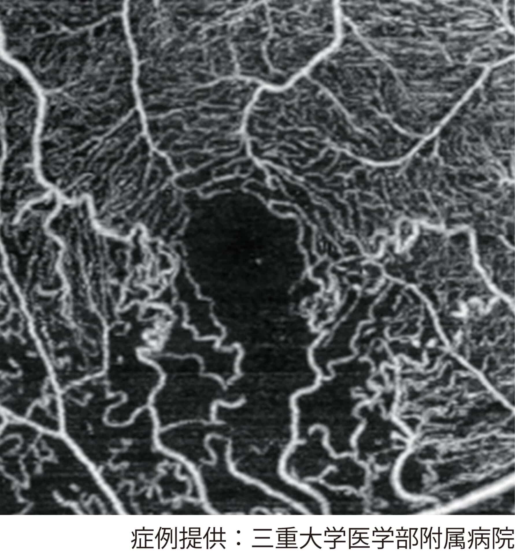
In addition to a fundus examination, we use OCT (optical coherence tomography) to check for swelling of the macula (macular edema), and OCT angiography (OCTA) to check, without contrast dye, the areas where blood flow has stopped (non-perfusion areas).
With ultra-widefield fundus photography, which is easy on the eyes because it needs little pupil dilation, we evaluate a wide area of the retina at once.
We show all the images on a monitor in front of you while we explain.
Treatment
For reduced vision caused by macular edema, the mainstay of treatment is intravitreal injection of an anti-VEGF drug.
When the areas where blood flow has stopped are wide, retinal laser photocoagulation may be performed to prevent new abnormal blood vessels (neovascularization) from forming.
In CRVO, regular checks are especially important to prevent and detect early a complication called neovascular glaucoma.
When surgery or hospital treatment is needed, we proceed in cooperation with the Department of Ophthalmology at Kyushu University and affiliated facilities.
Managing Your Whole Body Is Also Eye Treatment
Controlling blood pressure, blood sugar, and lipids is essential to prevent recurrence and onset in the other eye.
We look after both your eyes and your whole body in cooperation with your primary care physician.
If you are not yet seeing a physician, we may recommend that you do.
For Early Detection
Symptoms in one eye are easy to miss.
If you feel “my vision suddenly blurred” or “part of it is missing,” please consult us early rather than waiting.
Bleeding and swelling without symptoms can be found at regular fundus checks.

