![Takeru Eye Clinic | Takatori Shopping Street, Sawara-ku, Fukuoka City [Nishijin Station / Fujisaki Station]](https://takeru-eye.com/wp-content/uploads/2022/10/takeru_logo_for-WP-header.png)
Age-related macular degeneration comes in a “wet” type and a “dry” type. Geographic atrophy (GA) is the advanced stage of dry AMD, and in 2025 Izervay™ (an intravitreal injection) was approved and launched as the first treatment for it in Japan. If things look distorted or the center of your vision is hard to see, it is important to check each eye separately yourself and see an eye doctor promptly.
Geographic Atrophy (GA) and Izervay™, Japan’s First Treatment (Approved in 2025)
Geographic atrophy (GA) is the advanced stage of dry age-related macular degeneration, in which retinal tissue wastes away in a map-like shape and vision declines irreversibly. In Japan, about 100,000 people are estimated to be affected.
| Item | Details (based on primary sources) |
|---|---|
| Product name / generic name | Izervay™ intravitreal injection 20 mg/mL (avacincaptad pegol sodium) |
| Indication | Slowing the progression of geographic atrophy (GA) in dry age-related macular degeneration (the first treatment in Japan approved for this indication) |
| Approval and launch | Approved September 19, 2025; listed on the NHI drug price list November 12, 2025; launched November 27, 2025 |
| Dosing | Intravitreal injection once a month for the first 12 months, then once every 2 months |
| Positioning | It is a treatment that “slows” progression; it does not restore atrophied tissue or lost vision |
| Date checked | August 19, 2026 |
At our clinic we evaluate the presence and progression of geographic atrophy with a fundus examination and OCT, and for people who may be candidates for treatment we discuss and explain, including referral to a specialized facility. Tests also distinguish it from the wet type (the target of anti-VEGF treatment).
Primary sources:Astellas Pharma news release (Izervay™ launch, November 27, 2025)/PMDA Search for Prescription Drugs (latest package insert)
[Preventive Eye Care: When Things Look “Distorted”]
◯ Age-related macular degeneration is a disease in which the macula, the “center of vision,” is damaged.
◯ It is more common in people over 50 and is one of the leading causes of visual impairment in Japan as well.
◯ It is hard to notice unless you check each eye separately, so it can be found late.
✔ If you feel things look “distorted” or “the center is dark,” cover one eye and check.
✔ OCT (optical coherence tomography) and OCT angiography allow low-burden examination.
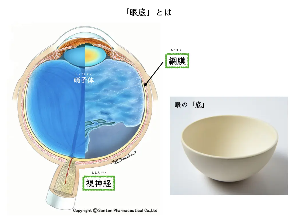
What Is the Macula?
The macula is the most important part of the retina for “seeing,” located at its center.
Reading letters, recognizing faces, and perceiving color are all functions of the macula.
When the macula is damaged, the center of the visual field becomes hard to see, greatly affecting daily life.
What Is Age-Related Macular Degeneration?
It is a disease in which waste products build up in the macula with aging and the macula gets damaged.
There are two main types.
- “Wet” type: abnormal blood vessels (choroidal neovascularization) grow under the macula and cause bleeding and swelling. It progresses relatively fast and is the type that is the main focus of treatment.
- “Dry” type: the tissue of the macula slowly thins. It progresses gradually, but regular follow-up is important.
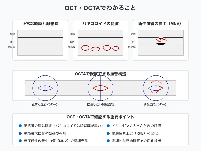
In recent years, the idea that the thickness of the choroid (the layer of blood vessels outside the retina) is involved in the disease process (pachychoroid) has also drawn attention, and our understanding of the disease deepens year by year.
Symptoms: “Distortion” and “Dark Center”
• Things look distorted (metamorphopsia): the bars of a paper screen or the grout lines of tiles look wavy
• The middle of the visual field is dark or missing (central scotoma)
• Reduced vision
When you look with both eyes, the other eye makes up for it, so it is hard to notice.
Every now and then, cover one eye and check whether a calendar or a grid pattern looks distorted.
Diagnosis: Low-Burden Imaging Tests
With OCT (optical coherence tomography) we see a cross-section of the macula, and with OCT angiography (OCTA) we check for abnormal blood vessels without contrast dye.
We also use ultra-widefield fundus photography, which needs little of the burden of mydriasis (pupil-dilating drops), and we show all the images on a monitor in front of the patient while we explain.
Treatment: It Has Changed Greatly in the Past 20 Years
Age-related macular degeneration was once a disease with no effective treatment, but photodynamic therapy (PDT) became available in 2003 and intravitreal injection of anti-VEGF drugs in 2008, and the way we think about treatment changed greatly.
Today, the mainstay of treatment for the wet type is intravitreal injection of anti-VEGF drugs.
When treatment is needed, we proceed in cooperation with the Department of Ophthalmology at Kyushu University and affiliated facilities.
Follow-up and regular checks after treatment can continue at our clinic.
Prevention and Lifestyle: There Are Things You Can Do
• Smoking is a clear risk factor for age-related macular degeneration. We recommend quitting.
• A balanced diet that includes green and yellow vegetables is recommended. Please also ask us about whether supplements are suitable for you.
• Make one-eye checks a habit. Noticing “distortion” early widens your treatment options.
For Early Detection
Changes before symptoms appear can be found with a fundus examination and OCT.
After age 50, we recommend regular fundus checks.

