![Takeru Eye Clinic | Takatori Shopping Street, Sawara-ku, Fukuoka City [Nishijin Station / Fujisaki Station]](https://takeru-eye.com/wp-content/uploads/2022/10/takeru_logo_for-WP-header.png)

The reason the eyes look clear and beautiful is
that the cornea is a transparent tissue that naturally has no blood vessels.
We can see the “iris” behind it through the transparent cornea, which is why eyes are called black or brown.
Americans and Europeans with blue irises have blue eyes, people from Northern Europe have green eyes, and so on.
When the cornea is inflamed, it is important for it to heal while staying transparent.
The body maintains homeostasis so that “angiogenesis” (new blood vessel growth) does not occur.
Oxygen is needed to keep the cornea transparent.
Long-term contact lens wear leaves the cornea short of oxygen
↓
The struggling corneal cells
• grow blood vessels from the corneal limbus (the border between the black and white of the eye)
• call in white blood cells
↓
VEGF, a protein (cytokine), is produced
↓
and they draw in blood vessels from the white of the eye (the conjunctiva) (angiogenesis)
Contact lenses: oxygen shortage makes blood vessels grow into the cornea
Corneal pannus (corneal neovascularization)
It occurs in people who wear contact lenses for long hours.
Under a microscope, blood vessels can be seen growing in from the periphery of the cornea.
It is described as occurring in 20% of contact lens wearers.1。
Unlike “eye redness,” which is dilation of the blood vessels of the white of the eye (conjunctiva), corneal pannus is characterized by blood vessels entering the cornea.
If the vessels do not reach the center (the pupil area), vision is not affected.
What is more important:
Low oxygen also leads to damage of the corneal endothelial cells2。
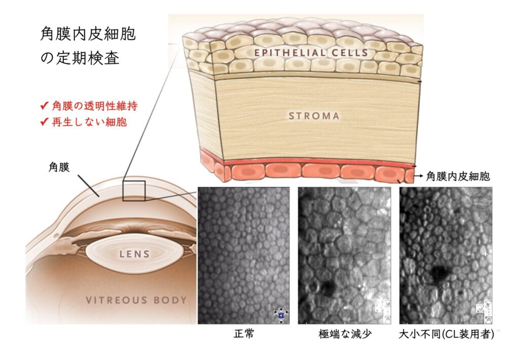
In daily practice, I pay close attention to whether the corneal endothelial cells are damaged.
The eyes of the Florida manatee, the model for mermaids
Blood vessels can be seen in the cornea of the Florida manatee, but they are limited to a very small part of the cornea (about 0.3% of its volume), and the effect on vision is said to be small.3。
The cause is that blood vessels grow all the way to the center of the cornea.
Genetically, the VEGF-related protein (sVEGFR1) has been found to be absent.4。
They swim through the sea without being able to see.
VEGF, which promotes angiogenesis
VEGF (vascular endothelial growth factor):
is known as an important protein (cytokine) in cancer metastasis, retinal angiogenesis, and more.
The drug-induced harm from oral thalidomide (teratogenicity / impaired growth of the limbs) is thought to be due to its action of inhibiting angiogenesis (the formation of new blood vessels).5。
(It has now made a comeback as a treatment for multiple myeloma.)
In basic research, there is a method of inducing angiogenesis in the cornea of animals.6。
It is an experimental method in which the body’s blood vessels are easy to see, and many drugs have been tested with it.
VEGF Treatments for this:
Steroids, immunosuppressants, and biologics (anti-VEGF antibodies, antibodies against inflammatory cytokines, and so on)
Anti-VEGF biologics such as Eylea®, Lucentis®, and Beovu® are used to treat age-related macular degeneration, diabetic macular edema, retinal vein occlusion, and neovascular glaucoma.
Anti-VEGF antibodies have been in clinical use for only about 10 years, but we still vividly remember how astonished everyone was by their dramatic effect.
How to prevent corneal pannus
○ Use highly oxygen-permeable contact lenses
Use silicone hydrogel contact lenses rather than HEMA lenses.
○ Shorten contact lens wearing time
Take your contact lenses out as soon as you get home
Treatment aims to control the inflammation with steroid eye drops.
Anti-VEGF biologics are not covered by insurance for corneal pannus.
To avoid corneal pannus (corneal neovascularization),
and also to avoid losing corneal endothelial cells,
I believe “prevention” is the most important thing.
(References)
1.Rolfsen, M.L., Frisard, N.E., Stern, E.M., Foster, T.P., Bhattacharjee, P.S., McFerrin Jr, H.E., Clement, C., Rodriguez, P.C., Lukiw, W.J., Bergsma, D.R., Ochoa, A.C., Hill, J.M., 2013. Corneal neovascularization: a review of the molecular biology and current therapies. Expert Review of Ophthalmology 8, 167–189. https://doi.org/10.1586/eop.13.8
2.Liesegang, T.J., 2002. Physiologic Changes of the Cornea with Contact Lens Wear. Eye & Contact Lens 28, 12–27.
Alipour, F., Khaheshi, S., Soleimanzadeh, M., Heidarzadeh, S., Heydarzadeh, S., 2017. Contact Lens-related Complications: A Review. J Ophthalmic Vis Res 12, 193–204. https://doi.org/10.4103/jovr.jovr_159_16
3.Harper, J.Y., Samuelson, D.A., Reep, R.L., 2005. Corneal vascularization in the Florida manatee (Trichechus manatus latirostris) and three-dimensional reconstruction of vessels. Veterinary Ophthalmology 8, 89–99. https://doi.org/10.1111/j.1463-5224.2005.00351.x
4.Ambati, B.K., Nozaki, M., Singh, N., Takeda, A., Jani, P.D., Suthar, T., Albuquerque, R.J.C., Richter, E., Sakurai, E., Newcomb, M.T., Kleinman, M.E., Caldwell, R.B., Lin, Q., Ogura, Y., Orecchia, A., Samuelson, D.A., Agnew, D.W., Leger, J.St., Green, W.R., Mahasreshti, P.J., Curiel, D.T., Kwan, D., Marsh, H., Ikeda, S., Leiper, L.J., Collinson, J.M., Bogdanovich, S., Khurana, T.S., Shibuya, M., Baldwin, M.E., Ferrara, N., Gerber, H.-P., Falco, S.D., Witta, J., Baffi, J.Z., Raisler, B.J., Ambati, J., 2006. Corneal avascularity is due to soluble VEGF receptor-1. Nature 443, 993–997. https://doi.org/10.1038/nature05249
5.D’Amato, R.J., Loughnan, M.S., Flynn, E., Folkman, J., 1994. Thalidomide is an inhibitor of angiogenesis. Proc. Natl. Acad. Sci. U.S.A. 91, 4082–4085. https://doi.org/10.1073/pnas.91.9.4082
6.Rogers, M.S., Birsner, A.E., D’Amato, R.J., 2007. The mouse cornea micropocket angiogenesis assay. Nature Protocols 2, 2545–2550. https://doi.org/10.1038/nprot.2007.368