![Takeru Eye Clinic | Takatori Shopping Street, Sawara-ku, Fukuoka City [Nishijin Station / Fujisaki Station]](https://takeru-eye.com/wp-content/uploads/2022/10/takeru_logo_for-WP-header.png)

Why use highly oxygen-permeable contact lenses
What are corneal endothelial cells?
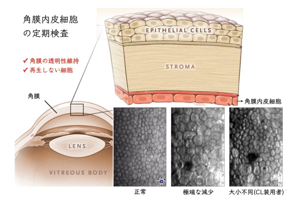
The transparent tissue on the surface of the eyeball is called the cornea, the clear front surface of the eye.
It is a very thin membrane, 0.5 mm thick at the center to 0.8 mm at the periphery:
the only tissue in the body without blood vessels, which keeps it beautifully clear.1。
And
the “corneal endothelial cells” that line the inside of the cornea are important formaintaining the transparencyof the cornea.
Features of corneal endothelial cells
✔ They have a pump function2
The cornea is bathed in water: tear fluid on the outside and aqueous humor on the inside.
Corneal endothelial cells push the inner “aqueous humor” out.
If this function is lost, water enters the clear cornea and it becomes swollen and soft (= bullous keratopathy).
✔ They do not regenerate3
They are always slightly damaged during intraocular surgery such as cataract surgery.
Even when their number decreases, they do not leave holes.
Each cell enlarges and compensates by filling the space at the back of the cornea.
✔ They decrease gradually with age4
For the reasons above, enlarged corneal endothelial cells are seen as people age.
The cells may also vary in size.
Corneal endothelial cells: actual cell counts
In healthy people, about 2,500 to 3,000 corneal endothelial cells are seen per 1 mm2 of cornea.
Down to about 2,000 cells/mm2 is usually not a problem.
At 400 cells/mm2 or fewer, the pump function fails and the cornea falls into a state of “bullous keratopathy.”
Vision is severely impaired and a corneal transplant becomes necessary.
Cells in the Eye That Decrease with Contact Lens Wear: Corneal Endothelial Cells
Why it is important to use highly oxygen-permeable contact lenses
While you wear contact lenses, the cornea becomes short of oxygen.
A cornea starved of oxygen may grow new blood vessels (corneal pannus).
» Oxygen shortage makes blood vessels grow into the cornea (corneal pannus)
What matters:
✔ If the oxygen shortage continues, corneal endothelial cells decrease
As ways to prevent a decrease in corneal endothelial cells,
○ Use highly oxygen-permeable contact lenses
○ Take care not to wear contact lenses for too long
are very important.
This helps prevent “bullous keratopathy,” which could develop decades from now.5。
(Reference)
1. Ambati, B.K., Nozaki, M., Singh, N., Takeda, A., Jani, P.D., Suthar, T., Albuquerque, R.J.C., Richter, E., Sakurai, E., Newcomb, M.T., Kleinman, M.E., Caldwell, R.B., Lin, Q., Ogura, Y., Orecchia, A., Samuelson, D.A., Agnew, D.W., Leger, J.St., Green, W.R., Mahasreshti, P.J., Curiel, D.T., Kwan, D., Marsh, H., Ikeda, S., Leiper, L.J., Collinson, J.M., Bogdanovich, S., Khurana, T.S., Shibuya, M., Baldwin, M.E., Ferrara, N., Gerber, H.-P., Falco, S.D., Witta, J., Baffi, J.Z., Raisler, B.J., Ambati, J., 2006. Corneal avascularity is due to soluble VEGF receptor-1. Nature 443, 993–997. https://doi.org/10.1038/nature05249
2. Bonanno, J.A., 2012. Molecular mechanisms underlying the corneal endothelial pump. Experimental Eye Research, Corneal Endothelium in Health and Disease 95, 2–7. https://doi.org/10.1016/j.exer.2011.06.004
3. Rao, G.N., Shaw, E.L., Arthur, E.J., Aquavella, J.V., 1979. Endothelial cell morphology and corneal deturgescence. Ann Ophthalmol 11, 885–899.
4. Schoessler, J.P., Orsborn, G.N., 1987. A theory of corneal endothelial polymegethism and aging. Curr. Eye Res. 6, 301–306. https://doi.org/10.3109/02713688709025182
5. Bourne, W.M., 2001. The effect of long-term contact lens wear on the cells of the cornea. CLAO J 27, 225–230.