![Takeru Eye Clinic | Takatori Shopping Street, Sawara-ku, Fukuoka City [Nishijin Station / Fujisaki Station]](https://takeru-eye.com/wp-content/uploads/2022/10/takeru_logo_for-WP-header.png)

“My internist says my thyroid numbers are stable, but every time I look in the mirror my face seems to be changing, and it worries me.”
“My eyes look like they are bulging, or my eyelids are swollen and I look like a different person.”
Quite a few patients come to an eye clinic with worries like these.
Thyroid eye disease (TED) is a condition in which inflammation can progress behind the eye even when internal medicine test values are normal.
In treating inflammation in ophthalmology, what is important is todo so without impairing the function of the tissues and cells themselves.
Inflammation of the eyeball must be treated while keeping the “transparency” that lets light through.
Likewise, if inflammation in the tissues around the eye leaves a “scar,” it seriously affects later visual function and facial appearance.
Once tissue scarring progresses and the tissue becomes stiff, it is extremely difficult to restore its original flexibility and shape.
Whether appropriate treatment can be given during the “active phase” of inflammation is therefore very important for the future.
Thyroid numbers and eye symptoms do not always match
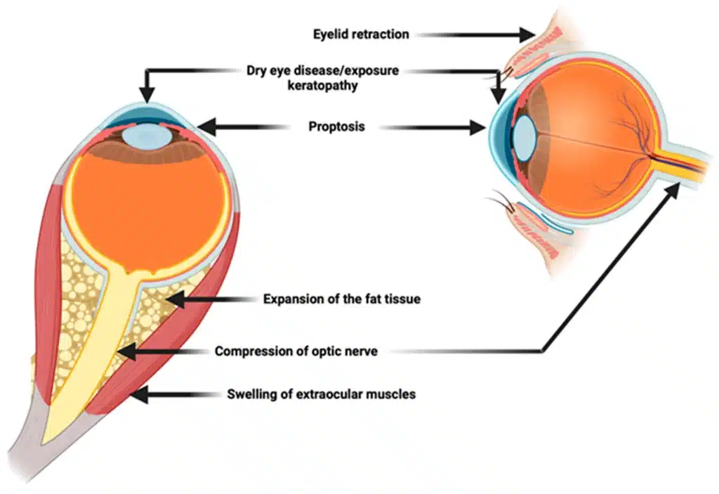
Thyroid eye disease (TED) is a condition in which the muscles and fat around the eye become inflamed in association with autoimmune thyroid disease such as Graves’ disease¹.
A large survey analyzing data from Japan puts the incidence at roughly 7 to 11 people per 100,000 people per year³ ⁴.
| Indicator | Figure / trend |
| Estimated number of patients in Japan | About 25,000 to 35,000 (including active and inactive disease) |
| Incidence | 7.3 to 11.1 per 100,000 person-years |
| Sex ratio | Overwhelmingly female, about 73 to 76% |
| Peak age (bimodal) | Women: early 40s and early 60s. Men: late 40s and late 60s |
To put these numbers in familiar terms,
among the crowd of about 2.5 full Fukuoka Dome stadiums (about 100,000 people), the number of people newly diagnosed in a year is about a baseball team’s worth (9 to 11 people).
It is not a large number, but it is a probability at which it would not be surprising for it to happen suddenly to any particular person.
The most important fact is thatthyroid hormone levels in blood tests and the progression of eye symptoms do not necessarily move together.
About 90% of TED patients have hyperthyroidism.
On the other hand, some people develop it when thyroid function is normal or low⁵ ¹.
While it is left alone because “the numbers are normal, so let’s wait and see,” the tissue behind the eye may be gradually degenerating and heading toward scarring.
In particular, people who smoke are known to have more severe inflammation and a weaker response to treatment¹ ⁹.
For this reason, quitting smoking is strongly recommended.
Main signs around the eye and their clinical significance
TED is not simply a disease in which the eyes swell.
Inflammation enlarges the tissue, which then hardens, causing functional and cosmetic problems such as the following.
- Eyelid retraction: the upper eyelid is pulled up and the white of the eye above the iris becomes visible² ¹.
- Proptosis (bulging of the eye): swelling of fat and muscle behind the eye pushes the eyeball forward².
- Double vision (diplopia): the extraocular muscles that move the eye become inflamed and then scarred and stiff, restricting eye movement and causing double vision⁸.
- Optic nerve compression: swollen tissue squeezes the optic nerve at the back of the eye, causing reduced vision and visual field defects. This is the condition that most needs vigilance¹.
If these symptoms are suppressed while inflammation is still in the “active phase,” the sequelae from scarring may be kept to a minimum.
A comprehensive check of the Clinical Activity Score (CAS) and its limits
The Clinical Activity Score (CAS) is used worldwide when doctors assess the current strength of inflammation.
At the examination, seven items are checked comprehensively, and if three or more apply, the disease is judged to be in the “active phase”⁵.
- Spontaneous pain: pain or a heavy feeling behind the eye while resting.
- Pain on eye movement: pain when moving the eyes up, down, left, or right.
- Eyelid redness: the skin of the eyelid is red.
- Conjunctival redness: the membrane covering the surface of the white of the eye is red.
- Eyelid swelling: the eyelids are swollen (tending to be worse in the morning).
- Swelling of the caruncle and plica semilunaris: the pink tissue near the inner corner of the eye is red and swollen.
- Conjunctival edema (chemosis): the membrane covering the white of the eye is swollen like jelly.
This scoring is an excellent index that can be done easily in the examination room.
On the other hand, it can lead to serious oversights.
This evaluation is based mainly on “findings on the surface, such as the eyelids and conjunctiva.”
As a result, it may underestimate inflammation progressing deep in the orbit⁶ ⁷.
If you overlook inflammation behind the eye while the surface is quiet, irreversible scarring can result.
Objective evaluation of deep inflammation using MRI
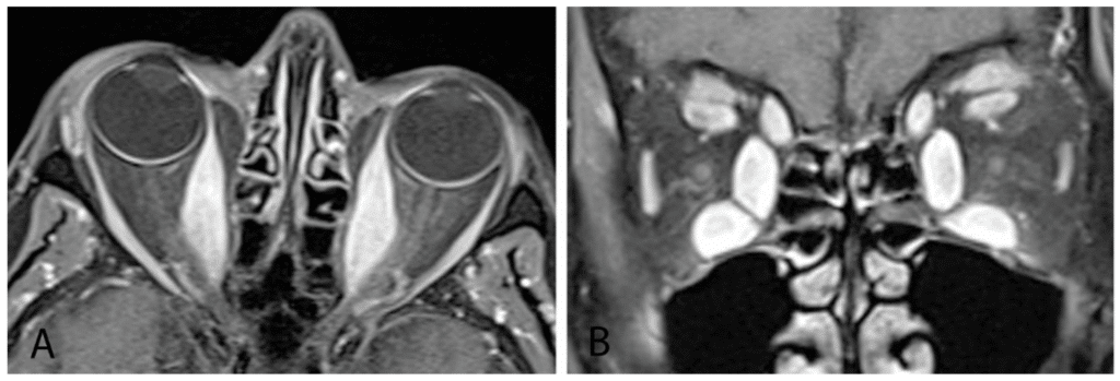
To fill the blind spot that arises from evaluating surface findings alone, and to lead to an accurate diagnosis,
evaluation with MRI (magnetic resonance imaging) is strongly recommended.
In particular, T2-weighted images, which clearly show tissue swelling from inflammation, are emphasized⁷.
Recent studies have reported that an objective index called the signal intensity ratio (SIR) helps improve diagnostic accuracy⁶.
It quantifies the brightness of the extraocular muscles compared with a reference such as brain tissue.
When the SIR is high, that is, when the muscles appear white and bright, it is judged that “active inflammation is present” even if the surface score is low, which is a basis for considering early treatment⁷.
Conversely, when the SIR is low,
it shows that inflammation has already quieted and the disease has moved into the “inactive phase” heading toward scarring.
Outlook for treatment: to minimize scarring
TED treatment proceeds in two stages, matched to the phase of inflammation.
Treatment in the active phase: calm inflammation quickly
The first stage of treatment is to calm inflammation quickly during the “active phase,” which is like a fire, and stop tissue degeneration.
Steroid pulse therapy and radiotherapy
Intravenous powerful anti-inflammatory treatment (steroid pulse) and low-dose radiotherapy to the orbit are established standard options⁵ ¹⁰. Their aim is to suppress inflammation and minimize damage to the optic nerve.
Drug therapy approved in Japan in 2024: teprotumumab(Tepezza®)
In September 2024, a biologic called teprotumumab (Tepezza®) was approved in Japan for active TED¹¹.
By directly inhibiting the receptor that triggers inflammation, it is an innovative option that can be expected to improve proptosis itself, which was difficult with conventional treatment¹¹ ¹².
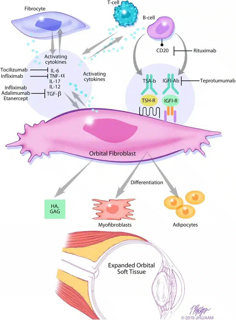
The goal at this stage is to calm inflammation quickly and prevent scarring.
Steroid pulse therapy and radiotherapy to the orbit are the standard options⁵ ¹⁰.
In addition, in September 2024, a biologic called teprotumumab was approved in Japan¹¹.
By directly inhibiting the receptor that triggers inflammation, it is an option that can be expected to improve proptosis itself¹¹.
Treatment in the inactive phase: surgical repair of remaining sequelae
After inflammation has completely settled (the inactive phase), the tissue hardens as “scar” (induration).
At this stage drug therapy is unlikely to help, so we use a surgical approach to restore the original function and appearance (functional recovery).
Thyroid eye disease surgery follows a strict order of priority.
Proceeding in this order, while adapting to each individual’s condition, is considered reasonable for balancing function and appearance¹⁰.
Orbital decompression
To bring a protruding eye back, the bone behind the eye is thinned or fat is removed to widen the space. Because this surgery greatly changes the position of the eye and how wide the eyelids open, it is generally performed first⁵.
Strabismus surgery
After the position of the eye is settled by decompression, the attachment points of the muscles that move the eye are adjusted to improve the remaining double vision (diplopia).
Eyelid surgery
Finally, the shape of the eyelids is adjusted. Adjusting the eyelids as the finishing step, after the position and movement of the eyes are settled, is considered the desirable order both functionally and cosmetically⁵.
By repairing step by step, one thing at a time and with care, it is possible to regain the quality of life that the disease took away.
Outlook: what early diagnosis of thyroid eye disease can bring
Thyroid eye disease is a disease in which the timing of diagnosis shapes the rest of life.
The worry that “my face has changed” is not merely a cosmetic matter. It can become a serious problem in social life.
But by combining a comprehensive examination with objective deep evaluation by MRI, it may be possible to stop inflammation before it turns into “irreversible scarring.”
To understand correctly which phase the current condition is in, and to keep future effects to a minimum:
we consider it important to receive an objective evaluation at the appropriate time.
References
- Shah SS, Stokkermans TJ, Patel BC. Thyroid Eye Disease. [Updated 2025 Nov 8]. In: StatPearls [Internet]. Treasure Island (FL): StatPearls Publishing; 2025 Jan-. Available from: https://www.ncbi.nlm.nih.gov/books/NBK582134/
- Tamhankar MA, Raza S, Brutsaert E, et al. The burden of illness in thyroid eye disease: current state of the evidence. Front Ophthalmol (Lausanne). 2025;5:1565762. Published 2025 Apr 17. doi:10.3389/fopht.2025.1565762
https://pmc.ncbi.nlm.nih.gov/articles/PMC12075187/ - Watanabe N, Kozaki A, Inoue K, Narimatsu H, Shinohara M, LoPresti MG. Prevalence, Incidence, and Clinical Characteristics of Thyroid Eye Disease in Japan. J Endocr Soc. 2023;8(1):bvad148. Published 2023 Nov 27. doi:10.1210/jendso/bvad148
https://pubmed.ncbi.nlm.nih.gov/38075564/ - Guide to the Management of Thyroid Eye Disease. Japan Thyroid Association and Japan Endocrine Society; Ministry of Health, Labour and Welfare research group on hormone receptor abnormalities, Committee for Clinical Priority Issue “Diagnostic criteria and treatment guidelines for malignant exophthalmos of Graves’ disease”
https://www.j-endo.jp/uploads/files/edu/koujyousengansyo_digest.pdf - Burch HB, Perros P, Bednarczuk T, et al. Management of thyroid eye disease: a Consensus Statement by the American Thyroid Association and the European Thyroid Association. Eur Thyroid J. 2022;11(6):e220189. Published 2022 Dec 8. doi:10.1530/ETJ-22-0189
Management of thyroid eye disease – Consensus Statement. 2022.
https://pmc.ncbi.nlm.nih.gov/articles/PMC9727317/ - Cai M, Yang J, Li X, Hu Y, Liao H and Xiong C (2025) MRI-based SIR quantitative biomarkers: a novel imaging diagnostic strategy for thyroid eye disease activity staging. Front. Endocrinol. 16:1650116. doi: 10.3389/fendo.2025.1650116
https://www.frontiersin.org/journals/endocrinology/articles/10.3389/fendo.2025.1650116/full - Ge Q, Zhang X, Wang L, et al. Quantitative evaluation of activity of thyroid-associated Ophthalmopathy using short-tau inversion recovery (STIR) sequence. BMC Endocr Disord. 2021;21(1):226. Published 2021 Nov 13. doi:10.1186/s12902-021-00895-3
https://pmc.ncbi.nlm.nih.gov/articles/PMC8590769/ - Strabismus in thyroid eye disease – EyeNews. 2024.
https://www.eyenews.uk.com/education/top-tips/post/strabismus-in-thyroid-eye-disease - Chrysoula Dosiou, Andrea Lora Kossler, Thyroid Eye Disease: Navigating the New Treatment Landscape, Journal of the Endocrine Society, Volume 5, Issue 5, May 2021, bvab034, https://doi.org/10.1210/jendso/bvab034
https://academic.oup.com/jes/article/5/5/bvab034/6174437 - Hoang TD, Stocker DJ, Chou EL, Burch HB. 2022 Update on Clinical Management of Graves Disease and Thyroid Eye Disease. Endocrinol Metab Clin North Am. 2022;51(2):287-304. doi:10.1016/j.ecl.2021.12.004
https://pmc.ncbi.nlm.nih.gov/articles/PMC9174594/ - TEPEZZA® (TEPROTUMUMAB) RECEIVES APPROVAL IN JAPAN FOR THE TREATMENT OF ACTIVE THYROID EYE DISEASE – PR Newswire. 2024.
https://www.prnewswire.com/news-releases/tepezza-teprotumumab-receives-approval-in-japan-for-the-treatment-of-active-thyroid-eye-disease-302257503.html - Tepezza (teprotumumab-trbw) receives approval in Japan for the treatment of active thyroid eye disease – Medical Update Online. 2024.
https://medicalupdateonline.com/2024/10/tepezza-teprotumumab-trbw-receives-approval-in-japan-for-the-treatment-of-active-thyroid-eye-disease-amgen/