![Takeru Eye Clinic | Takatori Shopping Street, Sawara-ku, Fukuoka City [Nishijin Station / Fujisaki Station]](https://takeru-eye.com/wp-content/uploads/2022/10/takeru_logo_for-WP-header.png)
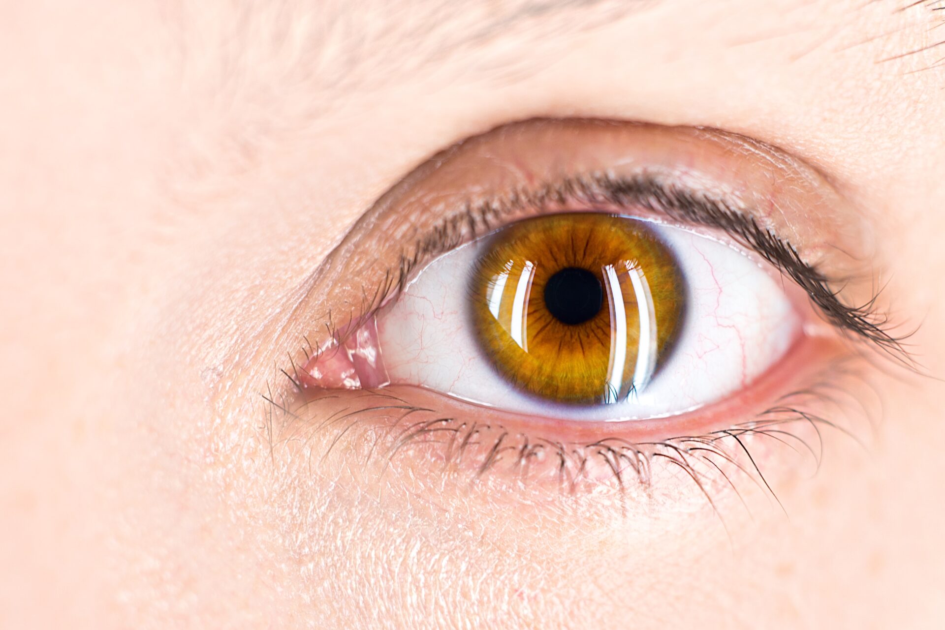
Why the black of the eye looks black:
Behind the clear, transparent “cornea” are the “iris” and “pupil,” also called the brown part of the eye.
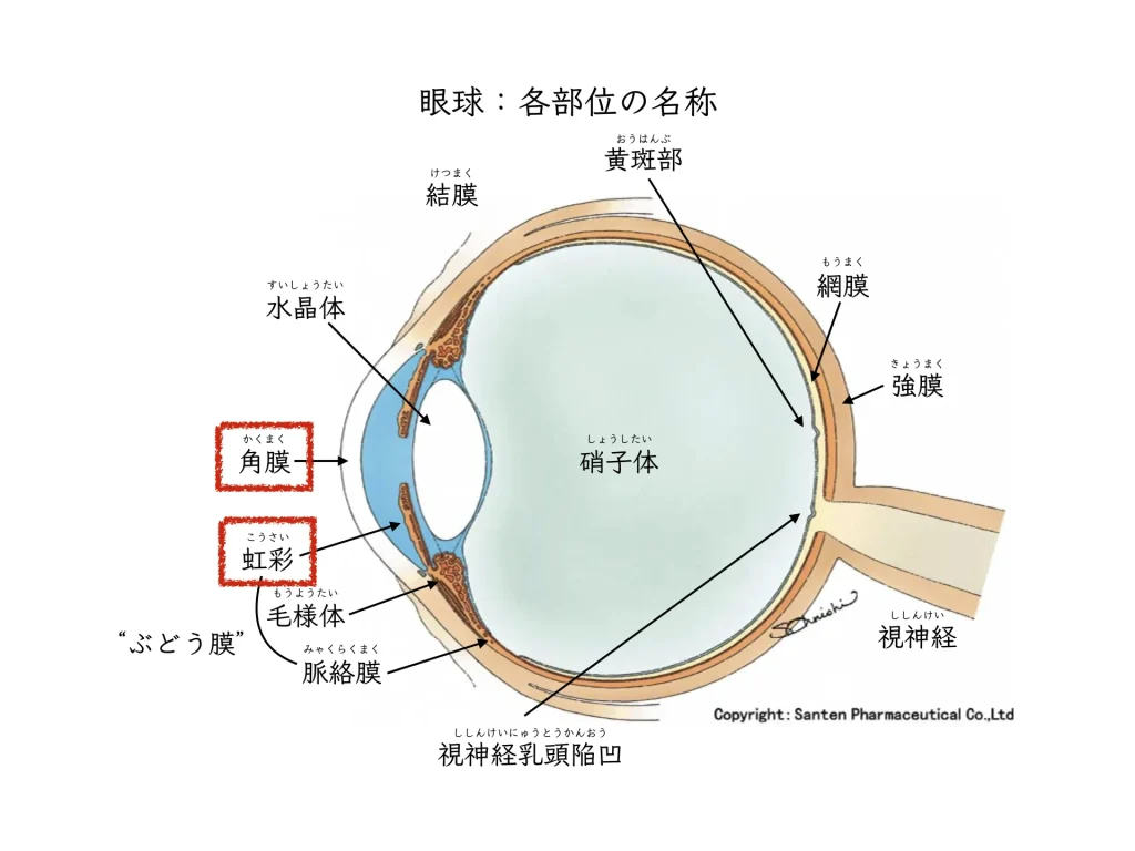
The cornea is the only tissue in the human body without blood vessels, yet it has a strong immune response.
This is because cells responsible for immunity are present there.
Keeping the cornea transparent is a “necessary condition” for seeing clearly.
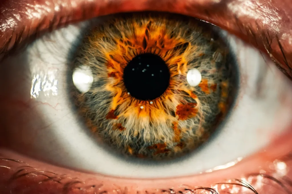
A White Spot on the Black of the Eye: Why It Happens, and How an Eye Doctor Judges and Treats It

When you look at your eyes in the mirror, you may see a white spot on the black of the eye.
Redness, pain, a gritty feeling, or discomfort often come with it.
Sometimes there is no pain.
Contact lens wearers often notice something wrong and come to see us.
In many cases, the true nature of it is“inflammatory cell infiltration of the cornea.”:
Some kind of inflammation has occurred, and cells have rushed to the transparent cornea.
The eye doctor uses a slit lamp microscope to look at
the “location” of the white spot (cell infiltration) on the black of the eye.
Within the cornea,
1. Near the center
2. The periphery (the edge of the black of the eye)
Depending on which of these it is, we infer the nature of the inflammation.
For cornealinflammation,it is very important that transparency is restored as it heals.
Sometimes it is urgent.
Is the white near the center of the black of the eye, or around it? Differences in the corneal immune response
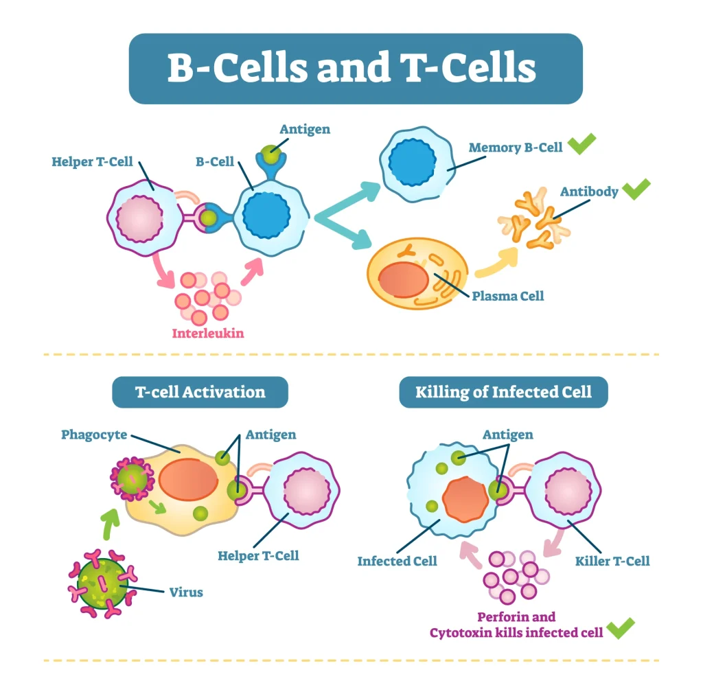
The periphery of the cornea (the limbus) is a part that easily mounts an immune response.
Many antigen-presenting cells (APCs) are present there, ready for inflammation.
Once inflammation occurs, T cells and B cells gather and form a cell cluster.
1. In the case of (inflammation near the center), we suspect “inflammation caused by microorganisms” that have escaped the immune response
2. In the case of (inflammation at the corneal periphery), we suspect “autoimmune inflammation” in which the body attacks its own cornea
is what we suspect.
1. Inflammation caused by microorganisms (corneal infection)
In many cases it is bacteria (germs): antibiotic eye drops and oral medicine are needed, and in some cases an intravenous drip too.
With fungi (molds and so on) and Acanthamoeba, it can become very severe.
When we judge that the microorganism’s activity has stopped, we consider steroid eye drops to reduce the cell infiltration.
The top priority is that the white spot disappears and transparency returns.
A Gram stain can also be done to identify the bacteria.
2. Autoimmune (non-infectious) inflammation
After confirming that it is not a corneal infection, steroid eye drops are started early.
It often heals with eye drops.
In the short term, we watch for the side effect of raised eye pressure.
When it is a peripheral corneal ulcer associated with rheumatoid disease, systemic steroids may be needed.
Steroids make infections worse.
That is why judging whether it is 1 or 2 is important.
You need to see an eye doctor as soon as possible.