
Myopia progresses as the depth of the eye (axial length) increases (that is, vision “gets worse”).1。
Once myopia becomes high myopia and moves on to pathologic myopia,
the risk rises of developing diseases that leave serious vision loss, such as myopic maculopathy, retinal detachment, and glaucoma.2, 3。
That is why preventive treatment is so important.4。
Over the past few years, myopia control (prevention) as an academic field has advanced greatly, and common sense in ophthalmology has changed.
At birth, the axial length is short.
Just as we grow taller, everyone’s axial length increases as they grow.
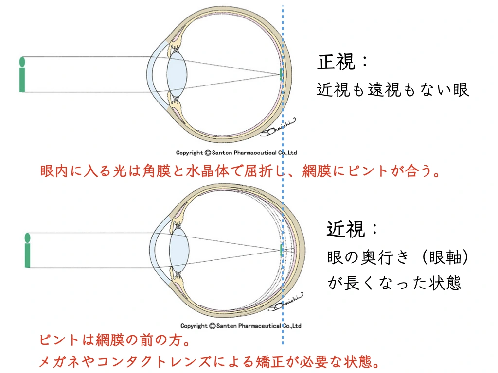
Why the axial length increases further (that is, why myopia progresses and vision “gets worse”):
• When the eye stays out of focus on the retina, the biological response that lengthens the eye becomes stronger.
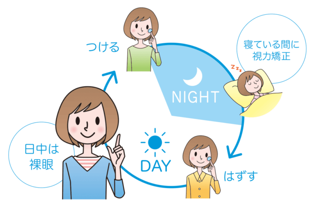
Orthokeratology is:
wearing special contact lenses during sleep to thin the center of the cornea.
→ During the day, light is focused across the whole retina, so you can go without glasses or contact lenses.
For myopia control:
Both low-dose atropine eye drops and orthokeratology have a large body of published statistical and scientific evidence of effectiveness.5, 6。
There is no clear answer yet as to why orthokeratology has a myopia-controlling effect.
The leading explanations to date:
○ Low-dose atropine eye drops:
“Pharmacological effect”7, 8
• Acts on the focusing muscle (ciliary muscle) and improves accommodation.
• Acts on α2-adrenergic receptors and resists stretching of the wall of the eye (retina and sclera).
○ Orthokeratology:
“Optical effect”9, 10
• Focusing light across the periphery (the whole) of the retina removes blur, so you can go through the day without glasses or contact lenses.
Combining the two can increase the myopia-controlling effect.
Combining low-dose atropine eye drops and orthokeratology increases the myopia-controlling effect
The following two synergistic effects
are thought to enhance the effect of myopia control (prevention):
✔ Low-dose atropine eye drops make the pupil slightly larger
✔ Orthokeratology creates a state in which light is focused across the whole retina during the day
On July 29, 2020, Scientific Reports (online) published the following findings:
Combining orthokeratology and low-dose atropine gave an additional myopia-controlling effect.
This is an introduction to the study results.
This is the first prospective controlled study (with follow-up) of the combined treatment.11。
[Participants]
Children aged 8 to 12 with myopia of -1.00 to -6.00 D,
80 of whom were able to undergo orthokeratology treatment
Orthokeratology / low-dose (0.01%) atropine group: 43 children
Orthokeratology-alone group: 37 children
* The orthokeratology lenses used were the domestically produced Breath-O Correct®.
* Before treatment, no significant differences were confirmed between the groups in age, sex, spherical equivalent, axial length, corneal endothelial cell count, eye pressure, or uncorrected vision.
The two groups were followed for 2 years (with a few dropouts),
and axial length was measured at 6, 12, 18, and 24 months to analyze the myopia-controlling (preventive) effect.
[Results]
In the orthokeratology / low-dose atropine group, axial length grew by 0.29 ± 0.20 mm over 2 years, significantly less than the 0.40 ± 0.23 mm in the orthokeratology-alone group, confirming a myopia-controlling (preventive) effect (P=0.03).
The effect was especially large in mild myopia of −1.00 to −3.00 D (0.30 mm vs. 0.48 mm, P=0.005), while no significant difference was seen in the subgroup with moderate myopia of −3.01 to −6.00 D (0.27 mm vs. 0.25 mm, P=0.74).
Low-dose atropine eye drops and orthokeratology: why the combination works
Two synergistic effects:
1. The pharmacological and optical effect of the slightly larger pupil produced by low-dose atropine
2. The optical effect of orthokeratology
With low-dose atropine eye drops,
the pupil diameter (size of the pupil) in bright light becomes slightly larger.
• 0.91 mm (ATOM2 study12)
• 0.49 mm (LAMP study13)
Low-dose atropine eye drops therefore have
an “optical effect”
in addition to the “pharmacological effect.”
* In practice, I pay attention to whether the child finds the light too bright.
The paper also states that light entering through the slightly enlarged pupil enhances the “optical effect” of orthokeratology.14。
(References)
1. Kimura, S., Hasebe, S., Miyata, M., Hamasaki, I., Ohtsuki, H., 2007. Axial Length Measurement Using Partial Coherence Interferometry in Myopic Children: Repeatability of the Measurement and Comparison with Refractive Components. Jpn J Ophthalmol 51, 105–110. https://doi.org/10.1007/s10384-006-0410-5
2. Asakuma, T., Yasuda, M., Ninomiya, T., Noda, Y., Arakawa, S., Hashimoto, S., Ohno-Matsui, K., Kiyohara, Y., Ishibashi, T., 2012. Prevalence and Risk Factors for Myopic Retinopathy in a Japanese Population: The Hisayama Study. Ophthalmology 119, 1760–1765. https://doi.org/10.1016/j.ophtha.2012.02.034
3. Flitcroft, D.I., 2012. The complex interactions of retinal, optical and environmental factors in myopia aetiology. Progress in Retinal and Eye Research 31, 622–660. https://doi.org/10.1016/j.preteyeres.2012.06.004
4. Morgan, I.G., Ohno-Matsui, K., Saw, S.-M., 2012. Myopia. The Lancet 379, 1739–1748. https://doi.org/10.1016/S0140-6736(12)60272-4
5. Chia, A., Lu, Q.-S., Tan, D., 2016. Five-Year Clinical Trial on Atropine for the Treatment of Myopia 2: Myopia Control with Atropine 0.01% Eye drops. Ophthalmology 123, 391–399. https://doi.org/10.1016/j.ophtha.2015.07.004
6. Chia, A., Chua, W.-H., Wen, L., Fong, A., Goon, Y.Y., Tan, D., 2014. Atropine for the Treatment of Childhood Myopia: Changes after Stopping Atropine 0.01%, 0.1% and 0.5%. American Journal of Ophthalmology 157, 451-457.e1. https://doi.org/10.1016/j.ajo.2013.09.020
7. Carr, B.J., Nguyen, C.T., Stell, W.K., 2019. Alpha2-adrenoceptor agonists inhibit form-deprivation myopia in the chick. Clinical and Experimental Optometry 102, 418–425. https://doi.org/10.1111/cxo.12871
8. Gallego, P., Martínez‐García, C., Pérez‐Merino, P., Ibares‐Frías, L., Mayo‐Iscar, A., Merayo‐Lloves, J., 2012. Scleral changes induced by atropine in chicks as an experimental model of myopia. Ophthalmic and Physiological Optics 32, 478–484. https://doi.org/10.1111/j.1475-1313.2012.00940.x
9. Kang, P., Swarbrick, H., 2011. Peripheral Refraction in Myopic Children Wearing Orthokeratology and Gas-Permeable Lenses. Optometry and Vision Science 88, 476–482. https://doi.org/10.1097/OPX.0b013e31820f16fb
10. Smith, E.L., Hung, L.-F., Huang, J., 2009. Relative peripheral hyperopic defocus alters central refractive development in infant monkeys. Vision Research 49, 2386–2392. https://doi.org/10.1016/j.visres.2009.07.011
11. Kinoshita, N., Konno, Y., Hamada, N., Kanda, Y., Shimmura-Tomita, M., Kaburaki, T., Kakehashi, A., 2020. Efficacy of combined orthokeratology and 0.01% atropine solution for slowing axial elongation in children with myopia: a 2-year randomised trial. Sci Rep 10, 12750. https://doi.org/10.1038/s41598-020-69710-8
12. Chia, A., Chua, W.-H., Cheung, Y.-B., Wong, W.-L., Lingham, A., Fong, A., Tan, D., 2012. Atropine for the Treatment of Childhood Myopia: Safety and Efficacy of 0.5%, 0.1%, and 0.01% Doses (Atropine for the Treatment of Myopia 2). Ophthalmology 119, 347–354. https://doi.org/10.1016/j.ophtha.2011.07.031
13. Yam, J.C., Jiang, Y., Tang, S.M., Law, A.K.P., Chan, J.J., Wong, E., Ko, S.T., Young, A.L., Tham, C.C., Chen, L.J., Pang, C.P., 2019. Low-Concentration Atropine for Myopia Progression (LAMP) Study: A Randomized, Double-Blinded, Placebo-Controlled Trial of 0.05%, 0.025%, and 0.01% Atropine Eye Drops in Myopia Control. Ophthalmology 126, 113–124. https://doi.org/10.1016/j.ophtha.2018.05.029
14. Chen, Z., Niu, L., Xue, F., Qu, X., Zhou, Z., Zhou, X., Chu, R., 2012. Impact of Pupil Diameter on Axial Growth in Orthokeratology. Optometry and Vision Science 89, 1636–1640. https://doi.org/10.1097/OPX.0b013e31826c1831