![Takeru Eye Clinic | Takatori Shopping Street, Sawara-ku, Fukuoka City [Nishijin Station / Fujisaki Station]](https://takeru-eye.com/wp-content/uploads/2022/10/takeru_logo_for-WP-header.png)

Rhegmatogenous retinal detachment is a disease in which a hole forms in the retina and the retina peels away.
- People with high myopia (that is, “very poor eyesight”)
- People with atopic dermatitis
- After an injury to the eye (trauma)
In the process of the retina detaching, inflammation spreads inside the eye (within the eyeball).
If inflammation inside the eye continues for a long time, it damages the tissue.
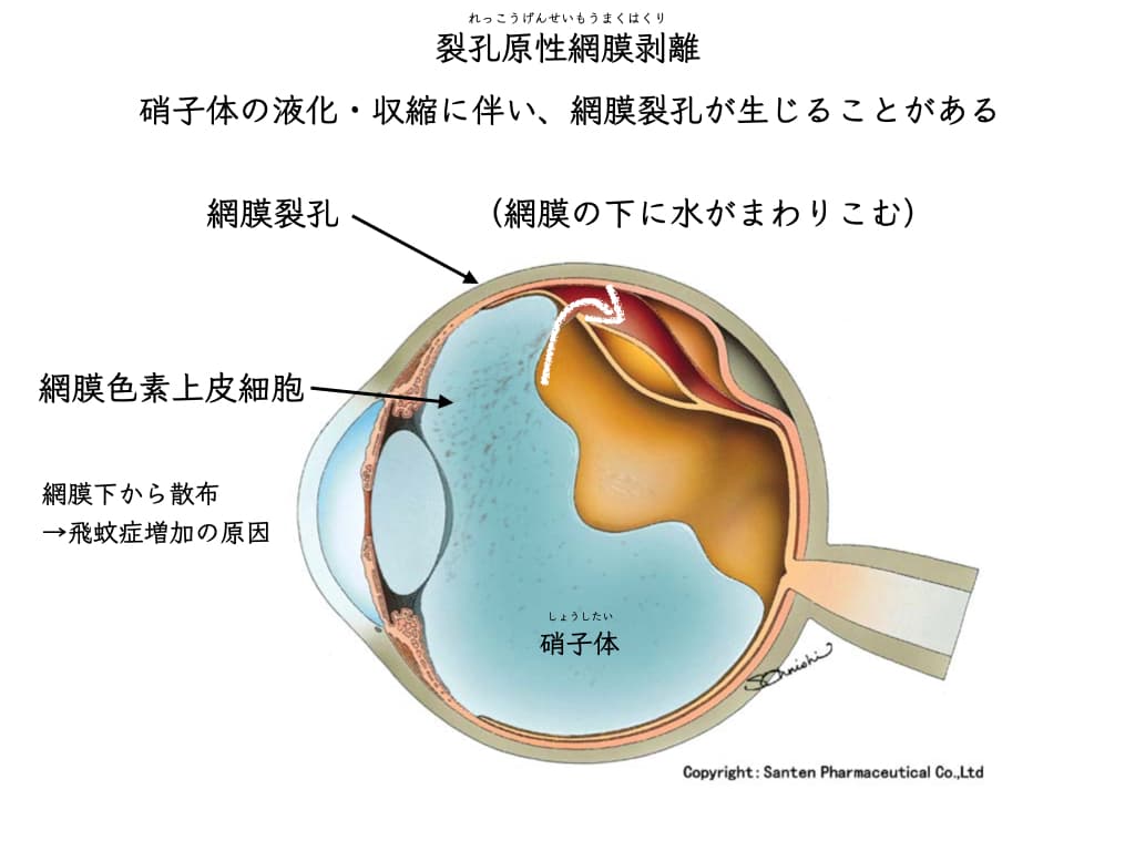
While the retina is detached, the cells that do the damage inside the eye are mainly the “retinal pigment epithelial cells.”
The longer the time since the retina detached, the more the retinal pigment epithelial cells spread inside the eyeball.
The treatment for rhegmatogenous retinal detachment is surgery.
(Once the retina starts to detach, there is no treatment other than surgery.)
In most cases, urgent surgery is needed.
The retina is damaged by retinal pigment epithelial cells scattered inside the eye,
and the function of a retina that was repaired by surgery (that is, properly reattached) remains reduced.
After the retinal detachment has been repaired, it can also cause a membrane to form at the center of the retina.
If the retina does not reattach well after surgery (redetachment), there is an even more troublesome condition:
it can progress to “proliferative vitreoretinopathy.”
“Inflammation inside the eye” and retinal detachment: the importance of early detection

Retinal detachment is a condition in which the “retina” “peels away”:
When a hole opens in the retina and it peels away, it is a “rhegmatogenous retinal detachment.”
There are other types of retinal detachment:
– Exudative retinal detachment (uveitis, intraocular tumors)
– Traction retinal detachment (diabetic retinopathy)
This discussion is limited to rhegmatogenous retinal detachment.
Causes, risk factors, and precautions for retinal tears
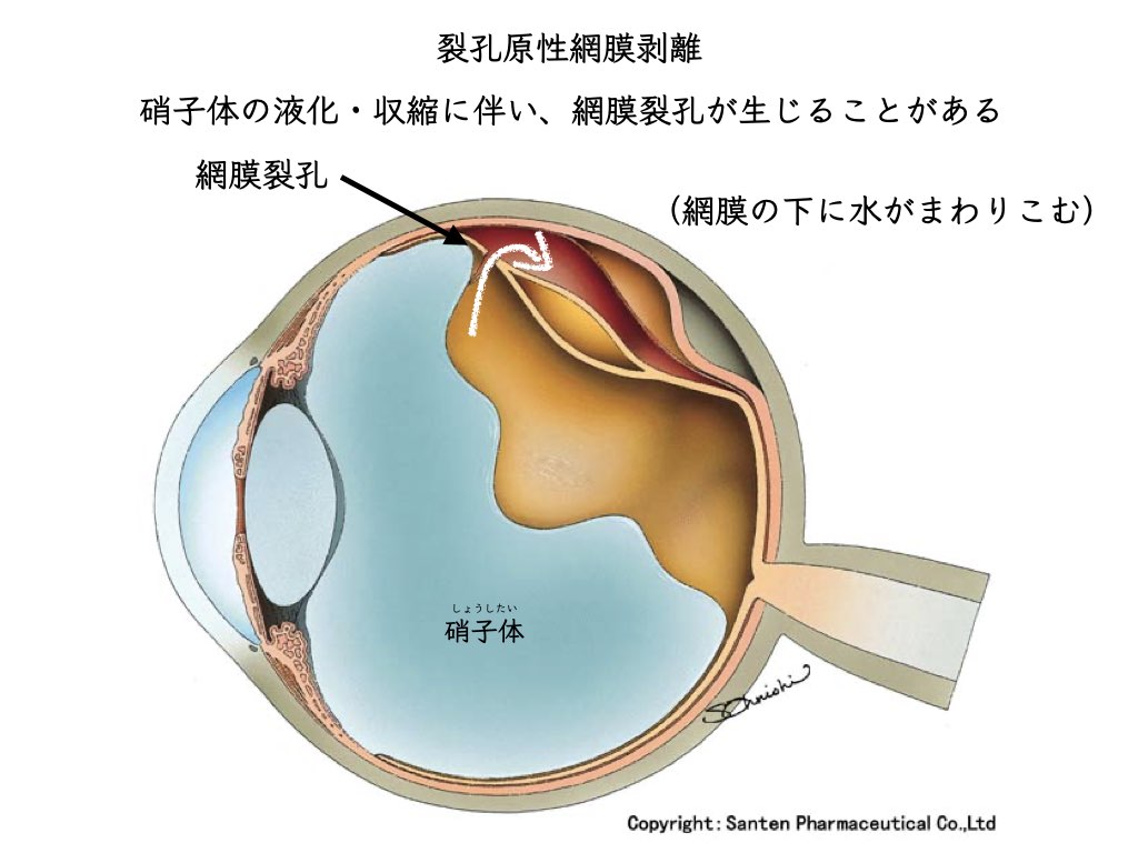
The gel inside the eye pulls on the retina and, at some moment, makes a hole in it.
Besides trauma,posterior vitreous detachmentis often the cause of retinal tears.
The “vitreous,” wrapped in a thin membrane, comes away (peels off) from the retina.
Unlike retinal detachment, posterior vitreous detachment also occurs in normal eyes (physiological).
The common cause of floaters is posterior vitreous detachment.
Floater symptoms may become stronger.
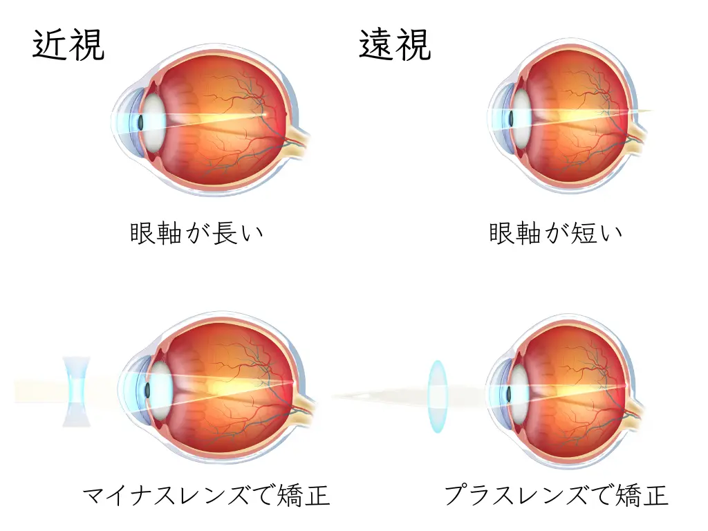
People who are highly myopic often have areas where the retina is thin.
High myopia, “very poor eyesight”: the depth of the eye (axial length) is long.

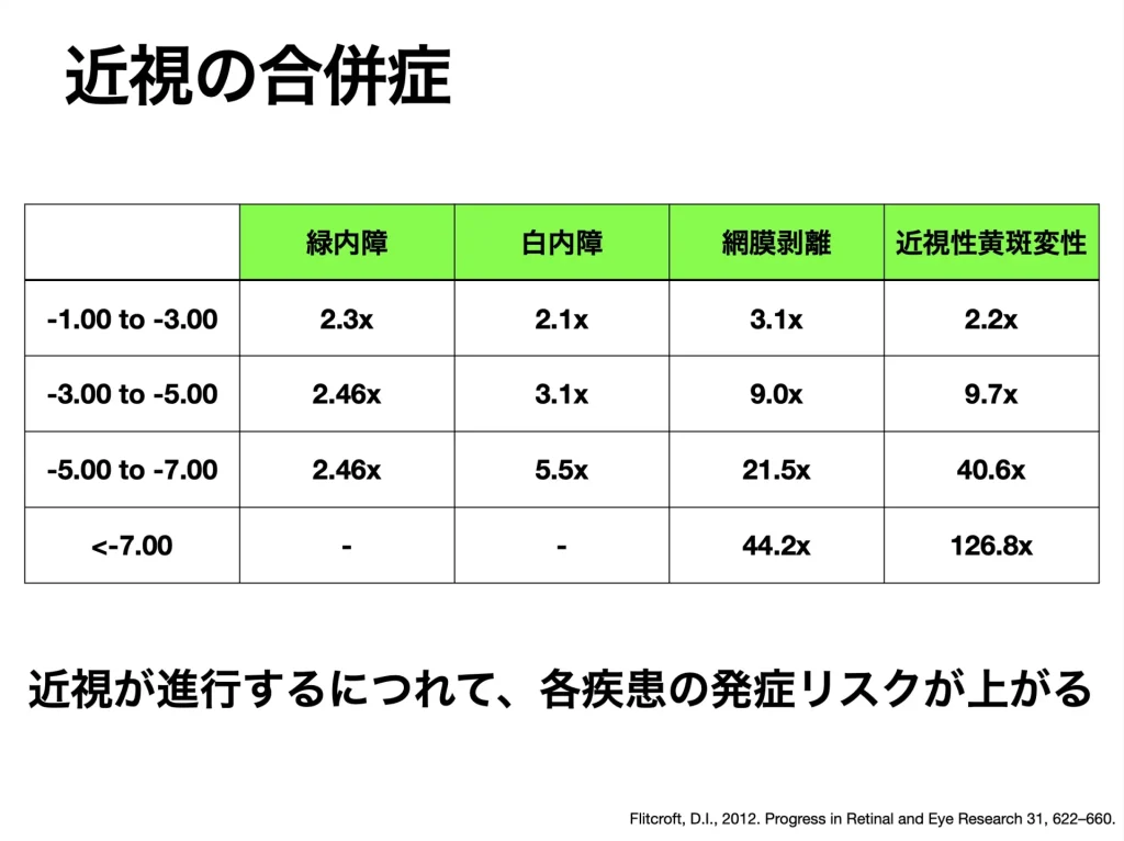
The periphery of the retina (the edge, the front part of the retina) may also be weakened,
and your ophthalmologist may already have pointed out “peripheral retinal degeneration.”
Peripheral degeneration? What should you do if you are not sure yet?
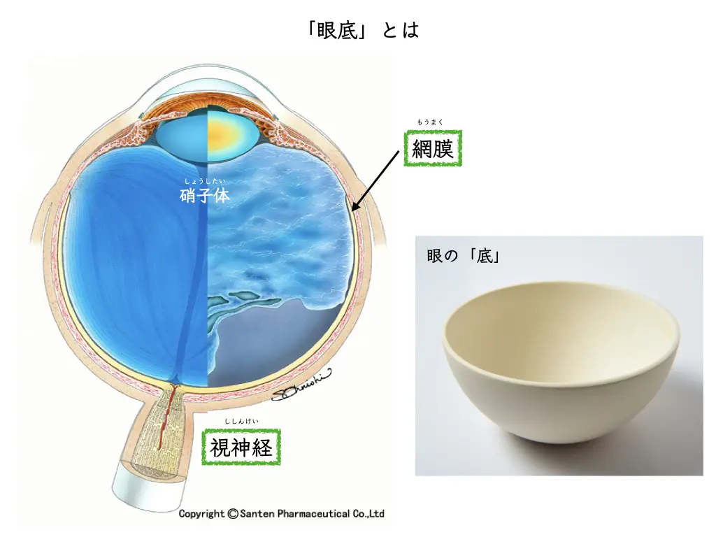
Ophthalmology has changed: we are now in an era when a fundus examination without dilating the pupil can reveal a great deal.
- How strong is your myopia (the refractive power)?
- Whether there is peripheral retinal degeneration or an asymptomatic (symptom-free) retinal tear
Knowing the condition of your own eyes lets you notice quickly when symptoms appear1
Cells that come out from behind the detached retina: retinal pigment epithelial cells
When the retina actually detaches:
Cells come out from the back of the retina and scatter inside the eyeball.
These are a group of cells called “retinal pigment epithelial cells”.
Retinal pigment epithelial cells release “inflammatory cytokines,” soluble proteins that promote inflammation2
After the retina detaches, inflammatory cytokines accumulate inside the eyeball and cause a bad condition (damage to the retinal tissue).
It has been found that a large amount of inflammatory cytokines is stored in the vitreous of an eye with retinal detachment3
Retinal and vitreous diseases are understood by thinking about them from the viewpoint of inflammation in the eye (ocular immunology):
(Inflammatory cytokines are also closely related to the pathology of COVID-19.)
In proliferative vitreoretinopathy, described below, an even higher concentration of inflammatory cytokines has also been found to accumulate.
These are thought to be inflammatory cytokines produced mainly by retinal pigment epithelial cells.
“Macular pucker” (epiretinal membrane) after retinal detachment surgery
After retinal detachment surgery, a state in which “things look distorted” may persist4
The cause of the epiretinal membrane (macular pucker, also called epiretinal membrane or macular epiretinal membrane) after retinal detachment is cell proliferation.
Inflammatory cytokines, the fluid component, increase retinal pigment epithelial cells and nerve-supporting cells (glial cells).5
As a result of the cell proliferation, a membrane forms on the surface of the retina.
An epiretinal membrane causes reduced vision and distorted vision (metamorphopsia: things look distorted).
Surgery to peel the membrane (vitrectomy) may be needed.
An epiretinal membrane does not lead to blindness, so it does not call for an urgent decision about surgery.
What is proliferative vitreoretinopathy?
The tissue change when a “scar” forms is called “fibrosis.”
A scar on the retina can lead to the formation of an abnormal membrane on its surface.
A central factor in fibrosis is the presence of inflammatory cytokines6
An abnormal membrane that has spread over a wide area of the retina tends to contract.
The contracting membrane can cause a further retinal detachment: “traction retinal detachment”
This is why proliferative vitreoretinopathy is called refractory (hard to treat).
A condition that needs vitrectomy:
Unlike an epiretinal membrane, the decision about surgery for proliferative vitreoretinopathy can sometimes be somewhat urgent.
Summary: “Inflammation inside the eye” and retinal detachment: the importance of early detection
After a hole opens in the retina (a retinal tear) and the retina detaches,
the “retinal pigment epithelial cells” scattered inside the eye increase over time.
Inflammatory cytokines produced by retinal pigment epithelial cells cause harmful changes inside the eye to advance over time.
As a result,
- the retina is damaged
The reason that, after retinal detachment surgery, vision seems “somehow harder to see” than in the other eye
may be that, even though the retina has returned to its original position after surgery, its function remains reduced - a macular pucker (epiretinal membrane) forms
it causes the symptom of distorted vision - the eye comes closer to the refractory condition of “proliferative vitreoretinopathy”
It raises the likelihood of needing another operation after retinal detachment surgery.
Using biologics against inflammatory cytokines in the body has changed the treatment of uveitis.
Treatment that suppresses VEGF inside the eye has also greatly changed the treatment of retinal and vitreous diseases.
Controlling inflammatory cytokines inside the eye may also lead to new treatments for retinal detachment.
References
- Flitcroft, D.I., 2012. The complex interactions of retinal, optical and environmental factors in myopia aetiology. Progress in Retinal and Eye Research 31, 622–660. https://doi.org/10.1016/j.preteyeres.2012.06.004
- Bernardo-Colón, A., Lerner, M., Becerra, S.P., 2022. Pigment epithelium-derived factor is an interleukin-6 antagonist in the RPE: Insight of structure-function relationships. Front Physiol 13, 1045613. https://doi.org/10.3389/fphys.2022.1045613
- Yoshimura, T., Sonoda, K.-H., Sugahara, M., Mochizuki, Y., Enaida, H., Oshima, Y., Ueno, A., Hata, Y., Yoshida, H., Ishibashi, T., 2009. Comprehensive Analysis of Inflammatory Immune Mediators in Vitreoretinal Diseases. PLoS ONE 4, e8158. https://doi.org/10.1371/journal.pone.0008158
- Hagler, W.S., Aturaliya, U., 1971. Macular puckers after retinal detachment surgery. Br J Ophthalmol 55, 451–457.
- Sato, T., Takeuchi, M., Karasawa, Y., Ito, M., 2021. Profiles of Cytokines Secreted by ARPE-19 Cells Exposed to Light and Incubated with Anti-VEGF Antibody. Biomedicines 9, 1333. https://doi.org/10.3390/biomedicines9101333
- Chaudhary, R., Scott, R.A.H., Wallace, G., Berry, M., Logan, A., Blanch, R.J., 2020. Inflammatory and Fibrogenic Factors in Proliferative Vitreoretinopathy Development. Translational Vision Science & Technology 9, 23. https://doi.org/10.1167/tvst.9.3.23