![Takeru Eye Clinic | Takatori Shopping Street, Sawara-ku, Fukuoka City [Nishijin Station / Fujisaki Station]](https://takeru-eye.com/wp-content/uploads/2022/10/takeru_logo_for-WP-header.png)

Hayari-me (epidemic keratoconjunctivitis) is very common this year, and at our clinic we feel that many patients have corneal clouding that lingers.
A child catches hayari-me, the parent who nursed the child catches it too, and many fine opacities appear in the parent’s cornea and do not clear easily.
Ophthalmologist acquaintances of mine have also been saying that “many patients have clouding that lingers.”
In this article, I first check with public data whether “it really is more common this year.”
Then I go through, in order, what corneal clouding is, how it is treated, and the matter of eye pressure that needs attention during treatment.
Key Points of This Article
Epidemic keratoconjunctivitis (hayari-me) cases are not unusually high nationwide this year.
Only Fukuoka Prefecture stands out as markedly high, and from late August it has been the prefecture with the most cases in Japan1,2.
Among nationwide detection reports, adenovirus type 54, which has been increasing since around 2023, accounts for most. However, the type responsible for the current outbreak in Fukuoka Prefecture itself has not been confirmed, and the characteristics of type 54 may be involved3,4.
As of September 2026, I could not find any report of a “new variant virus.”
Type 54 is a type known from the start to tend to leave clouding of the cornea (multifocal subepithelial infiltrates)4,5.
Steroid eye drops are used to treat the clouding, but the main purpose is to suppress inflammation that affects vision.
The general approach is to start with a weak steroid and adjust it while watching its effect and the eye pressure6.
The longer the treatment goes on, the more important it is to measure eye pressure regularly.
How Common Is Hayari-me This Year?
About 690 ophthalmology clinics across Japan report their patient numbers every week for hayari-me (epidemic keratoconjunctivitis)1.
The size of an epidemic is compared with the figure of “how many patients per clinic in one week” (the per-sentinel-site report count).
In Week 37 of 2026 (September 7–13), Fukuoka Prefecture’s figure was 4.812.
The national figure for the previous week (Week 36) was 0.65, so Fukuoka Prefecture is about 7 times higher2.
The figure for the same week last year was 1.23, so it is also 3 to 4 times last year’s level2.
By prefecture, Fukuoka Prefecture ranked first in Japan in Week 36 at 4.15, far ahead of second-place Fukushima Prefecture (2.08)8.
Of the 448 cases reported from ophthalmology sentinel sites nationwide, 108 (about 24%) were patients in Fukuoka Prefecture8.
In Fukuoka City alone, the figure has stayed at 2 to 3 in Weeks 34–379.
It has not reached the city’s “warning” threshold (8), but it is clearly higher than in the same period of 2022–2025 (roughly 0 to 1.5)9,10.
Within the prefecture, the Kurume City area and the health center districts of the Chikugo and Chikuho regions have exceeded the warning level (8 or more)2.
On the other hand, looking at the reports so far nationwide, 2026 is lower than last year.
It is also at a low level compared with 2016–2019, before the COVID-19 pandemic11.
In other words, the impression that “hayari-me is common this year” is true of Fukuoka Prefecture, not of the whole country.
Has the Causative Virus Changed?
The adenoviruses that cause hayari-me come in several types, including types 8, 37, 53, 54, 56, and 641.
According to the national pathogen surveillance, the most frequently detected hayari-me virus in 2025 was type 54, with 116 cases3.
This is a sharp increase from 2024 (45 cases)3. The type responsible for this year’s outbreak in Fukuoka Prefecture has not itself been confirmed, but from the nationwide trend, type 54 may be involved.
The Japanese Ophthalmological Society guideline (2025 edition) also states that type 54 “has been detected increasingly again since 2023, and attention to the spread of infection is needed”4.
So has the virus “mutated”?
As of September 2026, as far as I could check public reports in Japan and abroad, I could not find any report of a new type or variant strain of hayari-me virus.
The main player in the outbreak is type 54, an already known type first reported in Japan in 200912.
Why is it so common in Fukuoka this year?
The reason is not written in public materials.
To be honest, the clear reason is not known at present.
Characteristics of Type 54
With type 54 hayari-me, the initial conjunctivitis is of moderate severity, and pseudomembranes (white membranes that form on the inside of the eyelid) are relatively few.
Instead, corneal clouding (multifocal subepithelial infiltrates) occurs at a high frequency of 70–80%, and it is known that many people are left with clouding even after the illness has healed4.
In a 2018 report from ophthalmology clinics in Fukuoka Prefecture as well, clouding of the cornea was seen in 24 of 31 evaluable cases (about 77%)5.
The impression this year that “there is a lot of clouding and it lingers” may be explained by the inherent properties of type 54 rather than by a new variant strain.

What Is Corneal Clouding (MSI)?
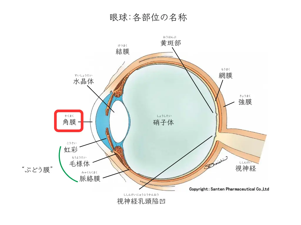
About a week after hayari-me begins, many small white dots may appear scattered over the cornea4.
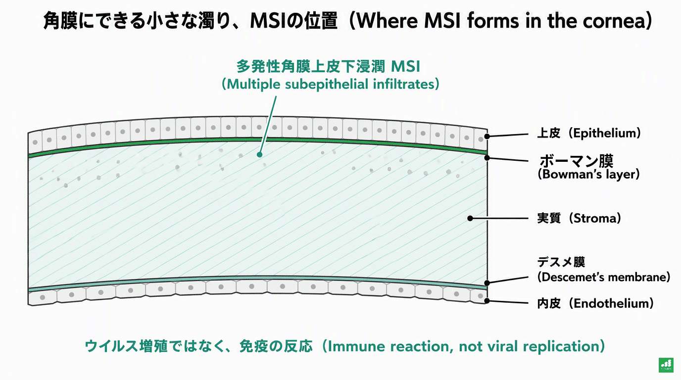
These are multifocal subepithelial infiltrates (MSI).
“Infiltration” refers to a state in which cells that cause inflammation have entered the tissue.
It is a state in which white blood cells have gathered in the most superficial part of the layer called the stroma, just beneath the surface of the cornea.
What matters is that MSI does not mean “the virus is multiplying.”
It is inflammation caused by the body’s immune system reacting to fragments of virus (antigen) left in the cornea4.
In animal experiments, it has been confirmed that even empty virus particles that cannot replicate can cause the same kind of inflammation in the cornea13.
In other words, MSI is an immune reaction that occurs “after” the infectious disease hayari-me.

Does the Clouding Go Away on Its Own?
In many cases it fades over several weeks to several months.
However, a certain number of people have it linger.
In data from 54 patients at a US university hospital, about 1 in 4 (25.9%) had symptomatic clouding that continued for more than 45 days14.
In German real-world data published in 2026 (146 eyes, an observational study that included treated patients), the proportion of eyes with remaining corneal clouding was 45% after 6 weeks, 39% after 6 months, and 9.5% after 1 year15.
In an older study that followed 72 patients with type 8 hayari-me for an average of 2.3 years, clouding remained in 47%16.
When clouding remains, even if visual acuity itself does not fall much, people may feel “glare,” “blurring,” or “reduced contrast.”
This is because the corneal surface becomes slightly irregular (irregular astigmatism), and reduced contrast sensitivity and increased aberrations have also been confirmed on testing17.
The Aim of Treatment and Choosing a Steroid Eye Drop
Steroid eye drops are used to treat MSI.
The first thing to be clear about here is the “aim” of treatment.
Steroid eye drops are not a drug that dissolves and removes the clouding.
They are a drug to suppress inflammation that is affecting vision and to ease glare and reduced vision6.
So if there is a little clouding but it is not troubling your vision, there is also the option of watching it without eye drops.
Conversely, if vision has dropped, or glare is interfering with work or driving, eye drops are used.
There are several kinds of steroid eye drops, differing in strength.
They are weak ones (such as fluorometholone) and strong ones (such as betamethasone and dexamethasone).
At our clinic, depending on each person’s condition, we often consider the order of starting with a weak steroid first and switching to a stronger one if the effect is insufficient6.
Because weak steroids penetrate less deeply into the eye, they are said to cause fewer side effects such as raised eye pressure and progression of cataract6.
In fact, it has been reported that even a weak steroid alone (0.1% fluorometholone) cleared the clouding in 70% of cases in a 6-month clinical trial18.
However, comparative trials that directly tested this “start with a weaker one” order are limited. There are studies comparing loteprednol and dexamethasone, but they compare the effect of the individual preparations and do not test the order itself of “starting weak and strengthening as needed.”
The decision to move to a stronger one when the effect is insufficient, or to use a strong one from the start for a short period, is made individually by the ophthalmologist, based on the degree of clouding, the effect on vision, age, and eye pressure.
Eye Pressure May Rise During Treatment
The side effect of steroid eye drops that needs the most attention is a rise in eye pressure.
If eye pressure is left raised, the optic nerve can be damaged, as in glaucoma.
Classic studies have shown that when a strong steroid eye drop is used for 4 to 6 weeks, eye pressure rises in about 1 in 3 people, and rises greatly (15 mmHg or more) in 1 in 2019,20.
Those more likely to have a rise are people with glaucoma or a family history of it, people with high myopia, people with diabetes, and children21.
In children, the response is known to be quicker and stronger.
In a trial in which a strong steroid (0.1% dexamethasone) was instilled in children under 10, eye pressure rose greatly in more than half, and in many it peaked in about a week22.
When a weak steroid (0.1% fluorometholone) was used in the other eye of the same children, a large rise occurred in only 1 of 1622.
Japanese research has confirmed that even in adults, eye pressure rises less easily with a weak steroid23.
If eye pressure rises, we respond by reducing the steroid, switching to a weaker one, or stopping it.
When MSI still needs treatment, there is the option of switching to another eye drop that does not raise eye pressure (an immunosuppressant eye drop)4. In Japan, its use for MSI is off-label, and whether to use it is decided individually after an examination.
Eye drops that lower eye pressure may also be used together temporarily.
In many cases, eye pressure returns to normal within a few weeks once the steroid is stopped24.
However, it may be harder to return after a long period of use, so regular measurement of eye pressure is essential24.
When Treatment Runs Long
Some people have clouding that does not clear easily and continue eye drops for months.
What is important here is not to “continue steroid eye drops without a clear purpose.”
The longer steroid eye drops are continued, the harder it may be for eye pressure to return to normal, and cataract may progress24.
Some people have been reported to have clouding become dense again (relapse), for example after treatment ends, and in specific trials this is about 15 to 16%18,25.
For that reason, the dosing frequency is gradually reduced before the drops are stopped (tapering).
Once vision has settled, we start reducing the frequency even if some clouding remains.
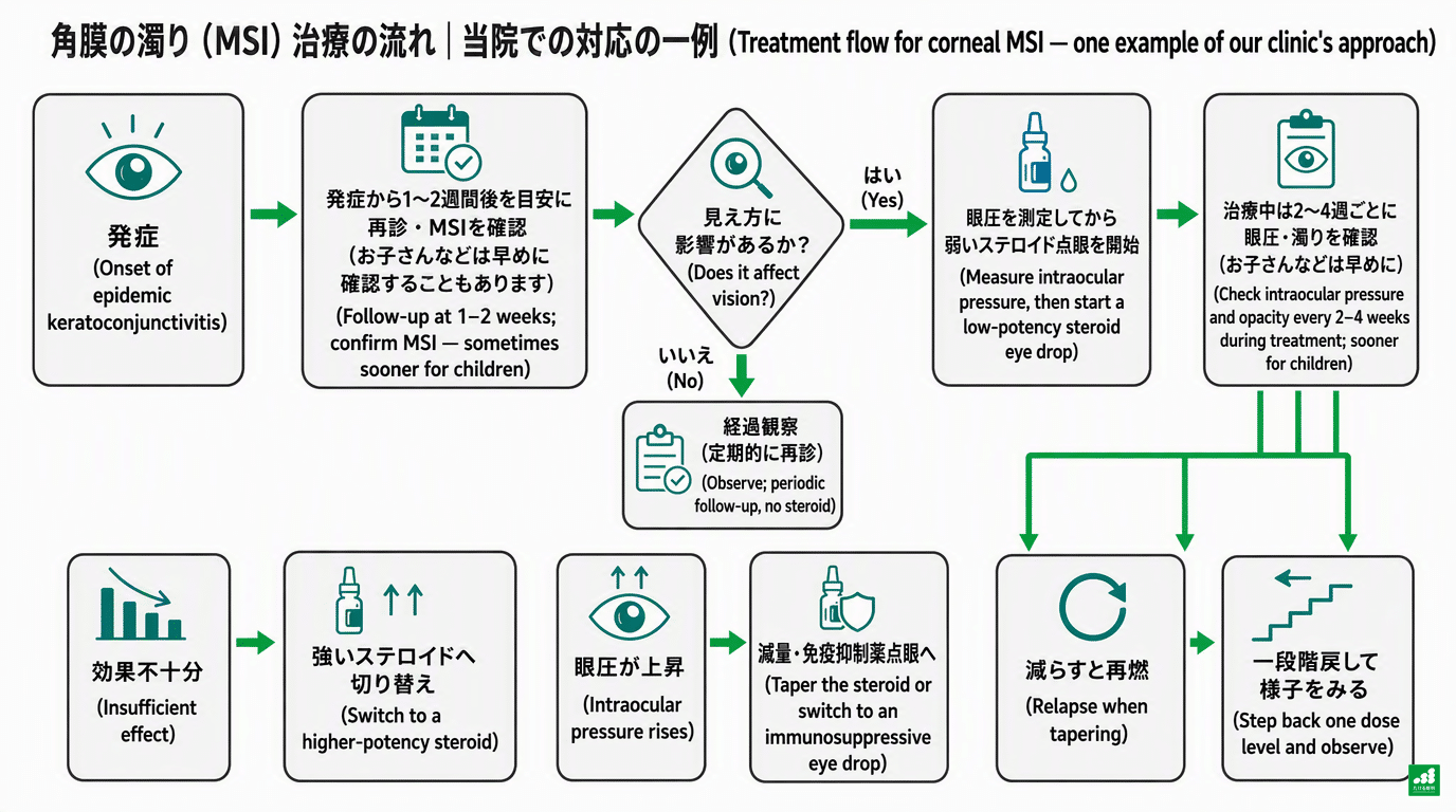
If it relapses after a reduction, we go back one step and watch.
If it relapses with each reduction, or if eye pressure rises, switching to an immunosuppressant eye drop is also proposed in the guideline of the Japanese Ophthalmological Society4.
While treatment continues, we check eye pressure and the state of the clouding about once every 2 to 4 weeks.
Please do not stop eye drops on your own judgment, nor keep using leftover eye drops for a long time.
What We Do at Takeru Eye Clinic (Takatori Shopping Street, Sawara Ward, Fukuoka City)
At our clinic, we ask hayari-me patients to return for a recheck about 1 to 2 weeks after onset.
It is important to check whether clouding has appeared in the cornea after the conjunctivitis symptoms (redness, discharge, etc.) have settled4.
If clouding has appeared, after confirming its effect on vision, we often start with a weak steroid eye drop.
Eye pressure is measured before starting the drops and about 2 weeks after starting.
For children, it is measured a little earlier.
After that, while the drops are continued, we measure eye pressure about once every 2 to 4 weeks.
When the effect is insufficient, when eye pressure has risen, or when it relapses on reduction, we respond by changing the type or strength of the eye drops.
In addition, hayari-me is designated a “Class 3 infectious disease” under the School Health and Safety Act. For that reason, school attendance is suspended until a doctor recognizes that there is no risk of infection26.
We also give guidance at the visit on when you can return to nursery school, school, or work.
What We Would Like Patients to Know
Corneal clouding after hayari-me is not an infection but an immune reaction. However, the presence or absence of clouding alone cannot be used to judge that the possibility of spreading it to others has gone.
In many cases it fades with time, but a certain number of people have it linger.
Steroid eye drops are a drug to suppress inflammation that affects vision, and are used while watching eye pressure and while reducing them.
If you have any concerns such as “I was given eye drops but I don’t know how long to use them,” “the glare continues,” or “I feel the clouding came back after I stopped the drops,” please ask us anything at your visit.
In More Detail: A Summary for Ophthalmologists (September 2026)
From here on is a more detailed summary of the medical evidence. Because the content is specialized, patients are welcome to skip it.
The treatment policies introduced in this article are general information based on the literature and guidelines of academic societies. The actual treatment is decided individually after an examination, so please consult us at your visit for details, including cost and indications.
1. Epidemiology: Fukuoka Prefecture’s Figures and the National Figures
The number of ophthalmology sentinel sites nationwide is about 680 to 690 based on actual measurements (Week 35: 573 cases / 0.84 = about 682)8.
The JIHS explanatory text still says “about 600 sites,” but the number of sentinel sites was changed in Week 15 of 202511.
In 2026 the national figure was 0.3 to 0.5 per sentinel site up to Week 33, lower than in 2025 (0.8–0.9) and also below the pre-COVID-19 period (2016–2020, 0.6–1.0)11.
The Week 34–37 per-sentinel-site report counts for Fukuoka Prefecture (4.23 / 4.88 / 4.15 / 4.81) are, in the prefecture’s weekly report, more than twice the 2025 peak (Week 42, 2.00)2.
Within the prefecture, the Chikugo and Chikuho areas are leading, and Fukuoka City is at 2 to 32,9.
Because Fukuoka City ended district-level publication in Week 52 of 2024, data for Sawara Ward cannot be followed9.
2. Virology: Resurgence of Type 54 and Whether There Has Been a “Mutation”
Among EKC-derived isolates in the JIHS pathogen detection information, type 54 accounted for 45 cases in 2023, 45 in 2024, 116 in 2025, and 34 in 2026 (up to Week 38), and has been the most frequent type since 20253.
According to the Fukuoka Prefectural Institute of Health and Environmental Sciences detection information, no EKC specimens were submitted from April 2025 to July 2026, so there is no typing data from within the prefecture.
Type 54 was detected by the Fukuoka block from pharyngoconjunctival fever specimens in May and July 2026, so circulation of type 54 within the prefecture can be confirmed indirectly.
No report of a novel or recombinant type could be found in 2025–2026 in IASR, local public health institute reports, PubMed, Eurosurveillance, EID, or ProMED.
Type 54 is a recombinant type evolutionarily close to type 8, was reported from Japan in 2009, and has been reported hardly at all overseas12,33.
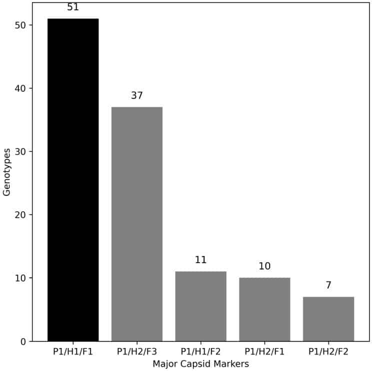
There is also a report that its slow growth in cell culture may contribute to a prolonged course27.
In a study of 250 cases in southern Kyushu, types 8, 37, and 54 were shown to have significantly more corneal complications and a longer duration of illness than types 53 and 5628.
The situation of “a lot of MSI that lingers” can be explained by the combination of the known properties of type 54 and the scale of this outbreak.
3. Choosing a Steroid: The Actual Evidence
I could not find any RCT that directly compared weak and strong steroids.
The basis for judgment is the following indirect data.
| Study | Design | Comparison | Result |
| Gouider 202118 | Prospective double-blind RCT, 72 eyes | 0.1% fluorometholone vs 0.5% cyclosporine | MSI resolution at 6 months 70% vs 47% (p = 0.068). Relapse 16% vs 9%. No eye pressure rise |
| Bhargava 201925 | Double-blind RCT, 45 cases each | 0.03% tacrolimus ointment vs 0.05% dexamethasone ointment, 6 months | Success rate 92.5% vs 85%. Eye pressure rise in 15.6% of the dexamethasone group (mean 1.24 months). Relapse 7.5% vs 15% |
| Koçluk 201729 | Bilateral (paired-eye) comparison | Loteprednol vs dexamethasone | No significant difference in speed of improvement. Relapse in both groups |
| Liu P 20237 | Meta-analysis (5 RCTs + 1 cohort, 286 cases) | Duration of acute-phase steroid | With 7 days there was no difference in MSI occurrence; with 21–28 days it decreased. In the chronic phase, compared with immunosuppressants, there was no difference in resolution rate at 6 months, and eye pressure rose significantly |
Source: each original paper (confirmed on PubMed). Because the subjects, drugs, and durations differ from study to study, treatments cannot be simply compared with one another.
The AAO PPP (2023 approved version) states: “consider the minimum effective dose of steroid if there is reduced vision or photophobia,” “taper slowly down to the minimum effective dose,” and “drugs with little intraocular penetration, such as fluorometholone and loteprednol, may cause less eye pressure rise and cataract”6.
The Japanese Ophthalmological Society Guideline 2025 says steroid eye drops are useful for MSI, but does not specify the preparation, concentration, duration, tapering method, or interval of eye pressure measurement4.
Therefore, the stepwise treatment of “starting with 0.1% fluorometholone and switching to 0.1% betamethasone if the effect is insufficient” can be called a reasonable expert proposal in line with the AAO’s view and the reality of Japanese clinical practice. However, superiority has not been shown in an RCT.
The acute phase is a separate matter: in a rabbit model, even a low-potency steroid such as 0.1% fluorometholone prolonged viral shedding when used for 3 days30.
CQ2 of Guideline 2025 gives a weak recommendation for acute-phase steroid eye drops only “under confirmed etiologic diagnosis, in severe cases, or in combination with iodine-based eye drops”4.
The same guideline also notes that 4.3% of cases reported as EKC included herpes simplex virus conjunctivitis, and cautions against starting steroids casually in the acute phase4.
4. Steroid-Induced Eye Pressure Rise: Frequency, Differences Between Drugs, and Reversibility
In the classic data of Armaly (1965) and Becker (1965), among healthy people who used 0.1% dexamethasone or betamethasone for 4 to 6 weeks, about 5% were high responders (a rise of more than 15 mmHg), about 30% were intermediate responders (6 to 15 mmHg), and about 65% were non-responders19,20.
Risk factors include POAG, a first-degree family history of POAG, high myopia, diabetes, connective tissue diseases, younger age, and a previous steroid response21.
Children: in a bilateral-comparison RCT using 0.1% dexamethasone 6 times a day for 4 weeks in children under 10, 56% were high responders and 37.5% intermediate responders, and 89% of the high responders peaked within 8 days. With 0.1% fluorometholone in the fellow eye, there was 1 high responder out of 16 (6%)22.
Differences between drugs (Japan): in 12 betamethasone high responders, the strength of eye pressure elevation was 0.1% betamethasone > 0.05% > tetrahydrotriamcinolone 1.25% > 0.02% betamethasone > 0.1% fluorometholone. With fluorometholone at 0.05% or below, there was no significant rise23.
Long term: in an 11-month RCT after DMEK, eye pressure rose in 22% of the 1% prednisolone acetate group vs 6% of the 0.1% fluorometholone group (p = 0.0005)31.
Reversibility: normally recovers in 1 to 4 weeks after stopping. In one case series, all cases normalized with use of less than 8 weeks, whereas with use over several years it did not return and treatment was required32. There are also reports that surgery was needed because drugs did not lower it (about 1 to 5%)24.
Summary of Management (expert opinion based on a review)24:
- Reduce or stop the steroid, or change to a low-potency drug (fluorometholone, loteprednol).
- If MSI treatment still needs to continue, switch to an immunosuppressant eye drop (Guideline 2025 CQ3: weak recommendation. In Japan, use of cyclosporine or tacrolimus eye drops for MSI is off-label)4.
- For lowering eye pressure, aqueous suppressants (beta-blockers, carbonic anhydrase inhibitors, alpha-2 agonists) are first choice. Many opinions advise avoiding prostaglandin analogues in inflamed eyes.
- A guide to eye pressure measurement: before starting, 2 weeks after starting (1 week after for children, relatives of POAG patients, and known high responders), every 4 to 6 weeks thereafter, and once after stopping. The AAO PPP calls for “regular eye pressure measurement” but does not give numerical intervals6.
5. Prolonged Treatment and When to Stop
No literature gives an upper limit for the duration of MSI treatment.
In German real-world data, MSI remained in 9.5% after 1 year, and it was reported that chronicity was seen more often in the group that started a steroid at the first visit, but causation has not been proven15.
The risks of long-term use are irreversible eye pressure elevation24, posterior subcapsular cataract (the threshold for eye drops is unknown; it tends to become a problem with use over years), and triggering of infection.
The thinking on stopping can be organized as follows (expert opinion).
- The goal is not the disappearance of the opacity but control of inflammation that affects visual function. Once corrected visual acuity and symptoms improve, start reducing even if opacity remains.
- Reduce in steps (4 times a day → 2 → 1 → every other day). If it relapses, go back one step.
- If there is “steroid dependence,” relapsing with every reduction, or if eye pressure rises, move to an immunosuppressant eye drop4.
- Watching for several months on a small maintenance dose of 0.1% fluorometholone once or twice a day is often done in Japanese clinical practice (expert opinion).
- Relapse after stopping is 15 to 16%18,25. If symptoms return, advise an early revisit.
References
- Japan Institute for Health Security (JIHS) Infectious Disease Information Site. Epidemic keratoconjunctivitis. Updated July 24, 2025. https://id-info.jihs.go.jp/infectious-diseases/epidemic-keratoconjunctivitis/index.html
- Fukuoka Prefecture Infectious Disease Surveillance Center. Fukuoka Prefecture Infectious Disease Surveillance Weekly Report, Reiwa 8, Week 37 (September 7–13, 2026). September 17, 2026. https://www.pref.fukuoka.lg.jp/contents/idwr2026.html (By health center district: http://www.fihes.pref.fukuoka.jp/~idsc_fukuoka/idwr/table_t02/14.html )
- JIHS. Pathogen Detection Information: Viruses isolated and detected from patients with epidemic keratoconjunctivitis (2022–2026). Created September 17, 2026. https://kansen-levelmap.mhlw.go.jp/Byogentai/Csv/data41j.csv (Explanatory page: https://id-info.jihs.go.jp/surveillance/iasr/iass/virus/adenovirus.html )
- Viral Conjunctivitis Clinical Practice Guideline Drafting Committee. Viral Conjunctivitis Clinical Practice Guideline (2025 edition). Nippon Ganka Gakkai Zasshi (J Jpn Ophthalmol Soc). 2025;129(12):1145-1183. doi:10.60330/nggz-2025-038. https://www.nichigan.or.jp/Portals/0/resources/member/guideline/viralconjunctivitis.pdf
- Uemura T, Migita H, Ueno T, et al. Clinical and virological analysis of epidemic keratoconjunctivitis caused by adenovirus type 54 in a regional ophthalmic clinic in Kyushu, Japan. Clin Ophthalmol. 2018;12:511-517. doi:10.2147/OPTH.S148264. https://pubmed.ncbi.nlm.nih.gov/29593376/
- Cheung AY, Choi DS, Ahmad S, et al; American Academy of Ophthalmology Preferred Practice Pattern Cornea/External Disease Committee. Conjunctivitis Preferred Practice Pattern. Ophthalmology. 2024;131(4):P134-P204. doi:10.1016/j.ophtha.2023.12.037. https://www.aao.org/education/preferred-practice-pattern/conjunctivitis-ppp-2023
- Liu P, Li G, Han M, et al. Efficacy and safety of ophthalmic preparations of glucocorticoids in patients with epidemic keratoconjunctivitis: a systematic review and meta-analysis. Ther Innov Regul Sci. 2023;57(3):476-483. doi:10.1007/s43441-022-00479-w. https://pubmed.ncbi.nlm.nih.gov/36414914/
- JIHS. Infectious Diseases Weekly Report (IDWR) provisional data, Week 36 of 2026: report counts and per-sentinel-site counts by disease and prefecture. Created September 9, 2026. https://id-info.jihs.go.jp/surveillance/idwr/provisional/2026/36/index.html
- Fukuoka City Public Health Center. Number of reported infectious diseases (sentinel reports), Reiwa 8, Week 37. Updated September 17, 2026. https://www.city.fukuoka.lg.jp/hofuku/hokensho/kansensho/kansenshojoho/chosa/teitenhoukoku.html
- Fukuoka City Public Health Center. Fukuoka City Infectious Disease Information (explanation of per-sentinel-site counts, etc.): threshold values for warning level. Updated October 23, 2025. https://www.city.fukuoka.lg.jp/hofuku/hokensho/kansensho/kansenshojoho/chosa/houkokukaisetu.html
- JIHS. IDWR 10-year comparison graph (weekly report): epidemic keratoconjunctivitis. https://id-info.jihs.go.jp/surveillance/idwr/graph/weekly/epidemic-keratoconjunctivitis/index.html
- Kaneko H, Suzutani T, Aoki K, et al. Epidemiological and virological features of epidemic keratoconjunctivitis due to new human adenovirus type 54 in Japan. Br J Ophthalmol. 2011;95(1):32-36. doi:10.1136/bjo.2009.178772. https://pubmed.ncbi.nlm.nih.gov/20530657/
- Chintakuntlawar AV, Zhou X, Rajaiya J, Chodosh J. Viral capsid is a pathogen-associated molecular pattern in adenovirus keratitis. PLoS Pathog. 2010;6(4):e1000841. doi:10.1371/journal.ppat.1000841. https://pubmed.ncbi.nlm.nih.gov/20419141/
- Butt AL, Chodosh J. Adenoviral keratoconjunctivitis in a tertiary care eye clinic. Cornea. 2006;25(2):199-202. doi:10.1097/01.ico.0000170693.13326.fb. https://pubmed.ncbi.nlm.nih.gov/16371782/
- Martin Lesan C, Flockerzi E, Löw U, Seitz B. Epidemic keratoconjunctivitis: treatment of subepithelial infiltrates and topographic parameters of the cornea in the postinfection course [in German]. Ophthalmologie. 2026;123(1):40-47. doi:10.1007/s00347-025-02347-y. https://pubmed.ncbi.nlm.nih.gov/41396362/
- Freyler H, Sehorst W. The fate of corneal infiltrations in cases of epidemic keratoconjunctivitis: a follow-up study over two and a half years [in German]. Wien Klin Wochenschr. 1976;88(11):341-343. https://pubmed.ncbi.nlm.nih.gov/997526/
- Aydin Kurna S, Altun A, Oflaz A, Karatay Arsan A. Evaluation of the impact of persistent subepithelial corneal infiltrations on the visual performance and corneal optical quality after epidemic keratoconjunctivitis. Acta Ophthalmol. 2015;93(4):377-382. doi:10.1111/aos.12496. https://pubmed.ncbi.nlm.nih.gov/25043311/
- Gouider D, Khallouli A, Maalej A, et al. Corticosteroids versus cyclosporine for subepithelial infiltrates secondary to epidemic keratoconjunctivitis: a prospective randomized double-blind study. Cornea. 2021;40(6):726-732. doi:10.1097/ICO.0000000000002589. https://pubmed.ncbi.nlm.nih.gov/33201059/
- Armaly MF. Statistical attributes of the steroid hypertensive response in the clinically normal eye. I. The demonstration of three levels of response. Invest Ophthalmol. 1965;4:187-197. https://pubmed.ncbi.nlm.nih.gov/14283012/
- Becker B. Intraocular pressure response to topical corticosteroids. Invest Ophthalmol. 1965;4:198-205. https://pubmed.ncbi.nlm.nih.gov/14283013/
- Phulke S, Kaushik S, Kaur S, Pandav SS. Steroid-induced glaucoma: an avoidable irreversible blindness. J Curr Glaucoma Pract. 2017;11(2):67-72. doi:10.5005/jp-journals-10028-1226. https://pubmed.ncbi.nlm.nih.gov/28924342/
- Kwok AK, Lam DS, Ng JS, et al. Ocular-hypertensive response to topical steroids in children. Ophthalmology. 1997;104(12):2112-2116. doi:10.1016/s0161-6420(97)30052-9. https://pubmed.ncbi.nlm.nih.gov/9400772/
- Kitazawa Y. Increased intraocular pressure induced by corticosteroids. Am J Ophthalmol. 1976;82(3):492-495. doi:10.1016/0002-9394(76)90500-6. https://pubmed.ncbi.nlm.nih.gov/961801/
- Razeghinejad MR, Katz LJ. Steroid-induced iatrogenic glaucoma. Ophthalmic Res. 2012;47(2):66-80. doi:10.1159/000328630. https://pubmed.ncbi.nlm.nih.gov/21757964/
- Bhargava R, Kumar P. Comparison of the safety and efficacy of topical tacrolimus (0.03%) versus dexamethasone (0.05%) for subepithelial infiltrates after adenoviral conjunctivitis. Indian J Ophthalmol. 2019;67(5):594-598. doi:10.4103/ijo.IJO_1352_18. https://pubmed.ncbi.nlm.nih.gov/31007215/
- Enforcement Regulations of the School Health and Safety Act (Ministry of Education Order No. 18 of 1958), Articles 18 and 19. e-Gov Law Search. https://laws.e-gov.go.jp/law/333M50000080018
- Tsukahara-Kawamura T, Hanaoka N, Konagaya M, Uchio E, Fujimoto T. Characteristic of slow growth in cell culture of adenovirus type 54 causing nationwide outbreak epidemic keratoconjunctivitis in Japan. Jpn J Ophthalmol. 2020;64(3):312-320. doi:10.1007/s10384-020-00727-2. https://pubmed.ncbi.nlm.nih.gov/32125553/
- Aoki K, Gonzalez G, Hinokuma R, et al. Assessment of clinical signs associated with adenoviral epidemic keratoconjunctivitis cases in southern Japan between 2011 and 2014. Diagn Microbiol Infect Dis. 2019;95(4):114885. doi:10.1016/j.diagmicrobio.2019.114885. https://pubmed.ncbi.nlm.nih.gov/31607514/
- Koçluk Y, Sukgen EA, Cevher S, Mat E. Symptomatic treatment of subepithelial infiltrates after viral conjunctivitis: loteprednol or dexamethasone? Ocul Immunol Inflamm. 2017;25(5):649-653. doi:10.3109/09273948.2016.1149593. https://pubmed.ncbi.nlm.nih.gov/27015587/
- Romanowski EG, Yates KA, Gordon YJ. Topical corticosteroids of limited potency promote adenovirus replication in the Ad5/NZW rabbit ocular model. Cornea. 2002;21(3):289-291. doi:10.1097/00003226-200204000-00010. https://pubmed.ncbi.nlm.nih.gov/11917178/
- Price MO, Price FW Jr, Kruse FE, Bachmann BO, Tourtas T. Randomized comparison of topical prednisolone acetate 1% versus fluorometholone 0.1% in the first year after Descemet membrane endothelial keratoplasty. Cornea. 2014;33(9):880-886. doi:10.1097/ICO.0000000000000206. https://pubmed.ncbi.nlm.nih.gov/25062336/
- Espildora J, Vicuna P, Diaz E. Cortisone-induced glaucoma: a report on 44 affected eyes [in French]. J Fr Ophtalmol. 1981;4(6-7):503-508. https://pubmed.ncbi.nlm.nih.gov/7299067/
- Dadkhah K, Dehghan S, Chodosh J, Zhang Q, Seto D. Evolution of Human Adenoviruses, a Double-Stranded DNA Viral Pathogen Documented Through Genomics and Bioinformatics and Viewed Through a Web Resource Database. Viruses. 2026;18(2):251. doi:10.3390/v18020251. https://doi.org/10.3390/v18020251