![Takeru Eye Clinic | Takatori Shopping Street, Sawara-ku, Fukuoka City [Nishijin Station / Fujisaki Station]](https://takeru-eye.com/wp-content/uploads/2022/10/takeru_logo_for-WP-header.png)

We place importance on finding keratoconus early.
When needed, we choose an appropriate contact lens that does not damage the corneal epithelium.
“Strong astigmatism,” “contact lenses don’t suit me,” “I still can’t see well even with contact lenses.”
Keratoconus is sometimes discovered from complaints like these.
Keratoconus care

What is keratoconus?
In keratoconus, the cornea (the clear front of the eye) bulges forward and causes irregular astigmatism.
Under the microscope, it may be only slightly visible.
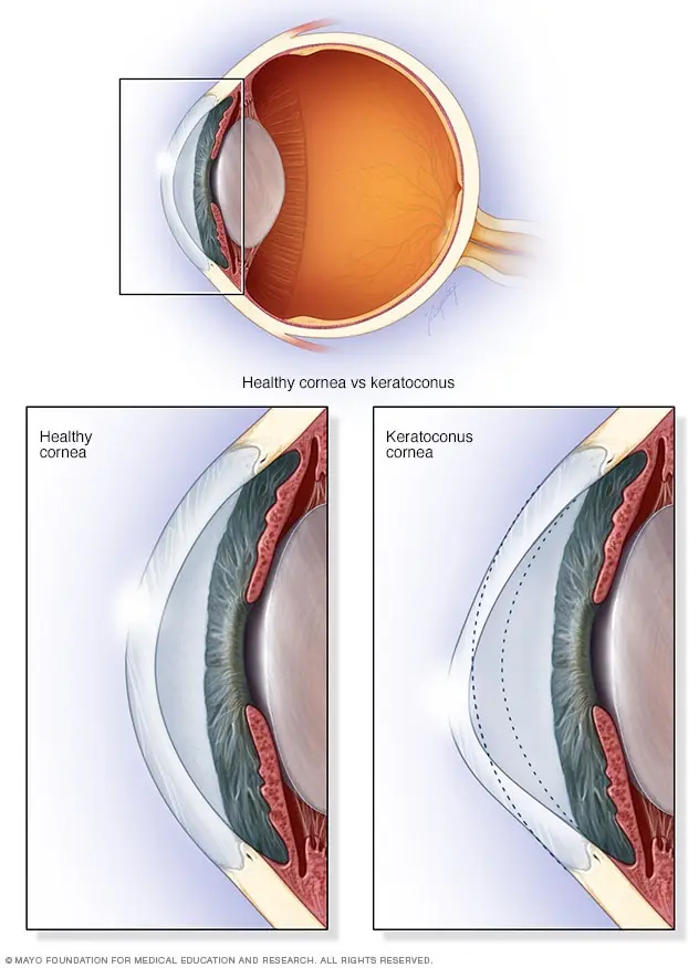
It is a progressive disease and does not involve inflammation.
Corneal topography (a corneal shape analyzer)helps in the diagnosis.
・Worsening irregular astigmatism
・Corneal clouding resulting from “acute hydrops”
can severely impair vision. 1, 2。

◯ Early detection, detailed examination, and follow-up of keratoconus using a corneal shape analyzer
◯ Prescription of special hard contact lenses matched to the shape of the keratoconic cornea (Rose K® lenses)
We provide care together with an orthoptist.
Prevalence of keratoconusis about 1 in 2,000:
It is less common than glaucoma (1 in 20 people aged 40 or older),
and more common than retinal detachment (1 in 10,000). 3。
(That is, keratoconus is 5 times as frequent as retinal detachment.)
Tests for people who visit because contact lenses don’t fit or vision does not improve:
Strong astigmatism may be found.
For people with strong astigmatism of −2 D or more, we screen with a corneal shape analyzer.
As a result, keratoconus is sometimes found.
Contact lens prescription for keratoconus
Soft contact lenses cannot correct the shape of a keratoconic cornea.
Therefore a hard contact lens prescription is needed.
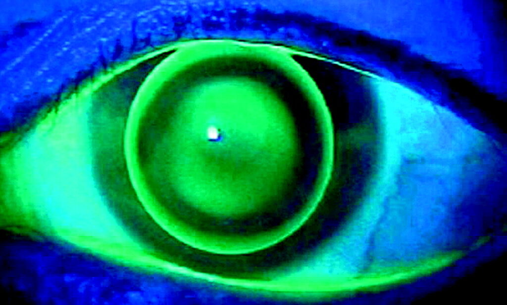
With ordinary hard contact lenses, the protruding part of the keratoconic cornea rubs against the lens,
which can cause a gritty foreign-body sensation and clouding at the rubbed spot:
A properly shaped contact lensis needed. 4。
“Rose K®” contact lenses for keratoconus
This lens was developed by New Zealand optometrist Paul Rose.
It is the only hard contact lens approved by Japan’s Ministry of Health, Labour and Welfare for keratoconus.
Background of development:
Development of the ROSE K® lens began in 1987 with Paul Rose of New Zealand.
He recognized that conventional contact lenses did not fit an abnormal corneal shape, leaving problems with comfort and vision improvement, and he sought a new lens for patients with keratoconus.
Technological evolution:
The ROSE K® lens was brought to market in New Zealand in 1990 and received approval for sale from the U.S. Food and Drug Administration (FDA) in 1995.
Advances in technology later brought new models such as the ROSE K2®, ROSE K2 IC®, and ROSE K2 PG® lenses, allowing a variety of corneal distortions to be accommodated.
IC: for Irregular Cornea (irregularity of the corneal epithelium)/PG: for Post Graft (after corneal transplant surgery)
Worldwide use:
Today, ROSE K® lenses are manufactured in 18 countries and distributed in more than 88 countries. This shows that they have grown into the most frequently prescribed brand of lens for irregular corneas in the world. As medical technology advances, more patients can benefit from comfort and improved vision.
Future outlook:
ROSE K® lenses continue to innovate in the field of lenses for irregular corneas and play an important role in improving patients’ quality of life. Further technological progress is expected to address new conditions and cases.

“Three-point touch”: we choose a lens that optimizes support at the protrusion and the periphery:
There are 960 trial lenses in total.
We choose among them following a protocol so that the lens suits the various grades of keratoconus.
“Selecting the optimal lens” based on a keratoconus lens-fitting methodology is reproducible and does not depend on the advanced lens-modification skills of an individual practitioner.
We select the appropriate base curve and lens periphery so that a very thin layer of tear fluid remains between the protrusion and the hard contact lens (“feather touch”).5。
Keratoconus: keeping up with the latest treatments
Keratoconus used to be a disease with very limited treatment options.
・Management of acute hydrops (steroid treatment)
・Indication for penetrating keratoplasty (full-thickness corneal transplant)
As a resident at a specialized corneal center (the Department of Ophthalmology at Yamaguchi University in 2001), I cared for many corneal transplant patients every week.
Postoperative complications: rejection (1 in 5 patients), loss of the graft requiring repeat surgery, and others
Surgery cannot be undone.
We would like to manage keratoconus with methods other than “surgery.”
A healthy cornea is clear and seems to have no immune reaction.
Rejection occurs after corneal transplantation because immune cells are also present in the cornea. 6。
Today new treatments are available, and early detection of keratoconus has become more important.
◯ “Corneal collagen cross-linking” (CXL) to slow the progression of keratoconus 7
◯ “Intrastromal corneal ring segments” (ICRS) to slow the progression of keratoconus 8
Neither is covered by insurance.
After discussing with the patient whether they are suitable candidates, we refer to or co-manage with the most appropriate corneal specialty center.
We also sometimes consult, using images, with corneal specialists in Japan and abroad who have expertise in specific areas.
Thank you for your continued trust.
(References)
1. Jhanji, V., Sharma, N., Vajpayee, R.B., 2011. Management of keratoconus: current scenario. British Journal of Ophthalmology 95, 1044–1050. https://doi.org/10.1136/bjo.2010.185868
2. Fan Gaskin, J.C., Patel, D.V., McGhee, C.N.J., 2014. Acute Corneal Hydrops in Keratoconus—New Perspectives. American Journal of Ophthalmology 157, 921-928.e1. https://doi.org/10.1016/j.ajo.2014.01.017
3. Ide T., 2019. How to find keratoconus in a clinic. Nihon no Ganka (Ophthalmology in Japan) 90(8): 1024-28.
4. Leung, K.K., 1999. RGP fitting philosophies for keratoconus. Clinical and Experimental Optometry 82, 230–235. https://doi.org/10.1111/j.1444-0938.1999.tb06653.x
5. Mandathara Sudharman, P., Rathi, V., Dumapati, S., 2010. Rose K Lenses for Keratoconus—An Indian Experience: Eye & Contact Lens: Science & Clinical Practice 36, 220–222. https://doi.org/10.1097/ICL.0b013e3181e5cd0b
6. Hamrah, P., Liu, Y., Zhang, Q., Dana, M.R., 2003. The Corneal Stroma Is Endowed with a Significant Number of Resident Dendritic Cells. Invest. Ophthalmol. Vis. Sci. 44, 581–589. https://doi.org/10.1167/iovs.02-0838
7. O’Brart, D.P.S., 2014. Corneal collagen cross-linking: A review. Journal of Optometry 7, 113–124. https://doi.org/10.1016/j.optom.2013.12.001
8. Miraftab, M., Hashemi, H., Hafezi, F., Asgari, S., 2017. Mid-Term Results of a Single Intrastromal Corneal Ring Segment for Mild to Moderate Progressive Keratoconus: Cornea 36, 530–534. https://doi.org/10.1097/ICO.0000000000001115
Photo by Raimond Klavins on Unsplash
» Do you know about keratoconus? Its frequency is five times that of retinal detachment

From Visually.