![Takeru Eye Clinic | Takatori Shopping Street, Sawara-ku, Fukuoka City [Nishijin Station / Fujisaki Station]](https://takeru-eye.com/wp-content/uploads/2022/10/takeru_logo_for-WP-header.png)

Cover one eye and check how things look.
Glaucoma causes noticeable symptoms only after it has progressed considerably.
(In other words, most people have no symptoms, such as missing areas in their vision.)


Even if the visual field of one eye is damaged, the visual field of the other eye compensates for it.
I check the condition of the fundus (optic nerve) almost instantly in nearly everyone who visits the clinic.
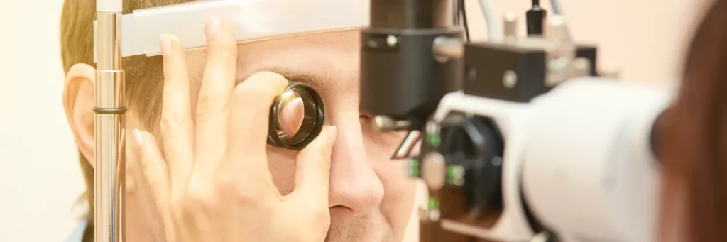
When using a slit-lamp microscope, I hold a small lens (Volk Super Field NC®) in front of the patient’s eye to bring the fundus into focus.
Early detection is very important: “The Importance of Preventive Medicine”
Early Detection of Glaucoma: How to Self-Check for Symptoms
Risk Factors for Glaucoma
• High myopia (being “nearsighted”)
• Age 40 or older
• Having a blood relative with glaucoma, and so on
Taking the above risk factors into account, I may proceed, as needed, with screening using OCT (optical coherence tomography) and Humphrey® static perimetry.
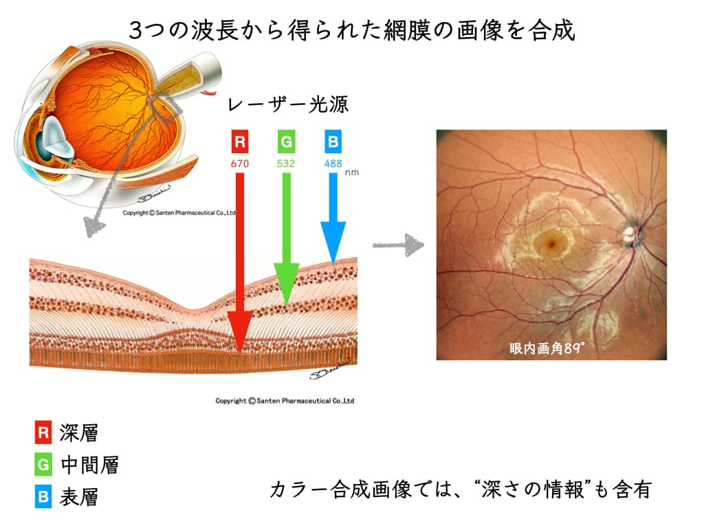
Image analysis using an RGB light source (SLO) can now also help lead to early detection of glaucoma.
I evaluate the thickness of the nerve fiber layer of the retina (fundus) and explain the results to you.
If you are 40 or older, you may also want to consider an Comprehensive Eye Examination (Ganka Dock).

Glaucoma Screening: How It Developed
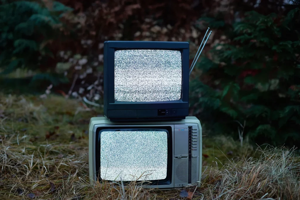
For a long time, methods have been developed that let people check for glaucoma themselves (screening).
○ Noise-Field Test
A system that uses the “snowstorm” static of old television sets, reported by the Department of Ophthalmology at the University of Tokyo in 19911.
In 1995, its effectiveness for self-checking visual field defects and visual field constriction was reported2 .
○ Frequency Doubling Technology(FDT)
Presented by Johns Hopkins University in Baltimore, USA, as a screening method for glaucoma3.
Using a medical device, it measures contrast sensitivity to striped patterns.
A simplified version can also be done with a digital device you already have (see below).
○ Suzuki Eye Check Chart
A chart developed by Dr. Taketoshi Suzuki, an ophthalmologist in Iwate Prefecture4.
In 2006, its effectiveness was verified at Tokyo Medical and Dental University5.
○ Clock Chart
A system developed by the research group of Dr. Chota Matsumoto at the Department of Ophthalmology, Kindai University6.
It lets you self-check for visual field defects and visual field constriction without covering one eye (CLOCK CHART binocular edition, or CCBE).
The latest findings on its effectiveness were updated in November 20197.
Glaucoma Self-Checks: How to Do Them in Practice(Three Methods)
Using a digital device (a computer monitor, tablet, or smartphone),
there are tests you can use to check yourself for glaucoma symptoms.
Here are some useful websites from Pfizer (free to use).
(1) Noise-Field Check
“Simple Noise-Field Check (Self-Check)”
https://www.ntg40.jp/Selfcheck
(2) Clock Chart
“Turn and Check: Visual Field Defect Check”
https://www.ntg40.jp/Selfcheck/Howtouse02
(3) Frequency Doubling Technology (FDT), Simple Version
Simple FDT Check
“How to Do the Sound-Matching Visual Field Check!”
https://www.ntg40.jp/selfcheck/howtouse03.html
The above are simple self-check tests for visual field defects and visual field constriction.
If you notice anything unusual, even slightly, please consult an ophthalmologist.
References
1. Shirato, S., Adachi, M., Hara, T., 1991. Subjective detection of visual field defects using home TV set. Jpn. J. Ophthalmol. 35, 273–281.
2. Adachi, M., Shirato, S., 1994. The usefulness of the Noise-Field Test as a screening method for visual field defects. Jpn. J. Ophthalmol. 38, 392–399.
3. Quigley, H.A., 1998. Identification of glaucoma-related visual field abnormality with the screening protocol of frequency doubling technology. Am. J. Ophthalmol. 125, 819–829. https://doi.org/10.1016/s0002-9394(98)00046-4
4. Suzuki T: Suzuki Eye Check Chart. Shikaku no Kagaku (Vision Science) 22: 133-135, 2001.
5. Murai, H., Kiyosawa, M., Mochizuki, M., Suzuki, T., 2006. Sensitivity and Specificity of New Eye Check Chart for Neuro-ophthalmological Diseases. Jpn J Ophthalmol 50, 383–386. https://doi.org/10.1007/s10384-006-0329-x
6. Matsumoto, C., Eura, M., Okuyama, S., Takada, S., Arimura-Koike, E., Hashimoto, S., Tanabe, F., Shimomura, Y., 2015. CLOCK CHART®: a novel multi-stimulus self-check visual field screener. Jpn J Ophthalmol 59, 187–193. https://doi.org/10.1007/s10384-014-0368-7
7. Ishibashi, M., Matsumoto, C., Hashimoto, S., Eura, M., Okuyama, S., Nomoto, H., Tanabe, F., Kayazawa, T., Numata, T., Kusaka, S., 2019. Utility of CLOCK CHART binocular edition for self-checking the binocular visual field in patients with glaucoma. Br J Ophthalmol 103, 1672–1676. https://doi.org/10.1136/bjophthalmol-2018-312845