![Takeru Eye Clinic | Takatori Shopping Street, Sawara-ku, Fukuoka City [Nishijin Station / Fujisaki Station]](https://takeru-eye.com/wp-content/uploads/2022/10/takeru_logo_for-WP-header.png)

Dry eye:
even when it differs from the image the word brings to mind, it is often judged to be dry eye.
The term “dry eye” tends to bring to mind the image of eyes that feel dry,
but in reality its definition and diagnostic criteria are more complex.
Dry eye is not simply “dry eyes”; various factors and symptoms are involved.
There are many meanings beyond “dry” and “dryness.”
The current diagnostic criteria (definition) for dry eye are as follows.1。
They were presented by the Japanese Ophthalmological Society in 2016.
(The word “dry” is not actually written in the Japanese name for dry eye.)
- If the tear film break-up time (BUT) is 5 seconds or less,
- and the patient has subjective symptoms (eye discomfort or visual disturbance), dry eye is diagnosed.
The 2019 criteria2aim to evaluate in more detail the patient’s symptoms and the condition of the eye when diagnosing dry eye.
This diagnostic criterion for dry eye2(definition) is explained here in plain terms.
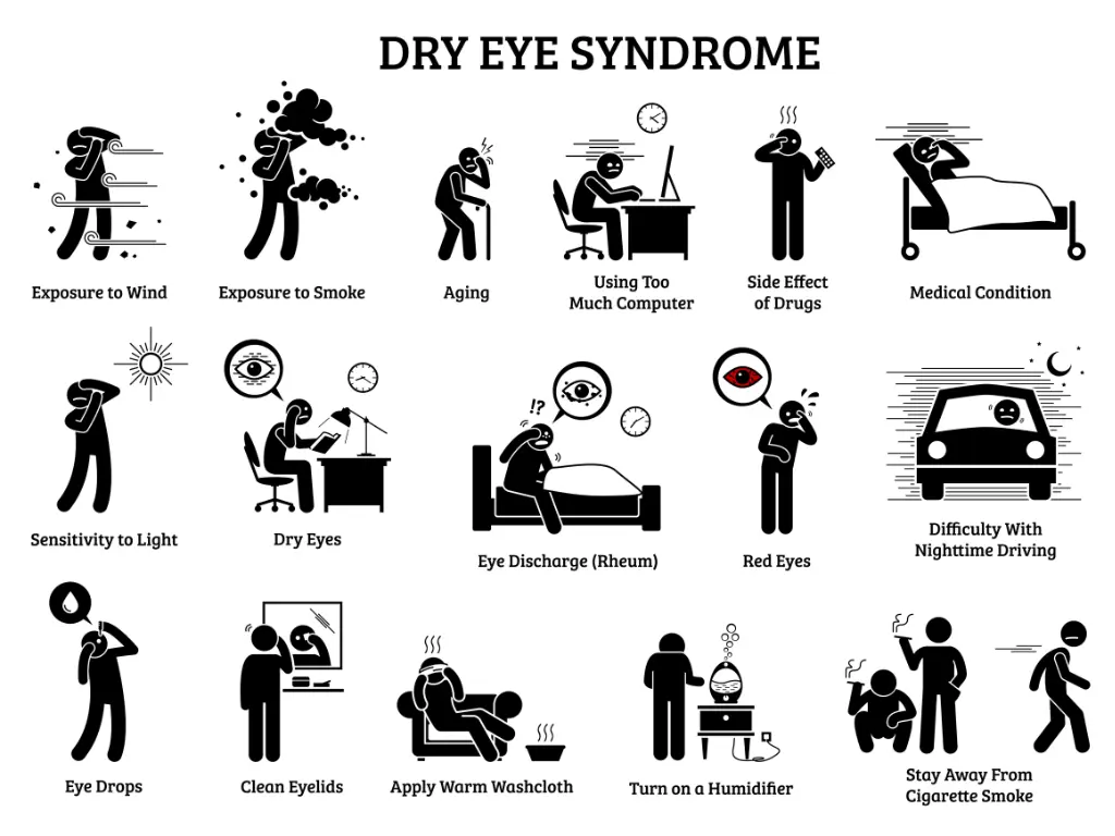
What Is “Dry Eye” Even When Your Eyes Do Not Feel “Dry”?
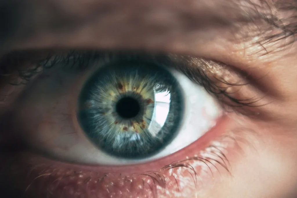
Two Kinds of Tears: What Are Basal Tears?
There are two ways tears are secreted.
- Emotional secretion: tears that come out in response to emotions
- Basal secretion: tears that are secreted constantly to keep the eyes healthy
Tears that come when you are happy, sad, or moved (emotional secretion),
and the tears involved in dry eye (basal secretion) have different properties.
In dry eye that does not feel “dry,” the condition of the basal tears is the problem.3。
Basal tears have a three-layer structure.
- Oil layer (lipid layer / surface film): prevents evaporation and helps tears spread evenly over the surface of the eye.
- Aqueous layer (water-soluble layer): keeps the eye clean and supplies nutrients.
- Mucin layer (mucous layer): spreads the tears evenly and protects the eye.
Basis for Judging Dry Eye

Two criteria are used.
1. Tear film break-up time (BUT)
When you blink, tears spread over the surface of the eye by surface tension. This is called the tear film (in Japanese, “namida no film”).4。
The stability of this “tear layer” can be checked (evaluated) easily at the eye clinic.
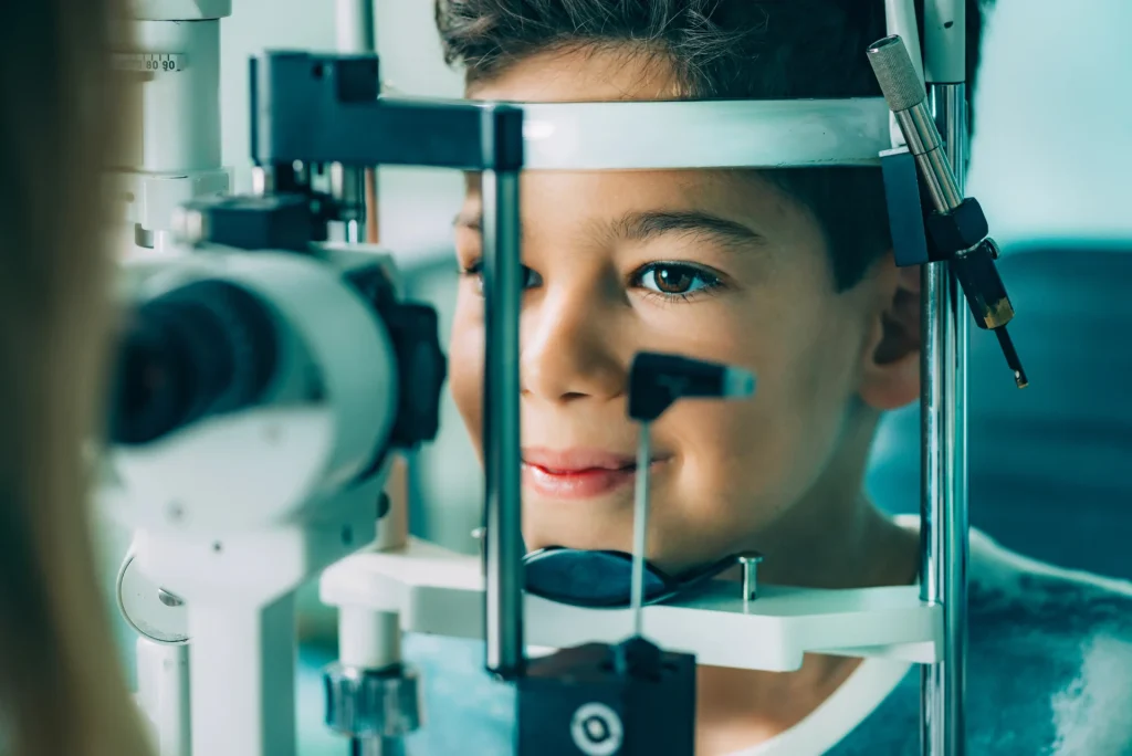
- A small amount of fluorescein dye strip is touched to the lower eyelid
- Blinking spreads the dye over the surface of the eye
- The ophthalmologist observes with the slit-lamp microscope that is always found in an eye clinic examination room
With this, we mainly look at the following points.
- Whether the tear film is spread neatly (whether the three-layer structure of the tears is maintained)
- How long until the tear layer breaks up (Break Up Time, BUT)
- Whether there are any injuries on the eye
2. Subjective symptoms (eye discomfort or visual disturbance)
We ask about subjective symptoms during the interview at the visit.
- Eyes feel dry
- Eyes feel gritty
- Eyes hurt
- Redness
- Eyes feel heavy
- Watery eyes
- Tears come out on their own
- Tears come out when going outside
- Eye discomfort means: dryness, a foreign-body sensation, pain, itching, and so on
- Visual disturbance means: reduced vision, blurring, reduced contrast sensitivity, and so on5
Most dry eye symptoms are something other than “dry eyes,” aren’t they?
If you have the symptoms above, you may meet the diagnostic criteria for dry eye.
Causes of and Measures for “Dry Eye” Even When Your Eyes Do Not Feel “Dry”
The three-layer structure of tears: what is the state of each layer?
This is how we think about it.
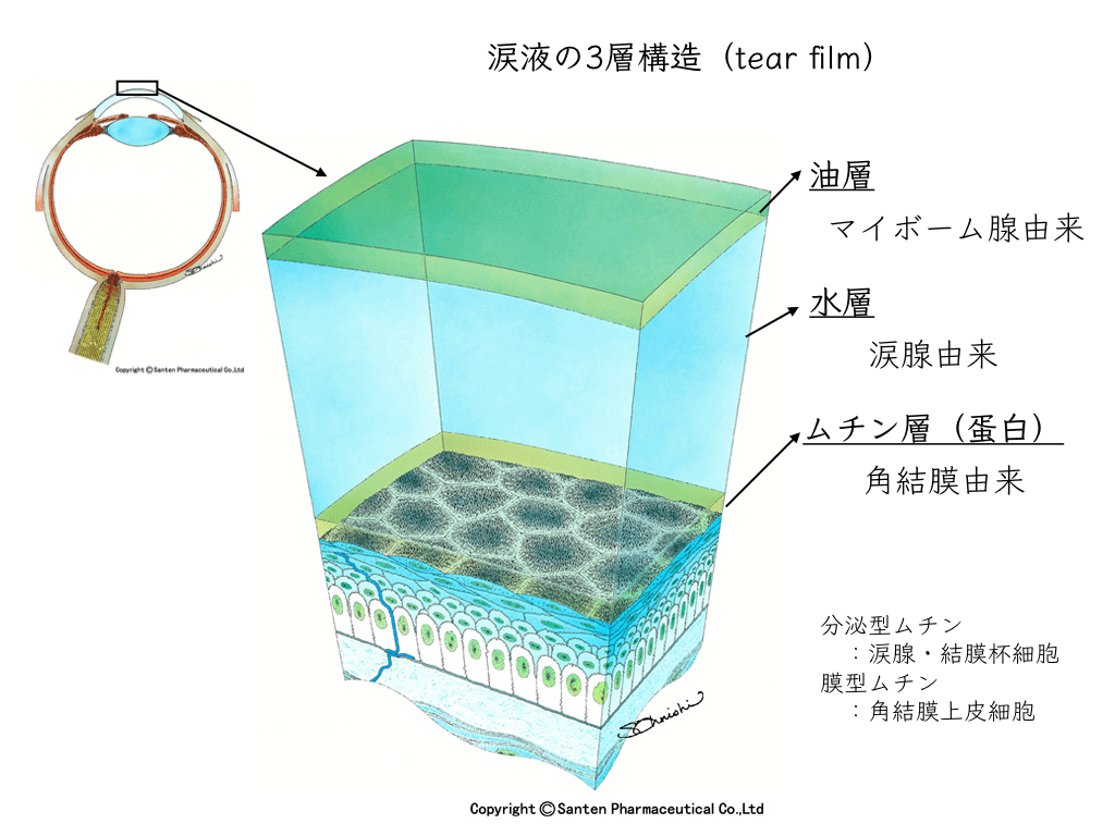
- Oil layer (lipid layer/ surface film): prevents evaporation and helps tears spread evenly over the surface of the eye.
Meibomian glandssecrete lipids (oil) that float on the surface of the tears and form a barrier against evaporation.
This allows tears to stay on the surface of the eye for a long time and keep it moist. - Aqueous layer (water-soluble layer): keeps the eye clean and supplies nutrients.
It consists of water secreted by the lacrimal gland and is the main component of the tears.
The aqueous layer washes the surface of the eye, removes foreign matter and bacteria, and supplies oxygen and nutrients to the cells of the eye. - Mucin layer(mucous layer): spreads the tears evenly and protects the eye.
It consists of mucus (mucin) secreted by the goblet cells of the conjunctiva and helps the tears spread evenly over the surface of the eye.
The mucin layer holds the tears on the surface of the eye, makes them glide smoothly, and protects the cornea and conjunctiva.
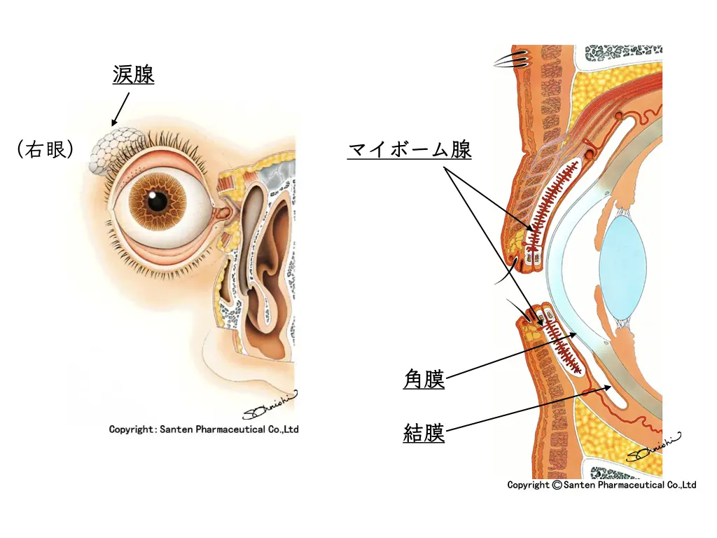
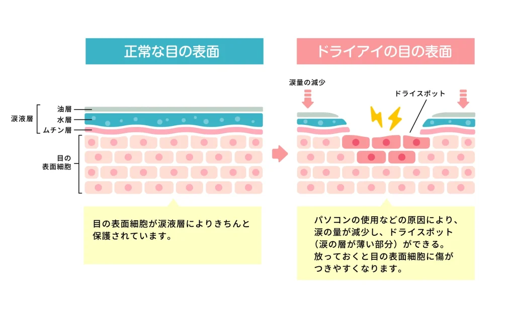
The three layers working together as the “tear film”
moisten and protect the surface of the eye and maintain comfortable vision.
By correctly understanding the role of each layer, the diagnosis and treatment of dry eye can be carried out more effectively.6。
The measures for each layer are as follows.
- Oil layer:Warming the eyeshelps the meibomian glands secrete oil and stabilizes the layer.
- Aqueous layer: supplemented with hyaluronic acid eye drops.
- Mucin layer: medications such as Diquas® and Mucosta® are used.
As an initial approach, the basic step is to calm inflammation on the surface of the eye.
In Europe and the United States, immunosuppressant eye drops (cyclosporine) are commonly used.
In Japan they are not covered by insurance, so we use steroid eye drops (Flumetholon®), which are used very often in ophthalmology.
Because it is diluted to 0.1%, it has the advantage of causing almost no side effects.
With Flumetholon®, complications such as increased eye pressure,steroid-induced glaucoma, and cataract formation are unlikely, but they can occur in steroid responders.7。
Eye pressure can rise very rarely, so it needs to be checked at the eye clinic from time to time.
“I feel like my eye pressure has gone up.”
Eye pressure increases that cause symptoms you can feel are almost nonexistent.
We check the actual numbers with the tonometers at the eye clinic.
There are mainly three ways to measure eye pressure.
A. Air-puff tonometer: the machine used first in eye exams
B. Goldmann applanation tonometer: used when eye pressure needs to be measured more accurately, such as in glaucoma or suspected glaucoma
C. A newer tonometer: iCare®

For people who dislike the air-puff tonometer or are very tense (which can give inaccurate values),
we use the newer tonometer, iCare®.
If that does not work well, we think about the next approach.
Punctal plugs may be considered.
For dry eye,IPL (Intense Pulsed Light) treatment needs to be considered carefully.
“Dry eye” even when your eyes do not feel “dry”:
We think of it mainly in terms of controlling inflammation of the ocular surface.
Discomfort After Cataract Surgery and Other Intraocular Surgery
- 見えるのは見える
- どうかある
- 違和感が残っている
After cataract surgery, when you can see but discomfort remains,
“dry eye” even without feeling “dry” may be a cause.
Increasing the stability of the tear film may be important.
Causes of Tired Eyes and Eye Strain
Along with tension of the ciliary muscle,
“dry eye” even without feeling “dry” is considered one of the causes.
We use an accommodation analyzerto judge the current state.
Dry Eye Diagnostic Criteria: Differences from Before 2016
Previously, the amount of tear secretion and damage to the corneal and conjunctival epithelium were included in the diagnostic criteria.
- The condition of the tears that always moisten the surface of the eye (the tear film)
- Whether there are injuries on the black and white parts of the eye
- Whether there are eye injuries
- The degree of the injuries
These items were removed in the 2016 revision.
Now the diagnostic criteria consist only of tear film instability (shortened BUT) and subjective symptoms.1,2。
With this change, tear-film-unstable dry eye came to be diagnosed appropriately.
The image the name “dry eye” gives and the actual condition are very often different.
Although it is not an official name,
“tear film instability syndrome”may help in understanding “dry eye.”
Dry Eye Diagnostic Criteria: Differences Between 2016 and 2019
The main differences between the 2016 and 2019 dry eye diagnostic criteria are as follows:
Clarification of the diagnostic criteria:
- 2016: Dry eye was defined as a “disease in which tear film stability is reduced,” with tear film break-up time (BUT) and subjective symptoms as key elements of diagnosis.
- 2019: While following the 2016 definition, the diagnostic criteria were further clarified, with specific evaluation methods and diagnostic indicators emphasized.
Importance of subjective symptoms:
- 2016: It was emphasized that subjective symptoms (eye discomfort or visual disturbance) are included in the diagnosis.
- 2019: Evaluation of subjective symptoms received more weight, and it was emphasized that the patient’s subjective experience plays an important role in diagnosis.
Comprehensiveness of the diagnosis:
- 2016: The focus was on reduced stability of the tear film.
- 2019: Not only tear film stability but also the health of the ocular surface and evaluation of inflammation were considered, taking a more comprehensive approach.
The 2019 criteria aim to evaluate patients’ symptoms and the condition of the eye in more detail when diagnosing dry eye.
This is thought to make it possible to choose more appropriate treatments for dry eye.
References
- Japan Dry Eye Society, 2016. Revision of the definition and diagnostic criteria for dry eye in Japan (2016 version). Journal of the Japanese Ophthalmological Society, 120(4), 284-293. p. 290. https://www.nichigan.or.jp/member/journal/guideline/detail.html?itemid=309&dispmid=909
- Dry Eye Clinical Practice Guidelines. Journal of the Japanese Ophthalmological Society 123: 489-592, 2019 https://www.nichigan.or.jp/member/journal/guideline/detail.html?itemid=315&dispmid=909
- Messmer, E.M., 2015. The Pathophysiology, Diagnosis, and Treatment of Dry Eye Disease. Dtsch Arztebl Int 112, 71–82. https://doi.org/10.3238/arztebl.2015.0071
- Zeev, M.S.-B., Miller, D.D., Latkany, R., 2014. Diagnosis of dry eye disease and emerging technologies. Clinical Ophthalmology 8, 581–590. https://doi.org/10.2147/OPTH.S45444
- Tsubota, K., Pflugfelder, S.C., Liu, Z., Baudouin, C., Kim, H.M., Messmer, E.M., Kruse, F., Liang, L., Carreno-Galeano, J.T., Rolando, M., Yokoi, N., Kinoshita, S., Dana, R., 2020. Defining Dry Eye from a Clinical Perspective. Int J Mol Sci 21, 9271. https://doi.org/10.3390/ijms21239271
- Pflugfelder, S.C., Stern, M.E., 2020. Biological Functions of Tear Film. Exp Eye Res 197, 108115. https://doi.org/10.1016/j.exer.2020.108115
- Morrison, E., Archer, D.B., 1984. Effect of fluorometholone (FML) on the intraocular pressure of corticosteroid responders. Br J Ophthalmol 68, 581–584.