![Takeru Eye Clinic | Takatori Shopping Street, Sawara-ku, Fukuoka City [Nishijin Station / Fujisaki Station]](https://takeru-eye.com/wp-content/uploads/2022/10/takeru_logo_for-WP-header.png)

After a blunt injury to the eye, the pupil may remain dilated.
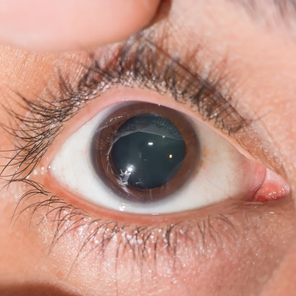
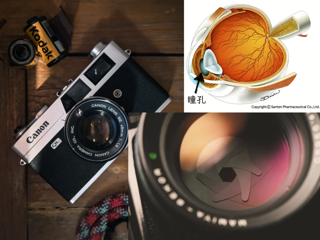
If you compare the eye to a camera,
the “pupil” corresponds to the “aperture.”
It adjusts the amount of light entering the eye or camera.
It acts like a gatekeeper, regulating how many rays of light enter the eye or camera.
David Bowie(1947–2014) had one green eye and one black eye:
As a child he was hit by a blowpipe dart, and his left pupil is said to have remained dilated.
He had anisocoria (unequal pupils).

As a result, David Bowie’s right eye looked green and his left eye looked black.
Right eye: the iris color typical of British people, green
(Almost all Asian people have brown irises)
Left eye: the color of the back of the eye, seen through the pupil that stayed open, is black
The symptom of traumatic mydriasis is “persistent glare after the eye has been hit”.
The pupil stays dilated: “mydriasis”

The cause is
・relaxation of the iris muscle“sphincter pupillae” or
・damage to the “parasympathetic nerve” that controlsthe movement of the sphincter pupillae
.
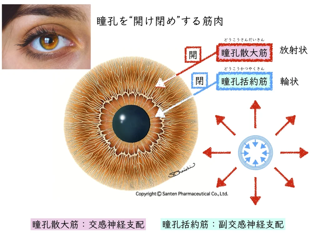

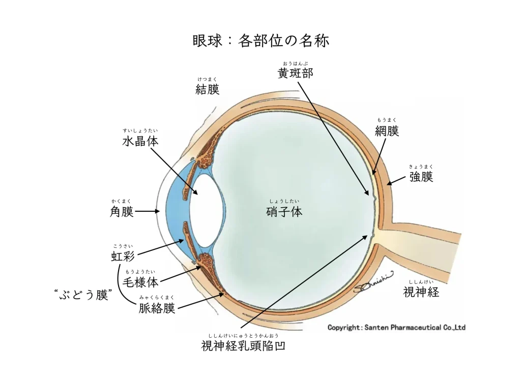
When an eye is injured, for example by being hit or struck by a ball,
・the iris (sphincter pupillae and dilator pupillae muscles)
・the iris nerves
・the ciliary body
can be damaged.
It is known that the pupil first becomes small and then dilates as the pupillary reaction declines.
Spasm of the ciliary body causes eye pain,
and it may then shift into a state of accommodative paralysis.
The ability to focusbecomes paralyzed.
When the ciliary body is examined histologically, inflammatory cells gather and tissue changes (scar formation and atrophy) are seen.1。
The muscle that adjusts focus becomes inflamed and ends up scarred.
Atropine eye drops instilled in the exam room paralyze the spasming ciliary body.
This helps reduce pain right after the injury.
References
1. Kawasaki A: Disorders of pupillary function, accommodation, and lacrimation. In Miller NR, Walsh FB, Hoyt WF (Eds.), Walsh and Hoyt’s clinical neuro-ophthalmology. Lippincott Williams & Wilkins: Philadelphia, pp. 739—805, 2005.