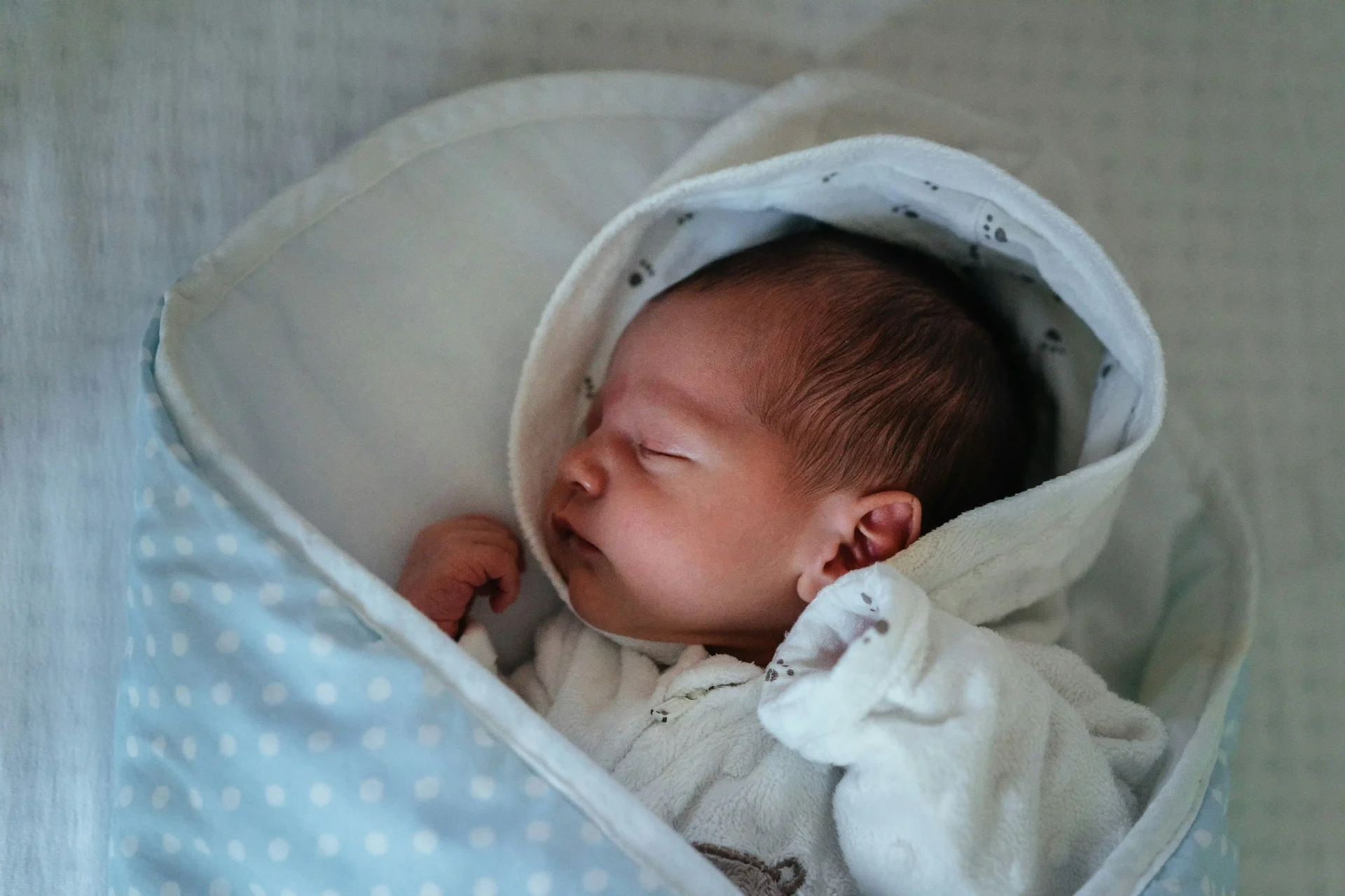
Babies see a great deal after birth, and in doing so they develop their ability to see.

Light entering the retina travels to the back of the head (the primary visual cortex in the occipital lobe of the cerebral cortex), where the ability to process visual information develops.
Information from the retina builds connections between nerve cells at their synapses.
“Amblyopia”is a term that describes
a condition in which, because of some factor during the growing years,
the ability to see has not developed properly.
(Note)
It is entirely different from low vision.
Causes of Amblyopia (Visual Development in Children)
When there is a marked refractive error (a problem with the eye’s power)
A newborn baby’s eye is short from front to back (axial length), which means farsightedness, and it lengthens as the child grows (which means nearsightedness).

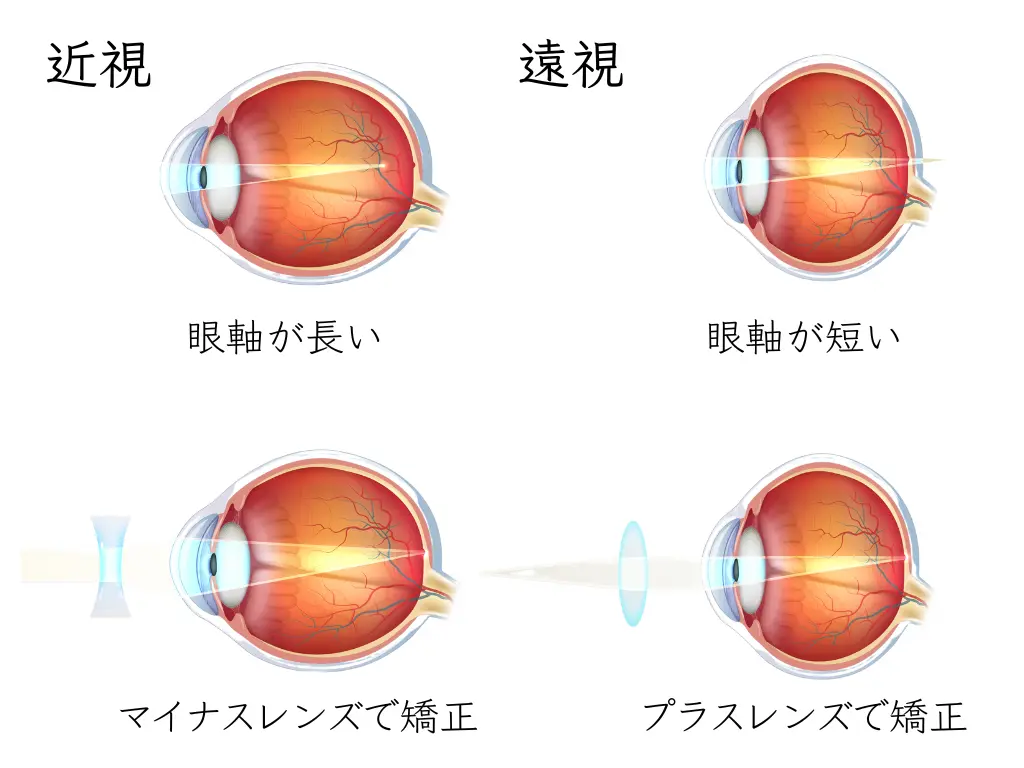
When there is mainly strong farsightedness, the image that falls on the retina stays blurred.
This is the cause of many cases of amblyopia.
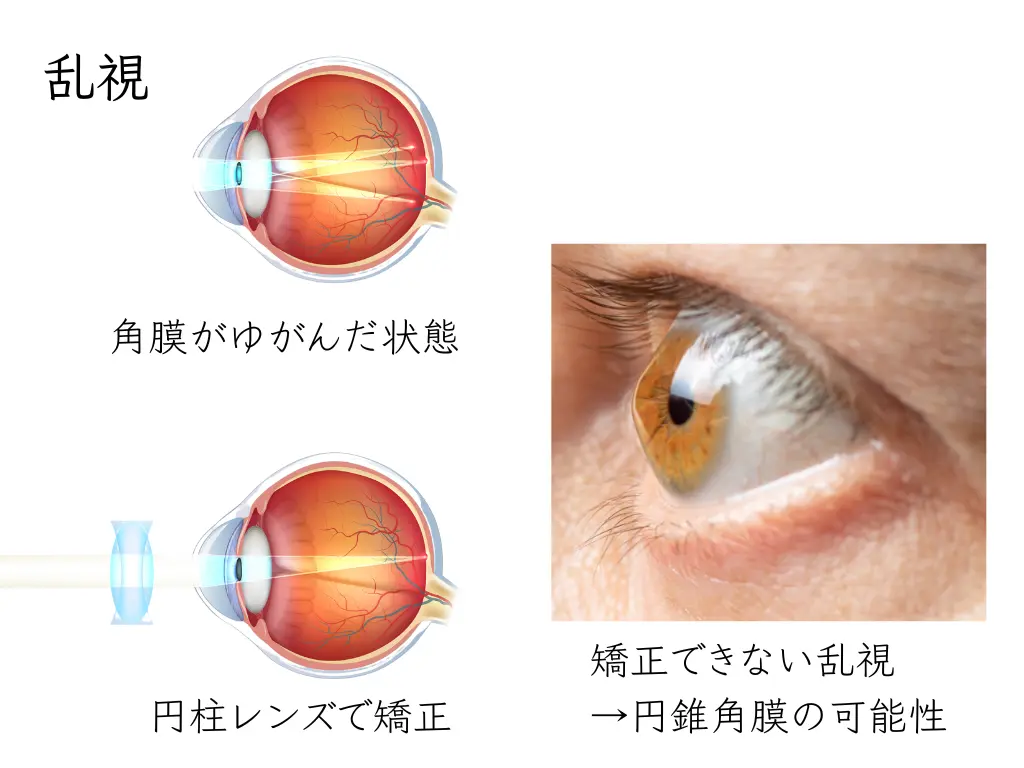
Strong distortion of the cornea (the clear front part of the eye), known as astigmatism,
or an eye that has lengthened too much (nearsightedness), can also cause amblyopia.
= Refractive amblyopia
When the eyes are misaligned
When we look at something, we aim the light at the center of the retina, a spot called the fovea.
Six muscles attached around the eyeball (the extraocular muscles) move both eyes so that the target is seen with the fovea.
✔ Extraocular muscles: superior rectus, inferior rectus, superior oblique, inferior oblique, medial rectus, lateral rectus
Eye movements are controlled by thin nerves that extend from the brain.
✔ Nerves involved in eye movement: trochlear nerve (superior oblique), abducens nerve (lateral rectus), oculomotor nerve (superior rectus, inferior rectus, inferior oblique, medial rectus)
Strabismus, in which one eye cannot move freely for some reason:
an eye that is not looking at things with its fovea may develop amblyopia.
= Strabismic amblyopia
When something blocks the line of sight
Clouding of the cornea: corneal opacity
Clouding of the lens: congenital cataract
A drooping eyelid: congenital ptosis
An eye patch placed on a child or baby
Any of these can lead to amblyopia.
= Form-deprivation amblyopia
The Time Limit for Treating Amblyopia and the Importance of Early Detection
As information from the retina arrives, nerves connect to one another at synapses.
During this process there is a “critical period” when development peaks.
By age 3 to 3½, about 70% of children reach the same level of vision as adults.
That is why finding amblyopia early is so important.
The time limit for treating amblyopia: treatment may still be possible until around age 8 to 10.
Children’s eye tests are performed bycertified orthoptists.
It may take several visits before a child gets used to the tests.
Achieving 1.0 vision (20/20) while wearing glasses,
and obtaining normal binocular vision (stereopsis and fusion),
are the goals of amblyopia treatment.
Amblyopia is sometimes missed at the 3-year-old checkup or the preschool checkup.
(From a video by the Japan Ophthalmologists Association)
“1 in 50 children has amblyopia. If it is found at the 3-year-old checkup, it can be treated.”
Amblyopia affects about 1 in 50 children, and it can be treated if it is found at the 3-year-old checkup. However, because there are no symptoms the child notices and testing at home is difficult, it is unfortunately often missed, or the window for treatment passes. The campaign therefore aims to spread “Don’t miss amblyopia” widely as a “new common sense” and to lead to early detection.
◆ [The key to treating amblyopia is the 3-year-old checkup] A child’s vision develops rapidly until about age 3 and is thought to be complete by about age 6 to 8. If amblyopia is found at the 3-year-old checkup, treatment can in most cases restore sufficient vision.
◆ [What is amblyopia?] It refers to a condition in which the development of vision is impaired by a refractive error such as farsightedness or astigmatism, or by strabismus. About 1 in 50 people are said to have it.
◆ [How to find amblyopia] The first step is to have a refraction test and a strabismus test at the 3-year-old checkup. Testing vision at home alone can miss amblyopia. Even if you cover one eye at a time and think both eyes see well, the eye that can see may in fact be “peeking.”
◆ [Treating amblyopia] The aim is to reach 1.0 vision with glasses by about age 8. After a refraction test, a strabismus test, and an examination by an ophthalmologist, treatment consists of wearing glasses and patching the stronger eye. The Japan Ophthalmologists Association promotes continuous eye care across all generations, and as part of this it has set up a “Committee on the Future of the 3-Year-Old Checkup” this fiscal year to focus on protecting children’s eyes.
Supervised by: Japan Ophthalmologists Association (public interest incorporated association)
In cooperation with: Department of Advanced Medical Information Technology Research, The Jikei University School of Medicine
©Crevo https://www.gankaikai.or.jp/