![Takeru Eye Clinic | Takatori Shopping Street, Sawara-ku, Fukuoka City [Nishijin Station / Fujisaki Station]](https://takeru-eye.com/wp-content/uploads/2022/10/takeru_logo_for-WP-header.png)

News about the spread of syphilis is widely reported.
Syphilis can also cause complications in the eyes.
In untreated syphilis, ocular inflammation can occur at any stage1.
Opportunities to see syphilitic uveitis have been gradually increasing.
A characteristic feature is that “any tissue” of the eye can be affected.
It presents with such a wide variety of clinical findings that its hallmark is, one might say, having no distinguishing features.
The Many Forms of Ocular Inflammation Caused by Syphilis
The Many Forms of Ocular Inflammation Caused by Syphilis ~ The Great Imitator (“Master of Mimicry”)
• Posterior uveitis
• Panuveitis
are known to be the most common manifestations2.

Others may include:
• Anterior uveitis
• Optic neuropathy
• Retinal vasculitis
• Interstitial keratitis
• Corneal opacity
• Cataract
• Glaucoma
• Epiretinal membrane
• Macular edema
• Optic atrophy
• Choroidal scarring
• Choroidal neovascularization

may also appear.
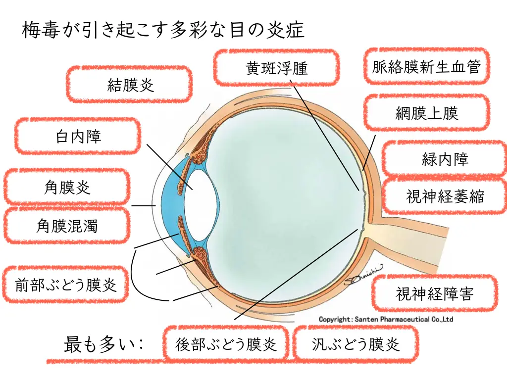
Syphilis is called the “great imitator” (very good at mimicking other diseases), and it is often difficult to distinguish from them.
Even for an ophthalmologist, it can be difficult to judge accurately whether ocular inflammation is caused by syphilis.
The eye symptoms of syphilis also appear in every possible form.
• Eye pain, redness
• Floaters (seeing specks drifting by)
• Light sensitivity (glare)
• Blurred vision
• Decreased vision, and so on
Along with various eye examinations,
syphilis is diagnosed from the results of blood tests and similar examinations.
The Cause of Syphilis: Infection with the Bacterium Treponema pallidum

Syphilis is caused by a bacterium called Treponema pallidum.
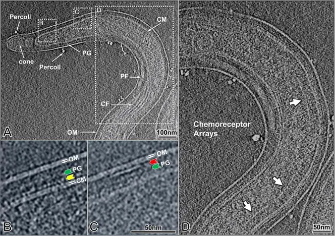
It is about 0.2 μm in diameter and 6–15 μm in length3.
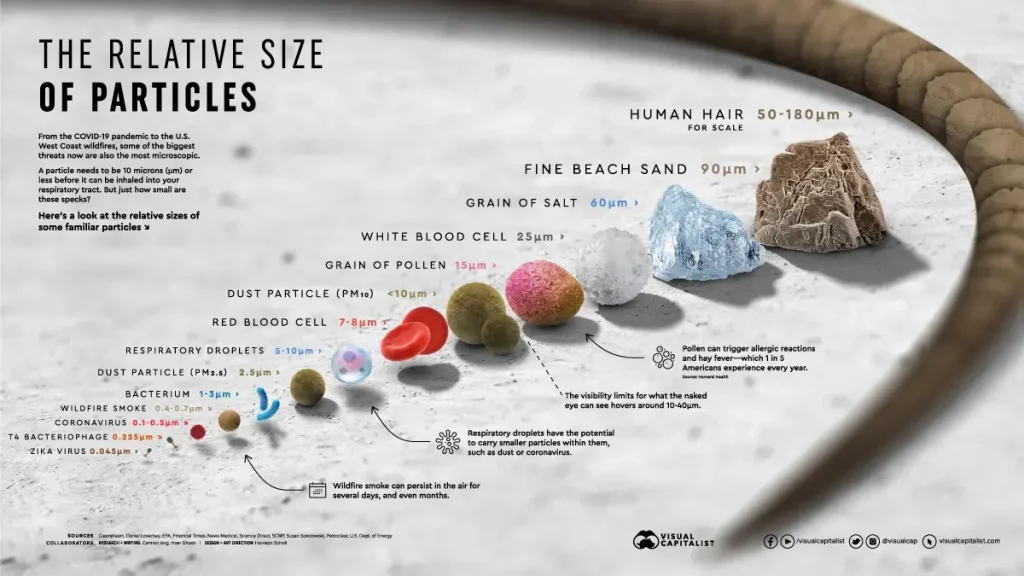

Its diameter is about the size of a virus
Its length is about the size of a red blood cell up to pollen
(far thinner than a human hair)
With this characteristic shape, it penetrates into tissues throughout the body.
Once inside, Treponema pallidum triggers a variety of immune responses.
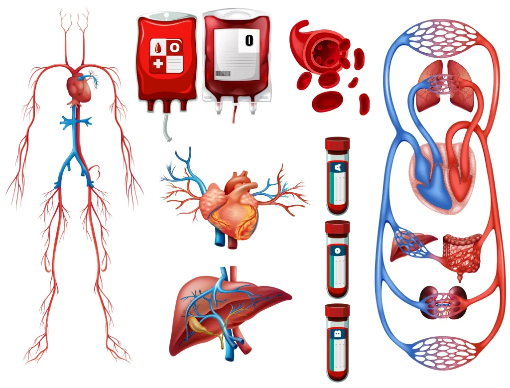
Treponema pallidum damages blood vessels.
Because the eye contains the finest blood vessels in the body, it is considered a particularly susceptible part.
Inflammation then arises everywhere in the body.
After inflammation, the tissues are damaged.
• Optic neuropathy
• Corneal opacity
• Cataract
• Glaucoma
• Epiretinal membrane
• Macular edema
• Optic atrophy
• Choroidal scarring
• Choroidal neovascularization
These appear as various sequelae.
Eye Symptoms When Syphilis Affects the Brain

When syphilis affects the brain, the pupils become extremely small.
(Argyll Robertson pupil4)
The size of the pupil can sometimes reveal syphilis.
Congenital Syphilis: Syphilis Passed from Mother to Fetus Through the Placenta

Inflammation of the black of the eye (the cornea), interstitial keratitis, is characteristic5.
As a result of keratitis, the eye may become cloudy and white: corneal opacity
Secondary glaucoma may also occur because of keratouveitis.
The Many Forms of Ocular Inflammation Caused by Syphilis: Over the Course of Time
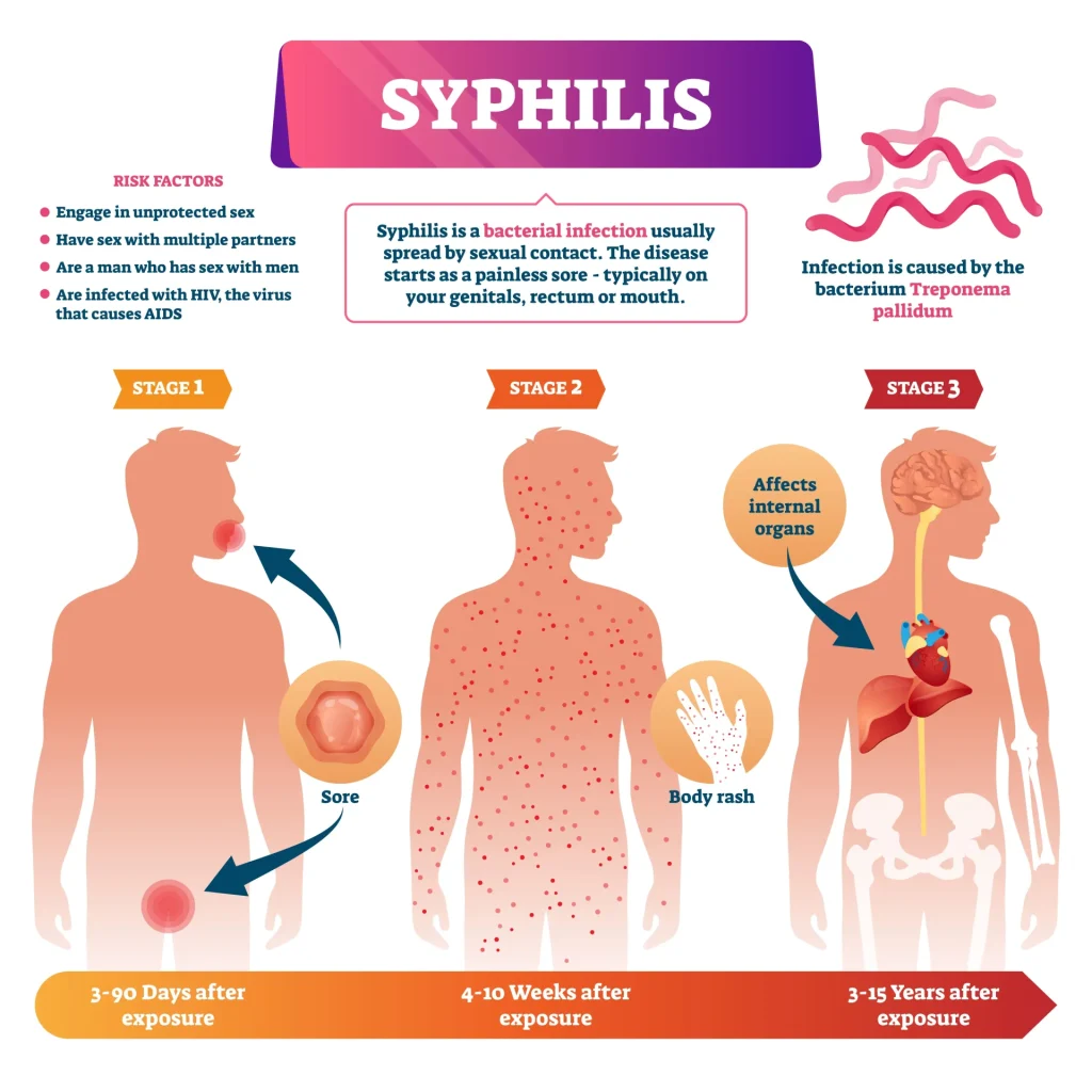

Primary Syphilis: Eye Symptoms
In the first stage, primary syphilis,
the sites where Treponema pallidum entered:
• Conjunctiva
• Eyelid
become the site of the lesion, the primary chancre (initial induration).
Infection is thought to occur through contact with secretions or through contaminated fingers.
Secondary Syphilis: Eye Symptoms
If syphilis is left untreated, the secondary stage begins 4–10 weeks later (1 to 2.5 months).
A syphilitic rash (roseola) can also develop on the eyelids, appearing as:
• Blepharitis
• Loss of eyelashes and eyebrows
In addition,
• Conjunctivitis
• Scleritis
• Keratitis
• Iridocyclitis
All of these are ocular inflammations of secondary syphilis.
As with other infectious uveitis,
anterior uveitis (iridocyclitis) usually presents as inflammation in only one eye.
Intraocular pressure can also rise considerably with the inflammation.

“Posterior uveitis,” in which inflammation spreads to the back of the eye, generally develops in the later stages of secondary syphilis.
Inflammatory lesions at multiple sites in the retina closely resemble “acute retinal necrosis”6.
“Acute retinal necrosis” is ocular inflammation caused by a herpesvirus, the varicella-zoster virus.
It leads to irreversible vision loss in a short time, so ophthalmologists greatly fear it.
In acute retinal necrosis, strong inflammatory lesions arise one after another from the peripheral retina, whereas
in “syphilitic retinal necrosis,” the inflammatory lesions are characteristically said to arise from the posterior pole of the retina7.
This is thought to reflect a difference in how the inflammation presents between a herpesvirus and the bacterium Treponema pallidum.
Acute meningitis can develop in 1–2% of patients who reach secondary syphilis8.
It is a brain complication of syphilis.
As mentioned above,
syphilitic inflammation can sometimes be recognized from an extremely small pupil.
(Argyll Robertson pupil)
Tertiary Syphilis: Eye Symptoms
Inflamed tissue turns into what is called scarring.
It occurs in the skin and is called a “gumma.”
The skin of the eyelids can also become a gumma.
Others:
• Blepharitis with loss of eyelashes
• Keratitis
• Iridocyclitis
• Vasculitis
• Chorioretinitis
• Retinal vein occlusion and retinal artery occlusion
• Retinal detachment with choroidal effusion
• Macular edema
• Optic neuritis
• Neuroretinitis
• Intermediate uveitis
• Pseudo-retinitis pigmentosa
The Three Signs of Congenital Syphilis
Signs of congenital syphilis, in which syphilis is passed from mother to fetus through the placenta:
Known as Hutchinson’s triad,
• Interstitial keratitis
• Hutchinson’s teeth (notched incisors)
• Hearing loss (sensorineural)
In addition, the following may also occur:
Pigmentary retinopathy
Secondary glaucoma
These may also occur together.

References
- Dutta Majumder, P., Chen, E.J., Shah, J., Ching Wen Ho, D., Biswas, J., See Yin, L., Gupta, V., Pavesio, C., Agrawal, R., 2019. Ocular Syphilis: An Update. Ocular Immunology and Inflammation 27, 117–125. https://doi.org/10.1080/09273948.2017.1371765
- Koundanya, V.V., Tripathy, K., 2023. Syphilis Ocular Manifestations, in: StatPearls. StatPearls Publishing, Treasure Island (FL).
- Liu, J., Howell, J.K., Bradley, S.D., Zheng, Y., Zhou, Z.H., Norris, S.J., 2010. Cellular Architecture of Treponema pallidum: Novel Flagellum, Periplasmic Cone, and Cell Envelope as Revealed by Cryo-Electron Tomography. J Mol Biol 403, 546–561. https://doi.org/10.1016/j.jmb.2010.09.020
- Robertson, D.A., 1869. Four Cases of Spinal Myosis; with Remarks on the Action of Light on the Pupil. Edinb Med J 15, 487–493.
- Keuning, M.W., Kamp, G.A., Schonenberg-Meinema, D., Dorigo-Zetsma, J.W., van Zuiden, J.M., Pajkrt, D., 2020. Congenital syphilis, the great imitator-case report and review. Lancet Infect Dis 20, e173–e179. https://doi.org/10.1016/S1473-3099(20)30268-1
- Tamesis, R.R., Foster, C.S., 1990. Ocular syphilis. Ophthalmology 97, 1281–1287. https://doi.org/10.1016/s0161-6420(90)32419-3
- Fu, E.X., Geraets, R.L., Dodds, E.M., Echandi, L.V., Colombero, D., McDonald, H.R., Jumper, J.M., Cunningham, E.T., 2010. Superficial retinal precipitates in patients with syphilitic retinitis. Retina 30, 1135–1143. https://doi.org/10.1097/IAE.0b013e3181cdf3ae
- Smith, G.T., Goldmeier, D., Migdal, C., 2006. Neurosyphilis with optic neuritis: an update. Postgrad Med J 82, 36–39. https://doi.org/10.1136/pgmj.2004.020875