![Takeru Eye Clinic | Takatori Shopping Street, Sawara-ku, Fukuoka City [Nishijin Station / Fujisaki Station]](https://takeru-eye.com/wp-content/uploads/2022/10/takeru_logo_for-WP-header.png)

“My eyes are red and painful,” “Light bothers me,” “My vision looks foggy”
These may be signs of a condition called uveitis.
Uveitis is by no means a rare disease.
Worldwide, 20 to 50 people per 100,000 develop it each year1, and in Japan it accounts for 3.2% of new patients seen at specialty eye clinics2, making it a relatively common inflammatory eye disease.
What Is Uveitis?

Uveitis is a disease in which inflammation occurs in the uvea, a tissue inside the eye.
The uvea consists of three parts, the iris, ciliary body, and choroid, and is rich in blood vessels, which makes it prone to inflammation. To use a camera as an analogy, it plays the role of an important component that adjusts the aperture and focus of the lens and regulates the light reaching the film.
When inflammation occurs, various symptoms appear, such as eye pain, redness, decreased vision, and sensitivity to light.

Incidence in the World and in Japan
Global Statistics
According to reliable epidemiological studies from around the world, the annual number of new uveitis patients is estimated at20 to 50 per 100,000 people3, 4.
The prevalence (the number of people living with the disease at a given point in time) is100 to 150 per 100,000 people5, but higher rates have been reported in some regions, such as 700 per 100,000 in Sweden6and 580 per 100,000 in Thailand7.
One of the major characteristics of uveitis is the marked difference between regions and ethnic groups.
The Situation in Japan in Detail
In Japan, a2016 nationwide surveyconfirmed that new uveitis patients accounted for3.2%of all new patients at specialty eye clinics2.
This shows that it is a relatively common disease in ophthalmology.
Ranking of Causes Identified in the 2016 Survey
- Sarcoidosis (10.6%)
- Vogt-Koyanagi-Harada disease (8.1%)
- Herpetic iritis (6.5%)
- Acute anterior uveitis (5.5%)
- Behçet’s disease (4.2%)
Compared with earlier surveys, sarcoidosis remains the most frequent cause, while Behçet’s disease continues to decline.
Classification by Cause and Characteristics
The causes of uveitis fall into two broad groups.
Non-infectious uveitis
is a group of diseases caused mainly by abnormalities of the immune system8and accounts for the majority of uveitis.
Sarcoidosis and Vogt-Koyanagi-Harada disease, the most common causes in Japan, belong here.
This is a condition in which the body’s own immune system mistakenly attacks the eye tissue it is meant to protect.
Infectious uveitis
About14 to 20%9of uveitis patients have infectious uveitis.
Pathogens such as bacteria, viruses, fungi, and parasites enter the eye and cause inflammation.
It can be described as an outside invader running amok inside the body.
Uveitis: The Reality That the Cause Is Often Unknown
Surprisingly,in more than one in three uveitis patients, the underlying disease cannot be identified.10。
Even after detailed testing, unclassifiable cases account for more than one third.
Why does this happen? Uveitis is a very complex disease, and more than 30 different diseases can cause it. The reality is that even modern medicine has not been able to clarify every cause.
The large number of cases with unknown cause is itself one of the characteristics of uveitis.
Trends by Age and Sex
Differences by Sex
In surveys at university hospitals and general hospitals alike, a tendency formore female patients11-13has been observed.
Women are known to develop uveitis more often, particularly the non-infectious forms related to autoimmunity.
Characteristics by Age
Overall, uveitis is seen most often in people in their40s and 50s, but the age of onset differs greatly depending on the disease2, 14.
For example:
- Sarcoidosis: onset peaks in the 20s and after the age of 50, and after 50 it is especially common in women
- Behçet’s disease: most common in the 20s and 30s
- Chronic iridocyclitis in children: most common between the ages of 2 and 5
Age and sex alone cannot establish a diagnosis, but they are important clues for physicians in identifying the disease.
Uveitis: Regional Differences Exist Even Within Japan
Regional differences have been confirmed even within Japan.
HTLV-1-associated uveitisis seen in the Kyushu region (including the area around Fukuoka) atabout 3 to 5 times the frequencyof the national overall rate15-17.
In addition,ocular toxoplasmosisalso occurs more frequently in the Kyushu region than the national average15.
Genetic factors, environmental factors, and lifestyle are thought to act in combination, but the detailed mechanisms remain a subject for future research.
Comparison of Incidence with Other Eye Diseases
Comparing the incidence of uveitis with that of other eye diseases makes its importance clearer.
Comparison with Retinal Detachment
Retinal detachment occurs at a rate of1 per 10,000 people per year(10 per 100,000 people per year)18.
In contrast, global statistics put new cases of uveitis at20 to 50 per 100,000 people per year4.
In other words,uveitis occurs about 2 to 5 times as often as retinal detachment.
Differences in Age Distribution
- Retinal detachment: a bimodal distribution with peaks in the 20s and in the 50s and 60s
- Uveitis: varies by disease, but overall is most common in the 40s and 50s
Retinal detachment is a relatively well-known eye disease, yet uveitis turns out to be far more frequent.
The Serious Impact of Uveitis on Vision
Uveitis is an important disease that accounts for10 to 15% of blindness worldwide.19
Without appropriate treatment, it can cause the following serious complications20, 21:
- Glaucoma (damage to the optic nerve from elevated eye pressure)
- Cataract (clouding of the lens)
- Macular edema (swelling of the central part of the retina)
- Retinal detachment (a condition in which the retina peels away)
However, with early detection and appropriate treatment, it is also a disease in which vision can be preserved in many cases.
Symptoms to Watch For and When to See a Doctor
If you have symptoms such as the following, uveitis may be the cause:
- Red eyes
- Eye pain
- Sensitivity to light
- Decreased vision
- Vision that looks foggy
- Objects look distorted
- Seeing black spots or something like bugs (floaters)
These symptoms can also occur in other eye diseases.
However, because uveitis can seriously affect vision as it progresses, please see an eye doctor early.
In addition, uveitis is sometimes discovered even without symptoms, through referral from another department.
This is because uveitis can arise in association with systemic diseases.
Uveitis: Outlook and Prognosis
Many patients probably want to know what will happen to them in the future.
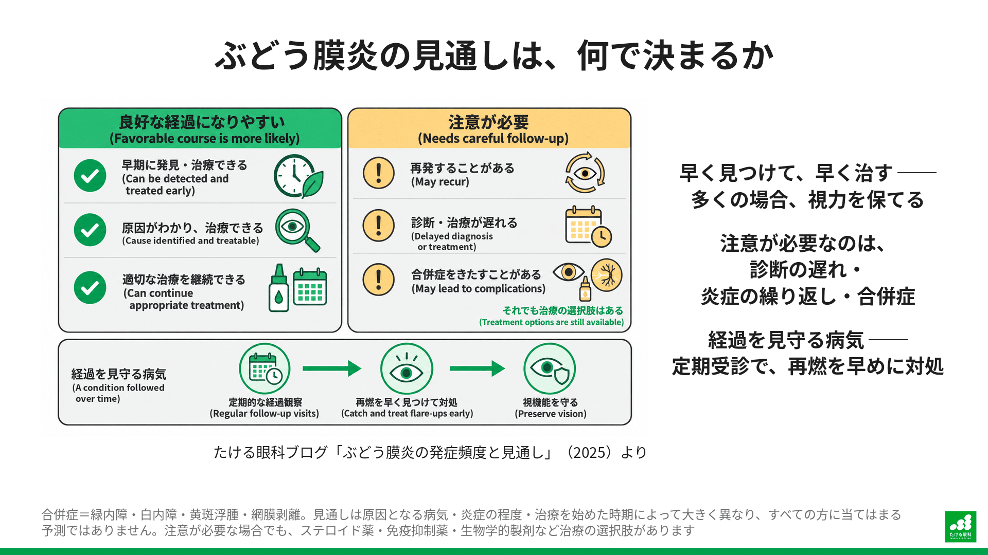
The prognosis of uveitis varies greatly depending on the underlying disease, the severity of inflammation, and the timing of treatment initiation.
Cases with a Favorable Course
- When an appropriate diagnosis and treatment are made early
- When the underlying disease is identified and effective treatment is available
- When the patient takes an active part in treatment
When these conditions are met, vision can be expected to be preserved or improved in many patients.
Cases That Require Caution
- When diagnosis or treatment is delayed
- When inflammation recurs repeatedly
- When serious complications occur
Even in such cases, there are now various treatment options.
Treatment tailored to each patient’s condition is possible, using corticosteroids, immunosuppressants, biologics, and other drugs.
The Importance of Long-Term Management
Uveitis is a disease that often follows a chronic course.
Like diabetes or hypertension, it may require long-term management.
With regular follow-up, recurrences of inflammation can be identified early and handled appropriately, which raises the likelihood of maintaining good visual function.
Medical Advances and New Treatments
Treatment of uveitis has advanced greatly in recent years.
In addition to conventional corticosteroids, new treatment options such as immunosuppressants and biologics have emerged. With these drugs, good results are now achievable even in cases that were previously difficult to treat.
Advances in diagnostic technology have also made more accurate and rapid diagnosis possible.
Imaging tests such as optical coherence tomography (OCT) and fluorescein angiography now allow the state of inflammation to be assessed in more detail.
The stages of treatment (eye drops, injections around the eye, oral medication, immunosuppressants, and biologics) and their respective side effects are explained in detail in a separate article.

A Message for Patients and Their Families
Even if you are diagnosed with uveitis, there is no need to give up.
With current medicine, many patients can preserve their vision and lead normal daily lives.
What matters, I believe, is to work with your doctor and stay patient and committed to treatment.
In fact, compared with 2001, when I became a resident, uveitis inflammation has become far easier to control.
Because uveitis can be associated with systemic disease, collaboration is often important not only with ophthalmology but also with internal medicine, rheumatology, and other departments.
(The above is general information. Please talk with your own doctor about your individual condition.)
Summary
Uveitis is by no means a rare disease.
The characteristics of its incidence can be summarized as follows2-4:
- Worldwide, 20 to 50 per 100,000 people develop it each year
- In Japan, it accounts for 3.2% of new patients at specialty eye clinics
- Sarcoidosis is the most common cause (about 10.6%)
- More than one in three cases has an unknown cause
- Slightly more common in women
- Most often seen in people in their 40s and 50s
- Occurs 2 to 5 times as often as retinal detachment
With early diagnosis and appropriate treatment, this is a disease in which vision can be preserved in many cases. If you have symptoms that concern you, I recommend seeing an eye doctor without hesitation.
Uveitis is a complex and diverse disease, but thanks to advances in modern medicine, far better treatment outcomes can now be expected than before.
With accurate knowledge and appropriate medical care, it is entirely possible to live a fulfilling life while being monitored regularly.

“My eyes are red,” “Light bothers me” — these may not be uncommon eye symptoms.
We explain the current state of uveitis clearly, using data.
Uveitis is by no means a rare disease. Both worldwide and in ophthalmology in Japan, it is a relatively common condition.
Global Annual Incidence of New Cases
20-50people
/ per 100,000
Share of New Eye Clinic Patients in Japan
3.2%
/ specialty eye clinics
Comparing uveitis with the well-known condition retinal detachment makes its high frequency clearer.
We can see that uveitis occurs about 2 to 5 times as often as retinal detachment.
Sonoda, K.-H., et al. 2021. Jpn J Ophthalmol 65, 184–190.
These are the proportions of causative diseases identified in the 2016 nationwide survey. Sarcoidosis accounts for the largest share.
Key point: the cause is unknown in more than one third of cases
Even after detailed testing, the cause cannot be identified in more than one in three uveitis patients. This shows the complexity of the disease.
The onset of uveitis shows tendencies by age and sex.
Onset Trends by Age
Across all uveitis the peak is in the 40s and 50s, but the age of onset differs greatly depending on the cause.
More Common in Women
In surveys at university hospitals and similar institutions, more patients tend to be women. This is especially marked in non-infectious uveitis related to autoimmunity.
Regional Differences
In the Kyushu region, HTLV-1-associated uveitis and ocular toxoplasmosis are reported more often than the national average, and regional differences are another characteristic.
10-15%
Share of worldwide blindness caused by uveitis
Without appropriate treatment, it can cause serious complications such as glaucoma, cataract, and macular edema. However,with early detection and appropriate treatment, vision can be preserved in many cases.
The following symptoms may be signs of uveitis. If even one applies to you, please see an eye doctor.
- ✓Red eyes
- ✓Eye pain
- ✓Sensitivity to light
- ✓Decreased vision
- ✓Vision that looks foggy
- ✓Distorted vision
- ✓Seeing black spots
References
- Kalogeropoulos, D., Asproudis, I., Stefaniotou, M., Moschos, M.M., Kozobolis, V.P., Voulgari, P.V., Katsanos, A., Gartzonika, C., Kalogeropoulos, C., 2023. The large Hellenic Study of Uveitis: epidemiology, etiologic factors and classification. Int Ophthalmol 43, 3633–3650. https://doi.org/10.1007/s10792-023-02772-5
- Sonoda, K.-H., Hasegawa, E., Namba, K., Okada, A.A., Ohguro, N., Goto, H., JOIS (Japanese Ocular Inflammation Society) Uveitis Survey Working Group, 2021. Epidemiology of uveitis in Japan: a 2016 retrospective nationwide survey. Jpn J Ophthalmol 65, 184–190. https://doi.org/10.1007/s10384-020-00809-1
- Wang, L., Guo, Z., Zheng, Y., Li, Q., Yuan, X., Hua, X., 2021. Analysis of the clinical diagnosis and treatment of uveitis. Ann Palliat Med 10, 12782–12788. https://doi.org/10.21037/apm-21-3549
- Acharya, N.R., Tham, V.M., Esterberg, E., Borkar, D.S., Parker, J.V., Vinoya, A.C., Uchida, A., 2013. Incidence and prevalence of uveitis: results from the Pacific Ocular Inflammation Study. JAMA Ophthalmol 131, 1405–1412. https://doi.org/10.1001/jamaophthalmol.2013.4237
- Joltikov, K.A., Lobo-Chan, A.-M., 2021. Epidemiology and Risk Factors in Non-infectious Uveitis: A Systematic Review. Front Med (Lausanne) 8, 695904. https://doi.org/10.3389/fmed.2021.695904
- Bro, T., Tallstedt, L., 2020. Epidemiology of uveitis in a region of southern Sweden. Acta Ophthalmol 98, 32–35. https://doi.org/10.1111/aos.14130
- Keorochana, N., 2020. Pattern and Outcome of Uveitis in a Tertiary Military Hospital in Thailand. Ocul Immunol Inflamm 28, 424–432. https://doi.org/10.1080/09273948.2019.1589527
- Thorne, J.E., Suhler, E., Skup, M., Tari, S., Macaulay, D., Chao, J., Ganguli, A., 2016. Prevalence of Noninfectious Uveitis in the United States: A Claims-Based Analysis. JAMA Ophthalmol 134, 1237–1245. https://doi.org/10.1001/jamaophthalmol.2016.3229
- Zhang, Y., Amin, S., Lung, K.I., Seabury, S., Rao, N., Toy, B.C., 2020. Incidence, prevalence, and risk factors of infectious uveitis and scleritis in the United States: A claims-based analysis. PLoS One 15, e0237995. https://doi.org/10.1371/journal.pone.0237995
- Liba, T., Gorenshtein, A., Leibovitch, L., Gepstein, R., Machinski, E., Segal, O., n.d. Epidemiological Characterization of Uveitis in Japan: a Systematic Review. Ocular Immunology and Inflammation 0, 1–10. https://doi.org/10.1080/09273948.2025.2452193
- Chan, C.-C., Goldstein, D.A., Davis, J.L., Sen, H.N., 2014. Gender and Uveitis. J Ophthalmol 2014, 818070. https://doi.org/10.1155/2014/818070
- de-la-Torre, A., Mejía-Salgado, G., Cifuentes-González, C., Rojas-Carabali, W., Cuevas, M., García, S., Rangel, C.M., Durán, C., Pachón-Suárez, D.I., Bustamante-Arias, A., 2024. Epidemiology, clinical features, and classification of 3,404 patients with uveitis: Colombian Uveitis Multicenter Study (COL-UVEA). Graefes Arch Clin Exp Ophthalmol 262, 2601–2615. https://doi.org/10.1007/s00417-024-06422-z
- González, M.M., Solano, M.M., Porco, T.C., Oldenburg, C.E., Acharya, N.R., Lin, S.C., Chan, M.F., 2018. Epidemiology of uveitis in a US population-based study. J Ophthalmic Inflamm Infect 8, 6. https://doi.org/10.1186/s12348-018-0148-5
- Yanai, R., Uchi, S.-H., Kondo, Y., Fujitsu, Y., Suzuki, K., Yoshimura, K., Kumagai, N., Egawa, M., Mitamura, Y., 2025. The epidemiology of uveitis: comparison of its causes and visual outcomes between three-tiered medical facilities in Ube city. Sci Rep 15, 8998. https://doi.org/10.1038/s41598-025-93223-x
- Takeda, A., Ishibashi, T., Sonoda, K.-H., 2017. Epidemiology of Uveitis, Caused by HTLV-1, Toxoplasmosis, and Tuberculosis; the Three Leading Causes of Endemic Infectious Uveitis in Japan. Ocul Immunol Inflamm 25, S19–S23. https://doi.org/10.1080/09273948.2016.1253851
- Takahashi, T., Ohtani, S., Miyata, K., Miyata, N., Shirato, S., Mochizuki, M., 2002. A clinical evaluation of uveitis-associated secondary glaucoma. Jpn J Ophthalmol 46, 556–562. https://doi.org/10.1016/s0021-5155(02)00549-x
- Hikita, S.-I., Sonoda, K.-H., Hijioka, K., Fujimoto, T., Ito, T., Ishibashi, T., 2012. [Incidence of uveitis in the northern Kyushu region of Japan –comparison between the periods of 1996-2001 and 2003-2008]. Nippon Ganka Gakkai Zasshi 116, 847–855.
- Saraf, S.S., Lacy, M., Hunt, M.S., Lee, C.S., Lee, A.Y., Chee, Y.E., 2022. Demographics and Seasonality of Retinal Detachment, Retinal Breaks, and Posterior Vitreous Detachment from the Intelligent Research in Sight Registry. Ophthalmol Sci 2, 100145. https://doi.org/10.1016/j.xops.2022.100145
- Wang, L., Guo, Z., Zheng, Y., Li, Q., Yuan, X., Hua, X., 2021. Analysis of the clinical diagnosis and treatment of uveitis. Ann Palliat Med 10, 12782–12788. https://doi.org/10.21037/apm-21-3549
- De Hoog, J., Ten Berge, J.C., Groen, F., Rothova, A., 2017. Rhegmatogenous retinal detachment in uveitis. J Ophthalmic Inflamm Infect 7, 22. https://doi.org/10.1186/s12348-017-0140-5
- Apivatthakakul, A., Tantraworasin, A., Rothova, A., Kunavisarut, P., Pathanapitoon, K., 2025. Prevalence and Risk Factors for Ocular Complications in New-Onset Uveitis: A Study From a Tertiary Referral Center in Northern Thailand. OPTH 19, 543–551. https://doi.org/10.2147/OPTH.S500339