![Takeru Eye Clinic | Takatori Shopping Street, Sawara-ku, Fukuoka City [Nishijin Station / Fujisaki Station]](https://takeru-eye.com/wp-content/uploads/2022/10/takeru_logo_for-WP-header.png)

A 6-year-old girl was brought to an eye clinic after telling her parents, “My eyes are red.”
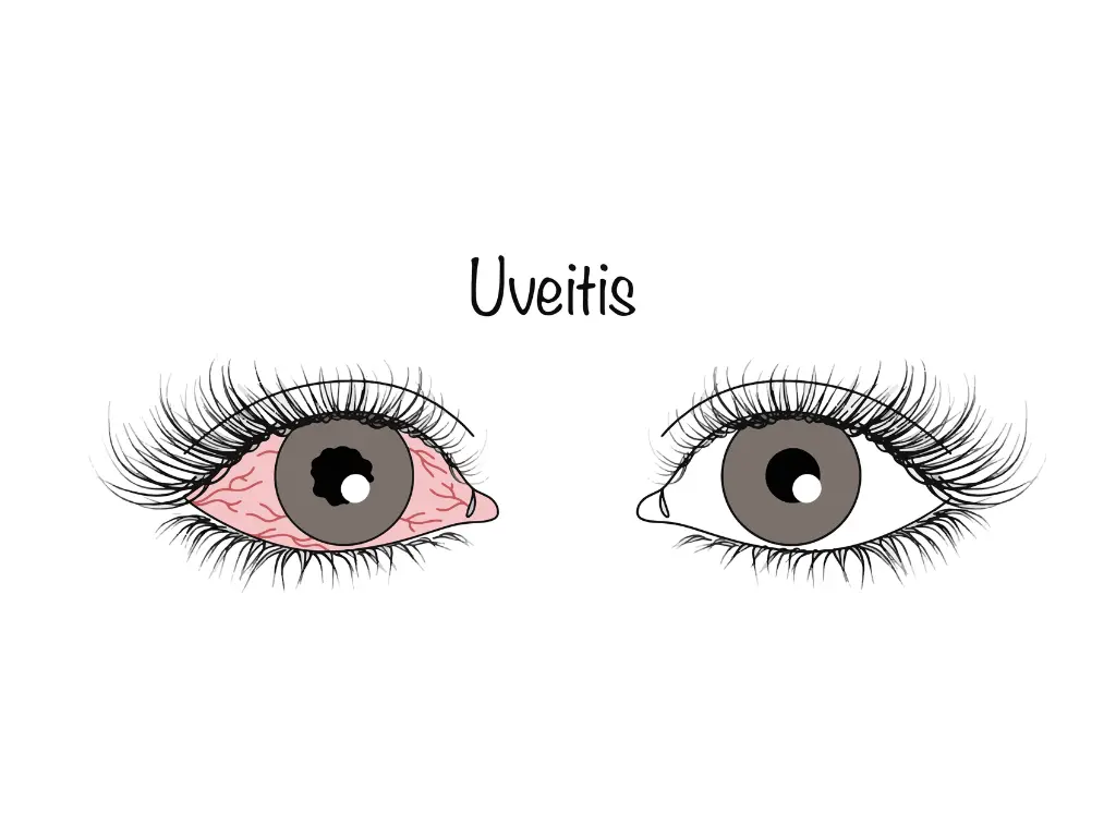
She did not complain of any pain at the examination, but slit-lamp microscopy showed inflammatory cells in the anterior chamber, and she was diagnosed with bilateral anterior uveitis.
Further workup revealed that she also had juvenile idiopathic arthritis (JIA).
Juvenile idiopathic arthritis (JIA: Juvenile Idiopathic Arthritis):
It was formerly called juvenile rheumatoid arthritis (JRA: Juvenile Rheumatoid Arthritis).
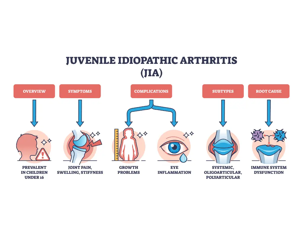
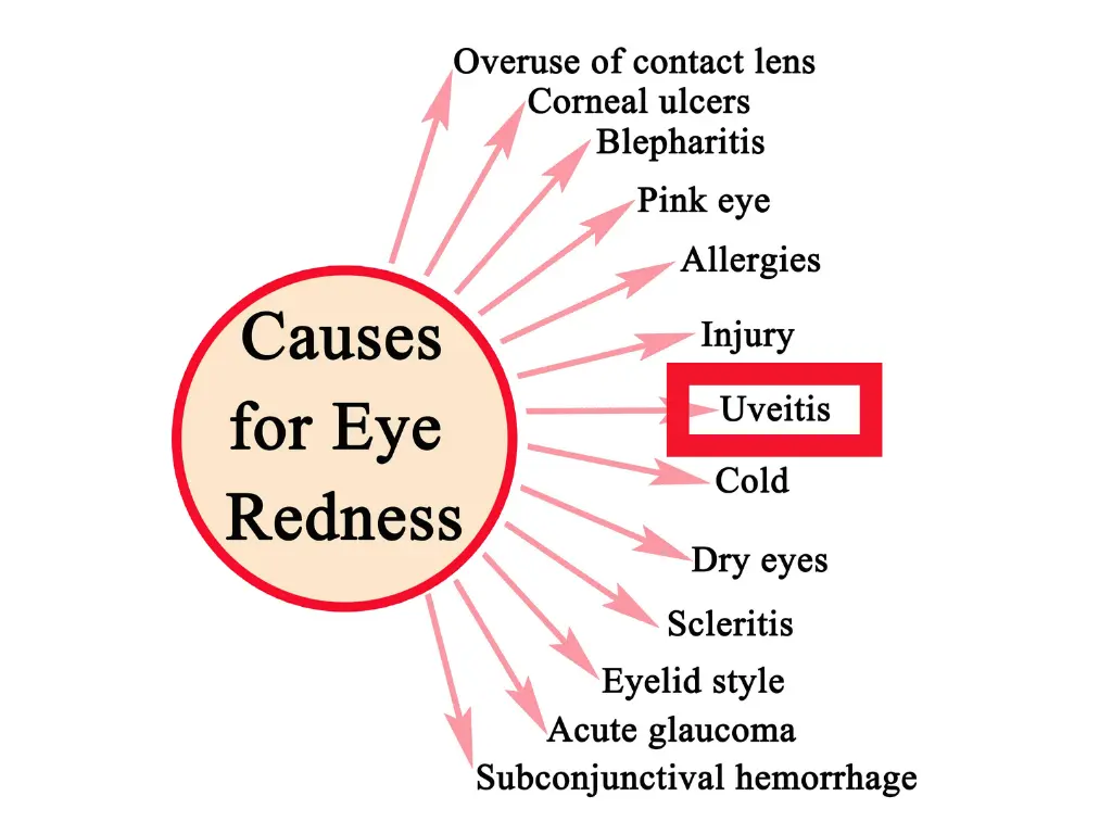
As in this case, uveitis in children often appears as one manifestation of a systemic disease.
Pediatric uveitis is a serious eye disease that can cause severe visual impairment if it is not properly diagnosed and treated.
Results of a 2024 study (Bani Khalaf)1reported that uveitis develops in about 20% of patients with JIA, and in many of them it takes the form of anterior uveitis with few symptoms.
In anterior uveitis there may be only mild irritation or slightly reduced vision, whereas in posterior segment inflammation there is little pain or redness, and symptoms such as floaters and decreased vision mainly progress slowly.
For this reason, focusing on anterior segment symptoms can lead to posterior segment inflammation being missed.
Because inadequate treatment can cause permanent visual impairment, a comprehensive eye examination including a detailed fundus examination is important.
Uveitis in Children: “Inflammation in the Eye That Reflects the Whole Body” — The Importance of Early Detection and Treatment

Uveitis in Children:Anatomical Classification and Characteristics
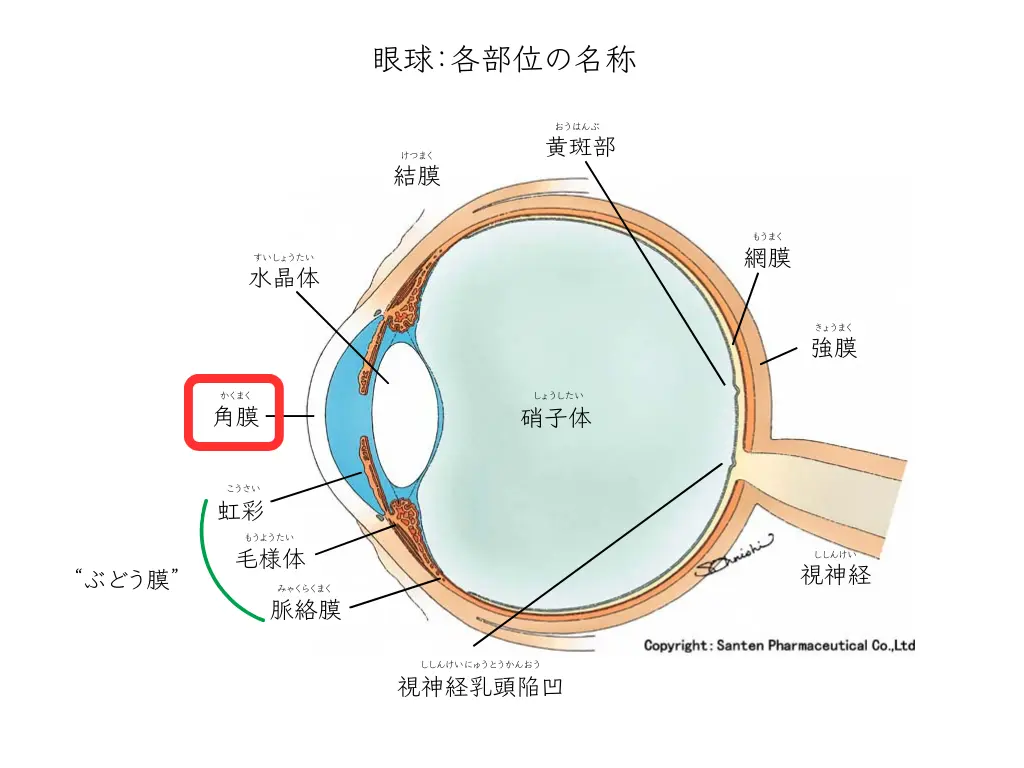
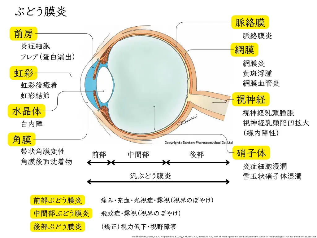
Uveitis is classified anatomically as follows (the figures in parentheses are the frequency in children):
- Anterior uveitis(about 57%):
Inflammation mainly of the iris and ciliary body; the most common type in children. Most JIA-associated uveitis takes this form - Intermediate uveitis(about 29%):
Mainly vitritis; when idiopathic it is also called pars planitis. It tends to begin at a young age and is often bilateral - Posterior uveitis:
Mainly inflammation of the retina and choroid. It is often caused by infections such as toxoplasmosis. Its frequency varies greatly by region - Panuveitis(about 16%):
Inflammation involving the entire eye. It is often seen in systemic diseases such as Behçet’s disease and Vogt-Koyanagi-Harada disease
These frequencies are known to vary greatly by region and population.
A study from Turkey reported 39% anterior uveitis and 29% intermediate uveitis2.
In North America and Northern Europe, JIA-associated anterior uveitis is the most common3.
In contrast, Behçet’s disease-associated uveitis is reported relatively often in the Mediterranean region, and Vogt-Koyanagi-Harada disease in Asia4, 5.
Characteristic features of uveitis in children include the following:
- Unclear clinical symptoms, which make delayed diagnosis likely
- Rapid progression and a tendency to develop complications
- The need for long-term follow-up
and others.
In JIA-associated uveitis in particular, the course is often asymptomatic, so regular screening examinations are considered important.
Uveitis in Children:Classification by Cause
Non-infectious uveitis
85-95% of uveitis in children is non-infectious6.
JIA-associated uveitis
JIA is considered the most common cause of non-infectious uveitis in children.
JIA-associated uveitis also varies greatly by region:
in Finland, 61% of children with uveitis7have JIA-associated uveitis,
and in the United States too, JIA is considered one of the leading causes of non-infectious uveitis in children
In Japan it is clearly less common than in Europe and North America, and the rate of uveitis among JIA patients isabout 6%9.
Sarcoidosis
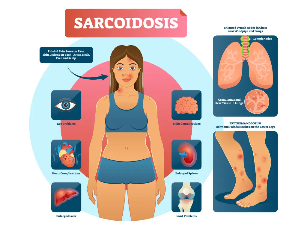
Sarcoidosis is also an important cause.
Sarcoidosis in children is a disease that causes granulomatous inflammation in multiple organs, and its symptoms differ by age.
In older children aged 5 and above, lung lesions are often the main feature,
whereas early-onset sarcoidosis, which develops before the age of 5 (non-hereditary sarcoidosis), and Blau syndrome, a familial autosomal dominant disease, are characterized by a triad of arthritis of the knees and wrists, skin lesions, and anterior uveitis10.
Lung lesions are usually not seen.
TINU syndrome
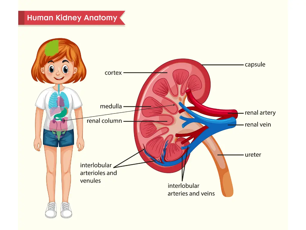
A kidney disease called tubulointerstitial nephritis and uveitis syndrome (TINU syndrome) can also present as uveitis.
TINU syndrome is seen mainly in girls in their teens.
It can present with bilateral uveitis together with systemic symptoms such as fever, loss of appetite, and weight loss.
80% of cases are anterior uveitis. Cases with posterior uveitis alone are rare.
The response to steroid treatment is good, but
the uveitis tends to recur, and 40% of patients experience a recurrence within one year11.
Other Systemic Diseases
Other systemic diseases, such as Vogt-Koyanagi-Harada disease, can also cause uveitis in children.
Uveitis:Distinguishing Infectious from Non-Infectious
Ophthalmologists distinguish infectious from non-infectious uveitis from a variety of findings. The following findings in particular provide important clues:
Characteristics of Iris Nodules
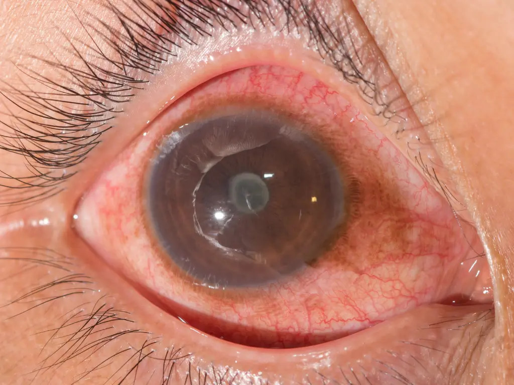
Small granular bumps are sometimes seen on the iris (the part of the eye that determines whether it looks brown or blue).
These small “lump-like” changes (nodules) may seem trivial at first glance, but they are in fact an important clue.
For example, they can be the “first visible sign” of a systemic disease such as sarcoidosis, Vogt-Koyanagi-Harada disease, or multiple sclerosis.
The color and character of iris nodules differ between infectious and non-infectious uveitis12.
- Infectious uveitis:
- Iris nodules have a soft, cream-colored or white appearance
- They do not respond to steroid treatment and tend to enlarge
- Non-infectious uveitis:
- Iris nodules are seen in sarcoidosis, VKH disease, multiple sclerosis, and others
- May be accompanied by mutton-fat keratic precipitates
(a finding seen by examining the back surface of the cornea, the “black of the eye”)
Pattern of Inflammation
- Infectious uveitis: Often begins in one eye, and the inflammation is localized
- Non-infectious uveitis: Often bilateral, and the inflammation tends to be widespread
Characteristics of Anterior Chamber Inflammatory Cells
- Infectious uveitis: Many large inflammatory cells, sometimes forming clumps
- Non-infectious uveitis: Relatively small inflammatory cells, scattered
Nature of Retinal Lesions
- Infectious uveitis: Many well-demarcated lesions, with relatively rapid progression
- Non-infectious uveitis: Many poorly demarcated lesions, with slow progression
One reason ophthalmologists carefully examine the inside of the eye with a slit-lamp microscope is to avoid missing such small changes.
If steroid treatment is given without noticing subtle changes, the condition worsens when the uveitis is in fact infectious.
Infectious Uveitis and “Masquerade Syndrome”
The most common causes of infectious uveitis are the herpesvirus group (herpes simplex, varicella-zoster virus, and cytomegalovirus)13.
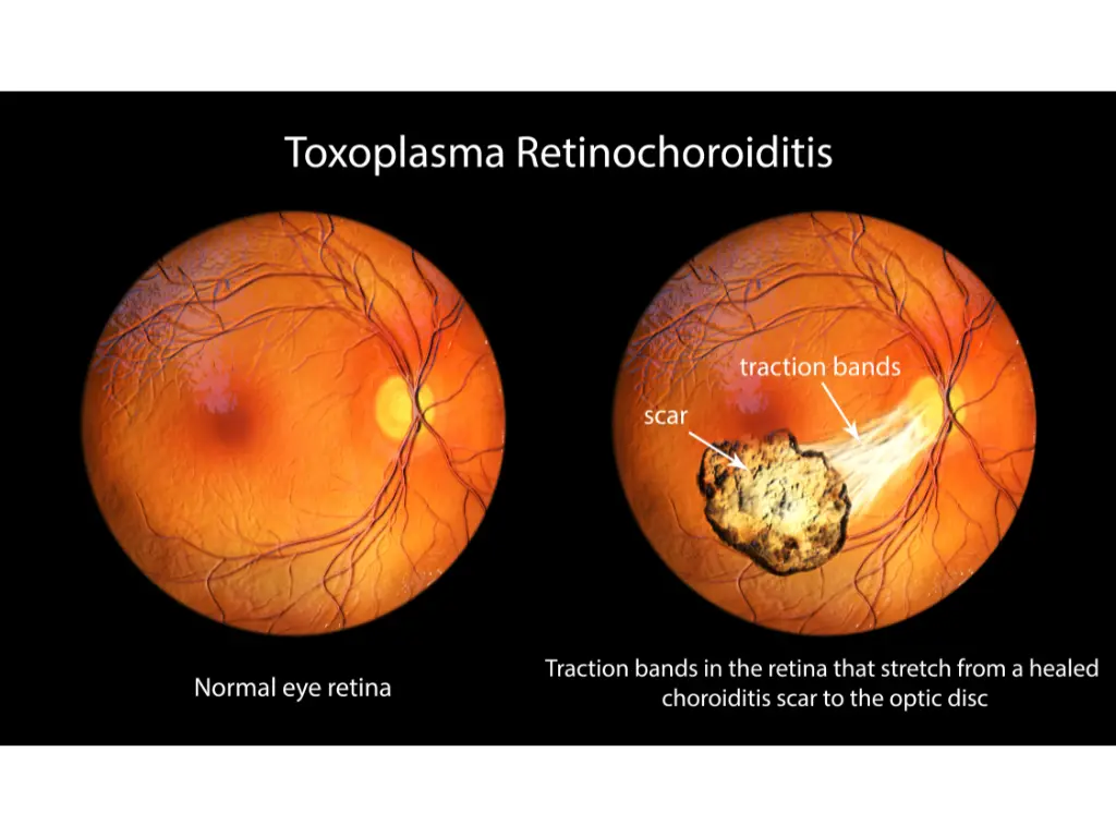
Toxoplasmosis and toxocariasis are also causes.
Toxoplasmosis is known to be transmitted through cat feces, raw or undercooked meat, and contaminated water14.
Sometimes what looks like uveitis at first glance actually conceals an entirely different disease.
This is the condition called “masquerade syndrome”15.
Typical examples are malignant tumors such as retinoblastoma and leukemia.
About 9% of retinoblastoma cases are initially judged to be uveitis16.
Unlike ordinary inflammatory uveitis, malignant tumors do not improve with standard anti-inflammatory treatment, which can instead lead to a delay in diagnosis.
Especially when the disease is unilateral and follows an atypical course, the possibility of “masquerade syndrome” must be kept in mind.
Uveitis in Children:The Importance of Early Detection and Screening
The most important characteristic of uveitis in children is that about half of cases may begin without symptoms.
Even when there is no visible change, severe visual impairment may be progressing silently, which calls for particular care17.
Screening Examinations
Appropriate screening requires the following:
- Performed by an ophthalmologist
- A detailed examination using a slit lamp (slit-lamp microscope) and various imaging devices
- Regular visits (especially within the first year after diagnosis in patients with JIA)
An examination at an ordinary eyeglass shop is not sufficient; an appropriate examination by a specialist is always needed.
Uveitis in Children: Treatment Approach
The treatment of uveitis in children aims to control inflammation reliably while taking the child’s growth into account.
The three pillars of treatment are, in my view, “controlling inflammation,” “preserving vision,” and “preventing complications.”
In JIA-associated uveitis in particular, about 70% of cases develop within one year of diagnosis, so appropriate treatment during this period is extremely important.
Stepwise Treatment
Treatment is started mainly with local therapy.
Steroid eye drops are used as the first-line drug and are effective in the acute phase of inflammation.
However, attention must be paid to the risk of cataract and glaucoma with long-term use.
Mydriatic drops are also used together to prevent posterior synechiae.
Systemic therapy is considered when local therapy is not sufficiently effective, in severe cases, and when both eyes are affected.
Oral steroids have a strong anti-inflammatory effect, but sufficient attention must be paid to side effects such as growth impairment.
Immunosuppressants and Biologics
In recent years, treatment with immunosuppressants and biologics has also become established18.
In JIA-associated uveitis in particular, methotrexate is positioned as the standard treatment.
When methotrexate is not sufficiently effective, biologics such as adalimumab are used, and good treatment outcomes have been reported19, 20.
Tocilizumab (Actemra®, an anti-IL-6 receptor antibody) is also expected to be effective21.
Management of Infectious Uveitis
In infectious uveitis, treating the causative infection takes priority.
Particularly important is the distinction between infectious and non-infectious uveitis described above.
This is because mistakenly giving steroid treatment can worsen symptoms when the uveitis is infectious.
The stages of uveitis treatment and their side effects, including in adults, are summarized in the following article.

Uveitis in Children:Complications and Their Risks
- Cataract
- The most common early complication (about 24%)
- Also occurs as a side effect of steroid treatment
- Glaucoma
- Elevated intraocular pressure is found in about 30% during follow-up
- Early detection is important for preserving visual function
- It can also occur as a treatment-related complication (steroid-induced glaucoma)
- Maculopathy
- The incidence of macular edema is 6-25%
- The most common cause of severe vision loss (35%)
- Can be prevented by early treatment
- Other Complications
- Band keratopathy: 15.7-29%
- The black of the eye (the cornea) becomes cloudy
- Posterior synechiae: 18-44%
- The iris and the lens stick together
- Hypotony: 3-10%
- Band keratopathy: 15.7-29%
Because of such complications, severe vision loss can occur in 25-30% of children with uveitis22-24.
When the above complications occur, surgical treatment may be considered after the inflammation has fully subsided.
In children in particular, careful attention is needed to the timing of surgery and postoperative management.
Uveitis in Children:Measures to Improve the Prognosis
In JIA-associated uveitis in particular, more than 70% of cases develop within one year of diagnosis, so regular examinations during this period are extremely important.
With early detection and appropriate treatment, many children can avoid serious complications.
For this reason, the following measures are recommended:
- Regular examinations by a uveitis specialist or an ophthalmologist
- Collaboration with pediatricians, rheumatologists, and others
- Continued follow-up regardless of the presence or absence of symptoms
- Appropriate explanation and education for the family
- Information sharing with school staff
This describes what can be done by a clinic run by a single physician.

References
- M. Bani Khalaf, I., Jain, H., Vora, N.M., ul Ain, N., Murtaza, F., Ram, M.D., Nankani, A., Motwani, J., n.d, 2024. A clearer vision: insights into juvenile idiopathic arthritis–associated uveitis. Proc (Bayl Univ Med Cent) 37, 303–311. https://doi.org/10.1080/08998280.2024.2305567
- Tuğal-Tutkun, İ., 2023. An Overview of Pediatric Uveitis. Turk Arch Pediatr 58, 363–370. https://doi.org/10.5152/TurkArchPediatr.2023.23086
- Rypdal, V., Glerup, M., Songstad, N.T., Bertelsen, G., Christoffersen, T., Arnstad, E.D., Aalto, K., Berntson, L., Fasth, A., Herlin, T., Ekelund, M., Peltoniemi, S., Toftedal, P., Nielsen, S., Leinonen, S., Bangsgaard, R., Nielsen, R., Rygg, M., Nordal, E., Nordic Study Group of Pediatric Rheumatology, 2021. Uveitis in Juvenile Idiopathic Arthritis: 18-Year Outcome in the Population-based Nordic Cohort Study. Ophthalmology 128, 598–608. https://doi.org/10.1016/j.ophtha.2020.08.024
- Joubert, M., Desbois, A.-C., Domont, F., Ghembaza, A., Le Joncour, A., Mirouse, A., Maalouf, G., Leclercq, M., Touhami, S., Cacoub, P., Bodaghi, B., Saadoun, D., 2023. Behçet’s Disease Uveitis. J Clin Med 12, 3648. https://doi.org/10.3390/jcm12113648
- Ozdal, P., Ozdamar, Y., Yazici, A., Teke, M.Y., Ozturk, F., 2014. Vogt-Koyanagi-Harada disease: clinical and demographic characteristics of patients in a specialized eye hospital in Turkey. Ocul Immunol Inflamm 22, 277–286. https://doi.org/10.3109/09273948.2013.856448
- Chang, M.H., Shantha, J.G., Fondriest, J.J., Lo, M.S., Angeles-Han, S.T., 2021. Uveitis in Children and Adolescents. Rheumatic Disease Clinics of North America 47, 619–641. https://doi.org/10.1016/j.rdc.2021.07.005
- Siiskonen, M., Hirn, I., Pesälä, R., Hautala, T., Ohtonen, P., Hautala, N., 2021. Prevalence, incidence and epidemiology of childhood uveitis. Acta Ophthalmologica 99, e160–e163. https://doi.org/10.1111/aos.14535
- Siiskonen, M., Hirn, I., Pesälä, R., Hautala, T., Ohtonen, P., Hautala, N., 2021. Prevalence, incidence and epidemiology of childhood uveitis. Acta Ophthalmologica 99, e160–e163. https://doi.org/10.1111/aos.14535
- Yasumura, J., Yashiro, M., Okamoto, N., Shabana, K., Umebayashi, H., Iwata, N., Okura, Y., Kubota, T., Shimizu, M., Tomiita, M., Nakagishi, Y., Nishimura, K., Hara, R., Mizuta, M., Yasumi, T., Yamaide, F., Wakiguchi, H., Kobayashi, M., Mori, M., 2019. Clinical features and characteristics of uveitis associated with juvenile idiopathic arthritis in Japan: first report of the pediatric rheumatology association of Japan (PRAJ). Pediatr Rheumatol Online J 17, 15. https://doi.org/10.1186/s12969-019-0318-5
- Kaufman, K.P., Becker, M.L., 2021. Distinguishing Blau Syndrome from Systemic Sarcoidosis. Curr Allergy Asthma Rep 21, 10. https://doi.org/10.1007/s11882-021-00991-3
- Legendre, M., Devilliers, H., Perard, L., Groh, M., Nefti, H., Dussol, B., Trad, S., Touré, F., Abad, S., Boffa, J.-J., Frimat, L., Torner, S., Seidowsky, A., Massy, Z.A., Saadoun, D., Rieu, V., Schoindre, Y., Heron, E., Frouget, T., Lionet, A., Glowacki, F., Arnaud, L., Mousson, C., Besancenot, J.-F., Rebibou, J.-M., Bielefeld, P., 2016. Clinicopathologic characteristics, treatment, and outcomes of tubulointerstitial nephritis and uveitis syndrome in adults: A national retrospective strobe-compliant study. Medicine (Baltimore) 95, e3964. https://doi.org/10.1097/MD.0000000000003964
- Myers, T.D., Smith, J.R., Lauer, A.K., Rosenbaum, J.T., 2002. Iris nodules associated with infectious uveitis. Br J Ophthalmol 86, 969–974.
- Chams, H., Rostami, M., Mohammadi, S.-F., Ohno, S., 2009. Epidemiology and Prevalence of Uveitis: Review of Literature 21. 2009 Iranian Journal of Ophthalmology 21(4):4-16
- Pinto-Ferreira, F., Caldart, E.T., Pasquali, A.K.S., Mitsuka-Breganó, R., Freire, R.L., Navarro, I.T., 2019. Patterns of Transmission and Sources of Infection in Outbreaks of Human Toxoplasmosis. Emerg Infect Dis 25, 2177–2182. https://doi.org/10.3201/eid2512.181565
- Grange, L.K., Kouchouk, A., Dalal, M.D., Vitale, S., Nussenblatt, R.B., Chan, C.-C., Sen, H.N., 2014. Neoplastic Masquerade Syndromes among Uveitis Patients. Am J Ophthalmol 157, 526–531. https://doi.org/10.1016/j.ajo.2013.11.002
- Shields, C.L., Ghassemi, F., Tuncer, S., Thangappan, A., Shields, J.A., 2008. Clinical spectrum of diffuse infiltrating retinoblastoma in 34 consecutive eyes. Ophthalmology 115, 2253–2258. https://doi.org/10.1016/j.ophtha.2008.07.003
- Kim, L., Li, A., Angeles-Han, S., Yeh, S., Shantha, J., 2019. Update on the management of uveitis in children: an overview for the clinician. Expert Rev Ophthalmol 14, 211–218. https://doi.org/10.1080/17469899.2019.1663731
- AlBloushi, A.F., Solebo, A.L., Gokhale, E., Hayouti, H., Ajamil-Rodanes, S., Petrushkin, H., 2022. Long-Term Outcomes of Pediatric Idiopathic Intermediate Uveitis. Am J Ophthalmol 237, 41–48. https://doi.org/10.1016/j.ajo.2021.11.003
- Tanatar, A., Aktay Ayaz, N., 2023. Biologic Therapies in Juvenile Idiopathic Arthritis. btd 19, 269–275. https://doi.org/10.4274/BMJ.galenos.2023.2023.3-20
- Ramanan, A.V., Dick, A.D., Jones, A.P., Hughes, D.A., McKay, A., Rosala-Hallas, A., Williamson, P.R., Hardwick, B., Hickey, H., Rainford, N., Hickey, G., Kolamunnage-Dona, R., Culeddu, G., Plumpton, C., Wood, E., Compeyrot-Lacassagne, S., Woo, P., Edelsten, C., Beresford, M.W., 2019. Adalimumab in combination with methotrexate for refractory uveitis associated with juvenile idiopathic arthritis: a RCT. Health Technol Assess 23, 1–140. https://doi.org/10.3310/hta23150
- Iannone, C., Marelli, L., Costi, S., Pellico, M.R., La Franca, L., Caporali, R., Miserocchi, E., 2023. Tocilizumab in Juvenile Idiopathic Arthritis Associated Uveitis, a Narrative Review. Children (Basel) 10, 434. https://doi.org/10.3390/children10030434
- Gautam Seth, N., Kaur, S., Yangzes, S., Jugran, D., Bansal, R., Gupta, V., Dogra, M.R., Suri, D., Singh, S., Singh, R., 2021. Ophthalmic Complications in Pediatric Uveitis. Ocul Immunol Inflamm 29, 1375–1380. https://doi.org/10.1080/09273948.2020.1762897
- Shivpuri, A., Turtsevich, I., Solebo, A.L., Compeyrot-Lacassagne, S., 2022. Pediatric uveitis: Role of the pediatrician. Front. Pediatr. 10, 874711. https://doi.org/10.3389/fped.2022.874711
- Chang, M.H., Shantha, J.G., Fondriest, J.J., Lo, M.S., Angeles-Han, S.T., 2021. Uveitis in Children and Adolescents. Rheumatic Disease Clinics of North America 47, 619–641. https://doi.org/10.1016/j.rdc.2021.07.005