![Takeru Eye Clinic | Takatori Shopping Street, Sawara-ku, Fukuoka City [Nishijin Station / Fujisaki Station]](https://takeru-eye.com/wp-content/uploads/2022/10/takeru_logo_for-WP-header.png)

Sometimes a child has “amblyopia,” in which the ability to see has not developed in only one eye.
During the eye test at school health checkups,
the child may not be able to cover one eye properly, and the results can be unstable.
There is a possibility that even the eye that cannot see produces a “good result.”
Missing amblyopia leads to a major disadvantage throughout the child’s future.
Amblyopia can be improved by early detection and treatment.
On the other hand, if amblyopia is missed, the chance for treatment is lost.
Accurately Identifying Unnoticed Amblyopia
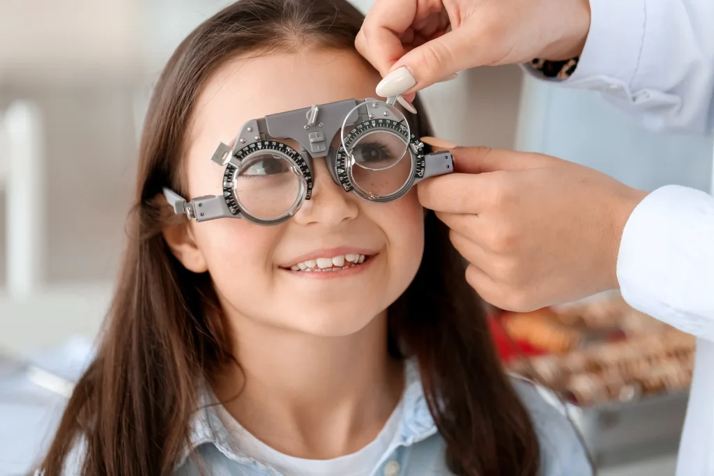
School Health: Vision Screening as a Health Survey
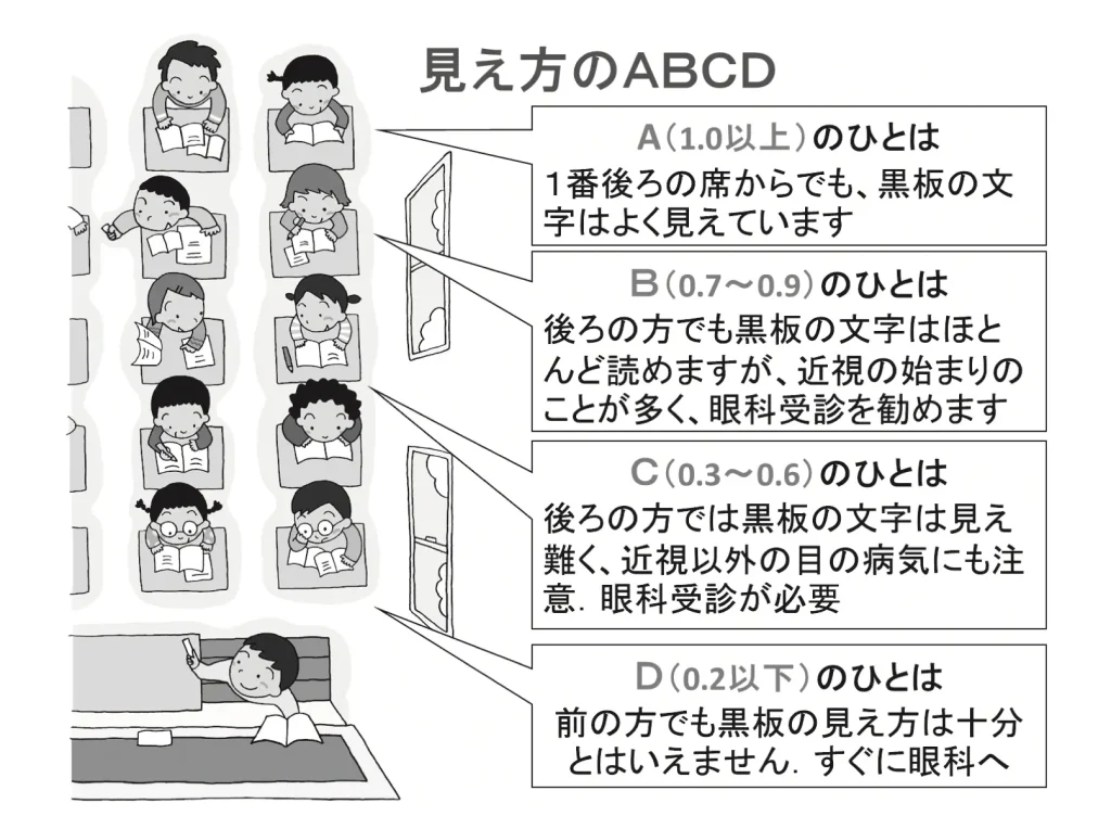
A school teacher (the school nurse-teacher) tests the children,
A: 1.0 or better
B:0.7〜0.9
C:0.3〜0.6
D: 0.2 or lower
and the grades are handed out.
(370 method vision measurement)
In school health, in order to carry out health examinations appropriately and smoothly, a health survey on growth, health status, and so on is to be conducted beforehand.
(School Health and Safety Act Enforcement Regulations, Article 11)
At elementary schools, junior high schools, high schools, and colleges of technology, it applies toall grades;、
at kindergartens and universities, it applieswhen deemed necessary. This was changed.
(Effective April 1, 2016)
― Examples of eye-related health survey items ―
○ Regarding vision, refraction, and accommodation
・Cannot see well (characters on the blackboard, in bad weather, etc.)
・How the child looks at things (squinting, looking up or sideways, going close to the TV, etc.)
・Use of glasses, contact lenses, etc.
・Eye strain (tires easily, cannot keep reading books, gets headaches)
A vision test is generally regarded as a preliminary screening test performed by school staff such as the school nurse-teacher.
An eye that cannot see could slip through a preliminary screening test.
Covering one eye at a time and checking whether the child can see
is an important thing that can be done at home.
When the brain’s “ability to see” has not developed = “amblyopia”
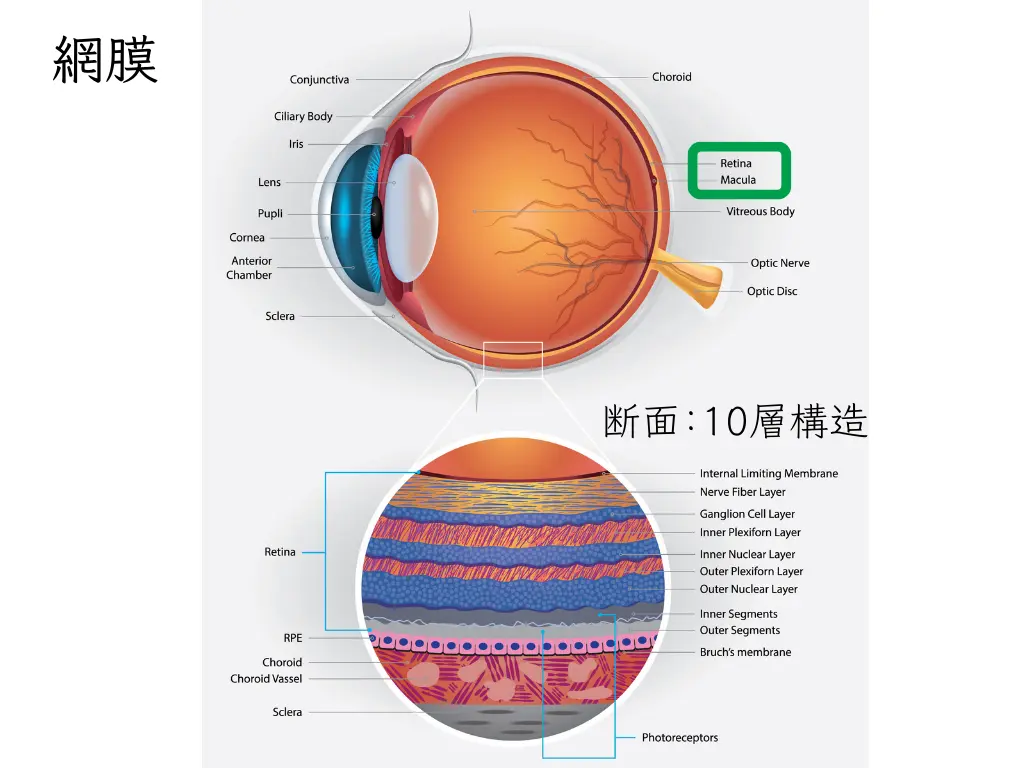
The retina is a membrane-like tissue made up of a collection of delicate nerves extending from the brain.
When magnified, it has a 10-layer structure.

- 1. Light is focused inside the eye and projected onto the retina
- 2. The information travels along the optic nerve and is passed to the occipital lobe of the brain (visual cortex)
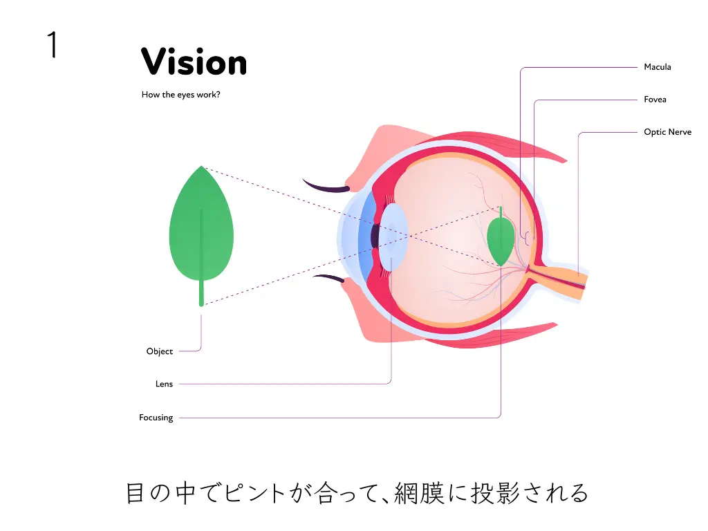
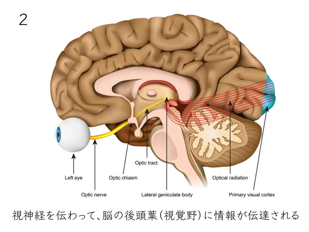
Seeing many sights after birth means continuing steps 1 and 2.
During that time, the brain’s “ability to see” develops.
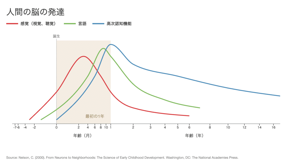
But
・Extreme refractive error (myopia, hyperopia, astigmatism)
・A child whose eye has been patched, creating a state in which the child cannot see
・Conditions in which light cannot reach the retina, such as congenital cataract or retinoblastoma
and so on:
for some reason,
・if the eye is not focused on the retina at step 1,
・or if light cannot reach the retina,
the image seen is not projected onto the retina normally.
In that case, step 2 is never reached.
The brain’s ability to see does not develop = a state of “amblyopia.”
The most common cause of amblyopia is refractive amblyopia.
Because of extreme refractive error (myopia, hyperopia, astigmatism), the eye is not focused on the retina.
Treatment for other causes of amblyopia (strabismic amblyopia, form-deprivation amblyopia) may also be needed.
Amblyopia Treatment Has a Time Limit
A child’s eyes develop rapidly from birth to age 3 and are nearly complete at around ages 6 to 8.
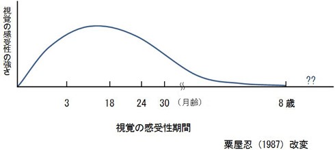
Early detection is the most important thing in amblyopia.
The brain of a child, which absorbs everything, has a time limit for growth.
If it is found early, we can work hard to develop the ability to see.
After appropriate testing, we prescribe glasses that focus light on the retina:
→ Covering the seeing eye (eye patching) is the most common treatment.
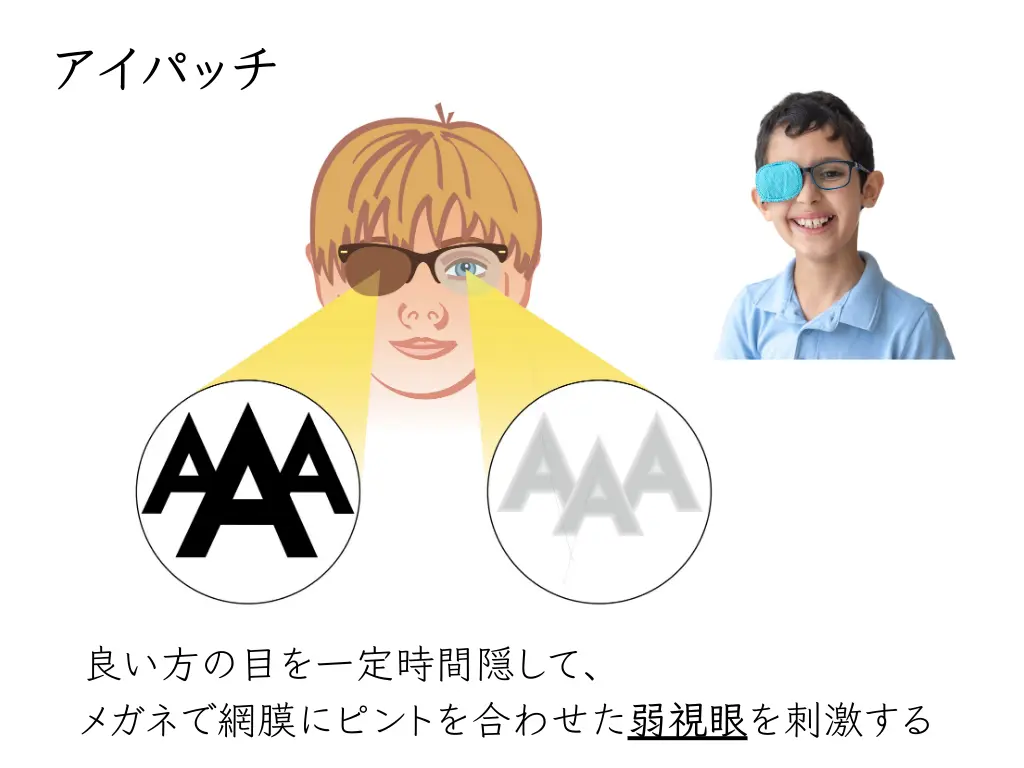
A small child wearing glasses may come as a big shock and feel sad.
The eye clinic does everything it can so that the child does not end up with “amblyopia,” a lifelong inability to see.
How We Test Whether Amblyopia Is Present
The tests are carried out bycertified orthoptists, who are used to testing children.
We cover one eye with a trial frame and measure the vision of each eye separately.
Sometimes we test using an eye patch.

If corrected visual acuity is not obtained, we check the retina (macula) for abnormalities.
With advances in testing equipment, abnormalities can now be detected quickly and without strain.
The tests are not dazzling and finish quickly, so children can often do them without problems.
The Difference Between Hyperopia and Myopia
Far? Near? Thinking about the meaning of the Japanese characters makes it confusing.
They are distinguished by the difference in the length of the eye (= axial length).
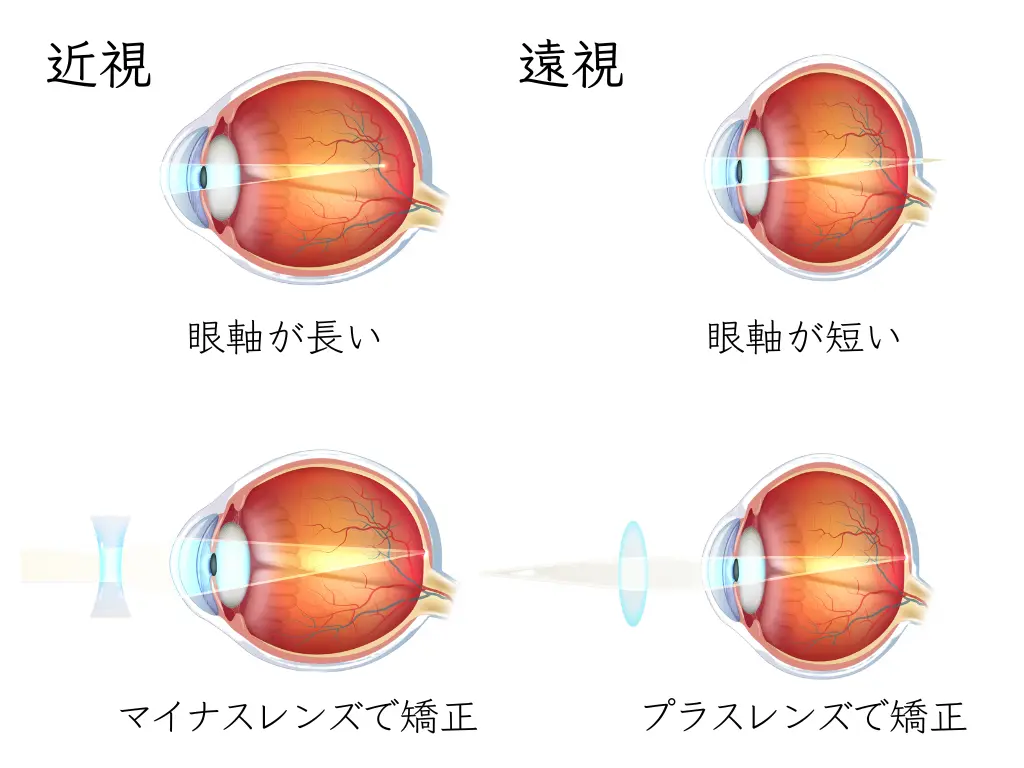
Short axial length = “hyperopia”
Long axial length = “myopia”
By measuring the axial length,we can tell whether the eye is a hyperopic eye or a myopic eye.
Hyperopia is corrected with plus lenses, and myopia with minus lenses.
Myopia Even with a Short Axial Length? How to Find Hidden Hyperopia
Children have a very strong ability to adjust focus, and a state of accommodative myopia may appear.
In this state, the accurate “refractive power of the eye” has not been measured.
To evaluate refraction accurately,
we take some time and test using cycloplegic drops (atropine, Cyplegin®).
・Axial length measurement
・Testing with cycloplegic drops
These can sometimes reveal hidden hyperopia.
Testing with cycloplegic drops dilates the pupil, so we also examine the fundus at the same time.
Astigmatism: Distortion of the Cornea
“The meaning of ‘astigmatism’ in daily life differs from the meaning of ‘astigmatism’ in ophthalmology.”
The meaning of astigmatism is
not “hard to see” or the like, but a state in which the cornea is distorted
: it simply refers to the shape of the cornea.
(There is also astigmatism caused by distortion of the lens.)
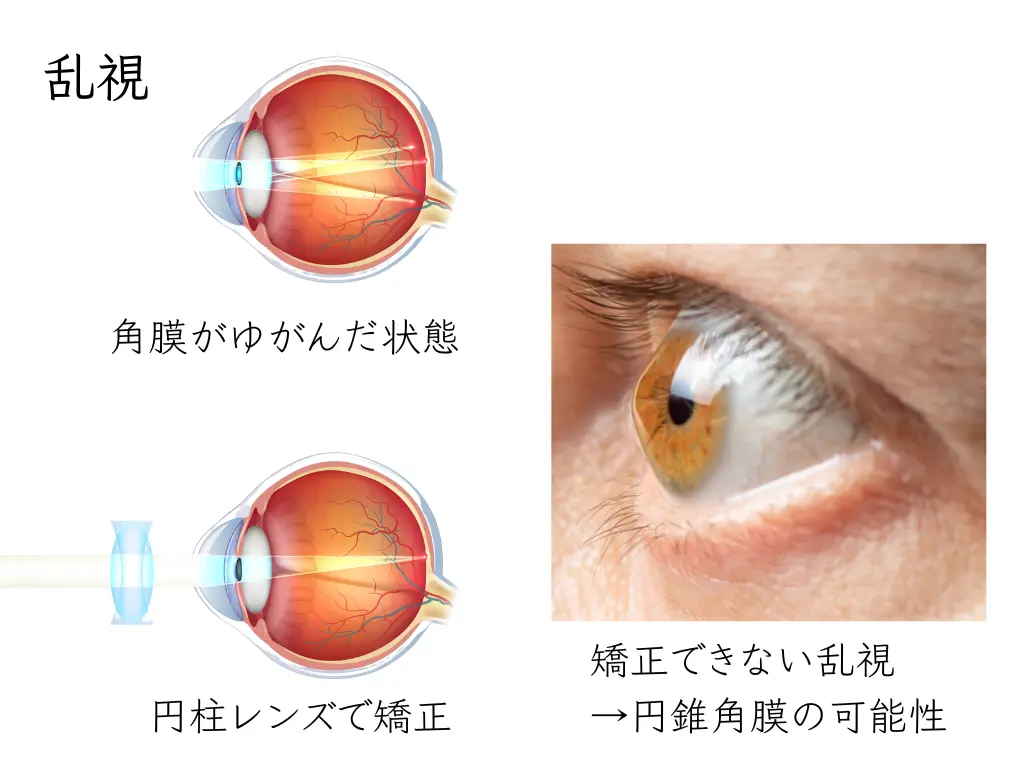
The cornea of a living eye is always slightly distorted, so everyone has astigmatism.
It is a condition that can be corrected with cylindrical lenses.
How to Find Out the Degree of Astigmatism and Whether Keratoconus Is Present
Refraction tests performed before the vision test
・Autorefractor
・Spot Vision Screener
give some idea of the state of astigmatism.
If the astigmatism seems strong,
we perform a test using a corneal topographer.
・Astigmatism that can be corrected with cylindrical lenses: “regular astigmatism”
・Cannot be corrected with cylindrical lenseskeratoconus: “irregular astigmatism”
((Other corneal diseases can also cause irregular astigmatism.)
These are distinguished.
If there is extremely strong “astigmatism,”
- light is focused inside the eye and projected onto the retina (step 1)
- and the information travels along the optic nerve and is passed to the occipital lobe of the brain (step 2),
but at step 1 the image may not be in focus, becoming a cause of amblyopia (refractive amblyopia).
This is the app for the “atropine course,” in which atropine is instilled at home and a test is done at the clinic on a later day.
We made it so that you won’t forget the eye drops. Please give it a try.
Atropine Eye Drop Reminder
Please set the period and times for the eye drops
Once you set the period, progress is displayed
How to use:
- Set the “start date,” “end date,” and the “morning and evening times.”
- Tap the green “Add to calendar” button.
- When the question “Download ‘atropine-schedule.ics’?” appears at the bottom of the screen, choose “Download.”
- When the download finishes, open the file from your browser’s download list and tap “Add all events” to finish.