![Takeru Eye Clinic | Takatori Shopping Street, Sawara-ku, Fukuoka City [Nishijin Station / Fujisaki Station]](https://takeru-eye.com/wp-content/uploads/2022/10/takeru_logo_for-WP-header.png)

Very many patients visit the eye clinic with the complaint of “red eyes.” The causes are varied: viruses, bacteria, allergies, and more.
Uveitis can also make the eyes red.
The judgment is made while imagining many possible diseases.
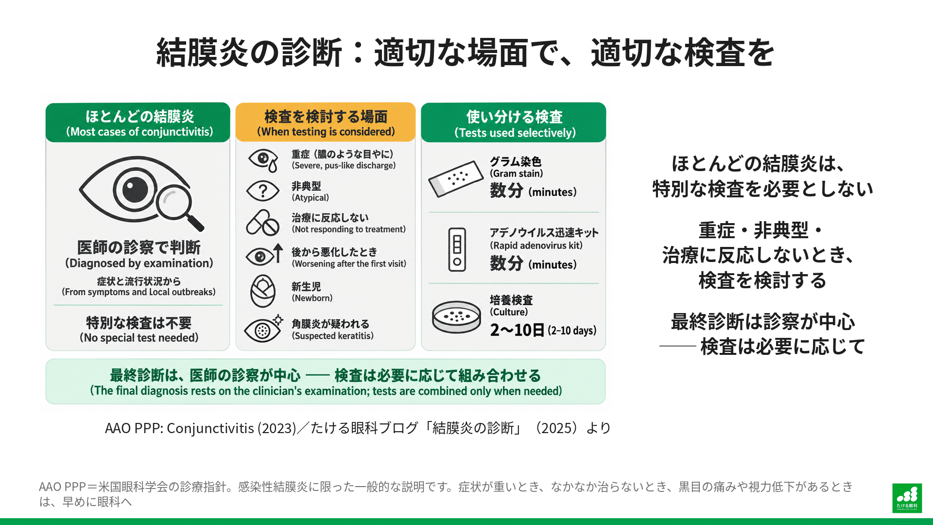
Most cases of conjunctivitis are judged by the doctor from the symptoms and the local epidemic situation, and no special tests are needed.
However, in certain situations—when symptoms are very severe, when it does not get better, or when it has affected the cornea (the black of the eye)—tests such as the “Gram stain” become an important clue for deciding on appropriate treatment.
This article is a general discussion limited to infectious conjunctivitis.
The Difficulty of Diagnosing Conjunctivitis: Early Symptoms and the Possibility of Worsening
“Even if symptoms are mild now, conjunctivitis symptoms often get worse later.”
Conjunctivitis often starts with very mild symptoms.
For example, the eyes may water a little or itch slightly, and you may not even notice it yourself.
However, these early symptoms can worsen over hours to days, leading to clear redness of the eye, discharge, pain, and in some cases even decreased vision.
Severe bacterial infections and adenovirus infections in particular tend to produce strong symptoms.
A disease that requires particular attention is gonococcal conjunctivitis.
It is rare, but it worsens very rapidly, so it is regarded as an ophthalmic emergency.
It is characterized by “hyperacute” symptoms appearing within 12 to 24 hours of onset: copious pus-like discharge, severe redness, and marked swelling of the eyelids.
If treatment is delayed, there is a high risk that the cornea (the black of the eye) will melt or perforate, and in the worst case blindness can result.
For this reason, when such symptoms appear, immediate diagnosis by a specialist (confirming the causative bacteria with a Gram stain) and treatment with systemic antibiotics such as ceftriaxone are necessary.
“Even for an experienced doctor, it is often difficult to determine the exact cause at the first visit.”
For that reason, even an experienced ophthalmologist is said to find it difficult to diagnose the exact cause at the first visit.
Why Not Test Every Case of Conjunctivitis?
Most acute conjunctivitis is caused by viruses and gets better on its own.
Even when bacteria are the cause, most cases are mild and are known to recover without antibiotics, although antibiotics can speed recovery.
For that reason, professional bodies such as the American Academy of Ophthalmology do not recommend routine testing of all patients with common conjunctivitis. Testing is considered in specific cases such as the following.
- Severe cases: copious pus-like discharge.
- Atypical cases: it looks different from common conjunctivitis.
- Cases that do not respond to treatment: no improvement even with prescribed medication.
- Newborns.
- Suspected keratitis: inflammation reaches the black of the eye and may seriously affect vision.
Choosing the Right Diagnostic Tool
There are several tests for diagnosing eye infections, each with strengths and weaknesses. The doctor chooses the best tool for the situation.
When the Gram Stain Shows Its Worth
In the “special situations” above, the Gram stain gives rapid results and is therefore very useful in deciding the initial treatment policy.
Sight-Threatening Severe Bacterial Keratitis
Bacterial keratitis, in which bacteria infect the cornea (the black of the eye), is judged to be a highly urgent disease, unlike conjunctivitis.
In this case, a Gram stain can estimate the presence and type of bacteria within minutes.
In fact, the Infectious Keratitis Practice Guideline of the Japanese Society for Ocular Infections also strongly recommends smear microscopy (such as the Gram stain) and culture for diagnosing bacterial keratitis.
A Hard-to-Find Pathogen: Microsporidia
In rare cases, a tiny parasite called “microsporidia” can cause keratitis.
In this diagnosis too, a smear examination that directly confirms the pathogen under a microscope is very important.
Another Rapid Test: The Adenovirus Rapid Diagnostic Kit
When epidemic keratoconjunctivitis (“hayari-me”) is strongly suspected, a diagnostic kit that rapidly detects adenovirus may be used.
These tests give results within minutes at the clinic, which helps confirm early whether adenovirus is the cause.
Once the diagnosis is confirmed, viral conjunctivitis can be distinguished from bacterial or allergic types, avoiding unnecessary antibiotic use. It also gives a guide to how long to stay away from work or school.
Kit sensitivity (the ability to detect disease) varies, but specificity (the ability to correctly identify people who do not have the disease) is very high at 96–98%, so a positive result is also highly reliable.
However, even if the test is negative, infection cannot be completely ruled out, especially in the early and late stages of symptoms.

Comparison of the Tests
| Test | Sensitivity (ability to detect disease) | Specificity (ability to correctly identify people without the disease) | Time required | Features |
| Gram stain | 36–75% | 83–98% | A few minutes | Rapid, and can strongly suggest the presence of bacteria and some parasites. However, sensitivity is moderate. |
| Rapid adenovirus test kit | About 90% (variable) | 96-98% | A few minutes | Useful for rapid diagnosis of epidemic keratoconjunctivitis (“hayari-me”). A negative result does not completely rule out infection. |
| Culture | 38–66% | 95–100% | 2–10 days | The “gold standard”: it can identify the causative bacteria accurately and show which antibiotic is effective. However, it takes time to get the result. |
| PCR | 25–88% | 93–100% | Several hours to 1 day | Excellent for detecting viruses and bacteria that are difficult to culture. |
Conclusion: The Right Test in the Right Situation
The Gram stain is not a test that every patient with a “red eye” must have.
Many everyday cases of conjunctivitis are diagnosed and treated by the doctor’s examination.
However, tests such as the Gram stain and the adenovirus rapid diagnostic kit are extremely valuable when bacterial keratitis is strongly suspected or severe conjunctivitis is a possibility, because they point the way to prompt and accurate initial treatment.
The final diagnosis is made mainly through the doctor’s clinical judgment, combined with these tests as needed.
References
- American Academy of Ophthalmology (2023), Preferred Practice Pattern: Conjunctivitis.
https://www.aao.org/education/preferred-practice-pattern/conjunctivitis-ppp-2023 - Azari, A.A. and Barney, N.P. (2013), ‘Conjunctivitis: a systematic review of diagnosis and treatment’, JAMA, 310(16), pp. 1721-1729.
https://pubmed.ncbi.nlm.nih.gov/24150468/ - Centers for Disease Control and Prevention (2021), Sexually Transmitted Infections Treatment Guidelines, 2021. https://www.cdc.gov/std/treatment-guidelines/gonorrhea-adults.htm
- EyeWiki (2024), Epidemic Keratoconjunctivitis.
https://eyewiki.org/Epidemic_Keratoconjunctivitis - Costumbrado, J., Ng, D.K. and Ghassemzadeh, S., ‘Gonococcal Conjunctivitis’, StatPearls [Internet], Treasure Island (FL): StatPearls Publishing.
https://www.ncbi.nlm.nih.gov/books/NBK459289/ - Gurnani, B. and Kaur, K. (2024), ‘Bacterial Keratitis’, StatPearls [Internet], Treasure Island (FL): StatPearls Publishing.
https://www.ncbi.nlm.nih.gov/books/NBK574509/ - Japanese Society for Ocular Infections, Infectious Keratitis Practice Guideline 3rd Edition Drafting Committee (2023), ‘Infectious Keratitis Practice Guideline (3rd edition)’, Nippon Ganka Gakkai Zasshi (J Jpn Ophthalmol Soc), 127(10), pp. 859-903.
https://www.nichigan.or.jp/member/journal/guideline/detail.html?itemid=672&dispmid=909 - Sabhapandit, S., Murthy, S.I., Garg, P., et al. (2016), ‘Microsporidial Stromal Keratitis: clinical features, unique diagnostic criteria, and treatment outcomes’, Cornea, 35(12), pp. 1569-1574.
https://pubmed.ncbi.nlm.nih.gov/27467044/ - Sambursky, R., Trattler, W., Tauber, S., et al. (2013), ‘Sensitivity and specificity of the AdenoPlus test for diagnosing adenoviral conjunctivitis’, JAMA Ophthalmology, 131(1), pp. 17-22.
https://pubmed.ncbi.nlm.nih.gov/23307204/ - Kam, K.Y.R., Ong, H.S., Bunce, C., et al. (2015), ‘Sensitivity and specificity of the AdenoPlus point-of-care system in detecting adenovirus in conjunctivitis patients’, British Journal of Ophthalmology, 99(9), pp. 1186-1189.
https://pubmed.ncbi.nlm.nih.gov/25824258/ - Moshirfar, M., et al. (2019), ‘Biological Staining and Culturing in Infectious Keratitis: Controversy in Clinical Utility’, Medical Hypothesis, Discovery & Innovation in Ophthalmology, 8(3), pp. 145-151.
https://pubmed.ncbi.nlm.nih.gov/31598516/ - Sharma, S. (2012), ‘Diagnosis of infectious diseases of the eye’, Eye (London), 26(2), pp. 177-184.
https://pubmed.ncbi.nlm.nih.gov/22094299/ - Sheikh, A., Hurwitz, B., van Schayck, C.P., McLean, S. and Nurmatov, U. (2012), ‘Antibiotics versus placebo for acute bacterial conjunctivitis’, Cochrane Database of Systematic Reviews, (9), CD001211.
https://pubmed.ncbi.nlm.nih.gov/22972049/