![Takeru Eye Clinic | Takatori Shopping Street, Sawara-ku, Fukuoka City [Nishijin Station / Fujisaki Station]](https://takeru-eye.com/wp-content/uploads/2022/10/takeru_logo_for-WP-header.png)

Do lines that should be straight look wavy? Do tile patterns or window frames seem distorted? If any of this sounds familiar, the cause may be a condition called an “epiretinal membrane.”
“Epiretinal membrane,” “preretinal membrane,” “macular epiretinal membrane,” “macular preretinal membrane,” “macular pucker,” and “cellophane maculopathy”
All of these are terms that refer to almost the same condition. Only the names differ; what is happening inside the eye is the same.

An epiretinal membrane (macular epiretinal membrane) is a condition in which a thin, cellophane-like membrane forms on the surface of the retina, the light-sensing nerve layer at the back of the eye. In many cases it progresses slowly, and urgent treatment is rarely needed. However, if symptoms interfere with daily life, surgery is a treatment option, and improvement in visual function can be expected.
In this article, I explain in detail what an epiretinal membrane (macular epiretinal membrane) is, why it occurs, and how to deal with it.
What Is an Epiretinal Membrane (Macular Epiretinal Membrane)?
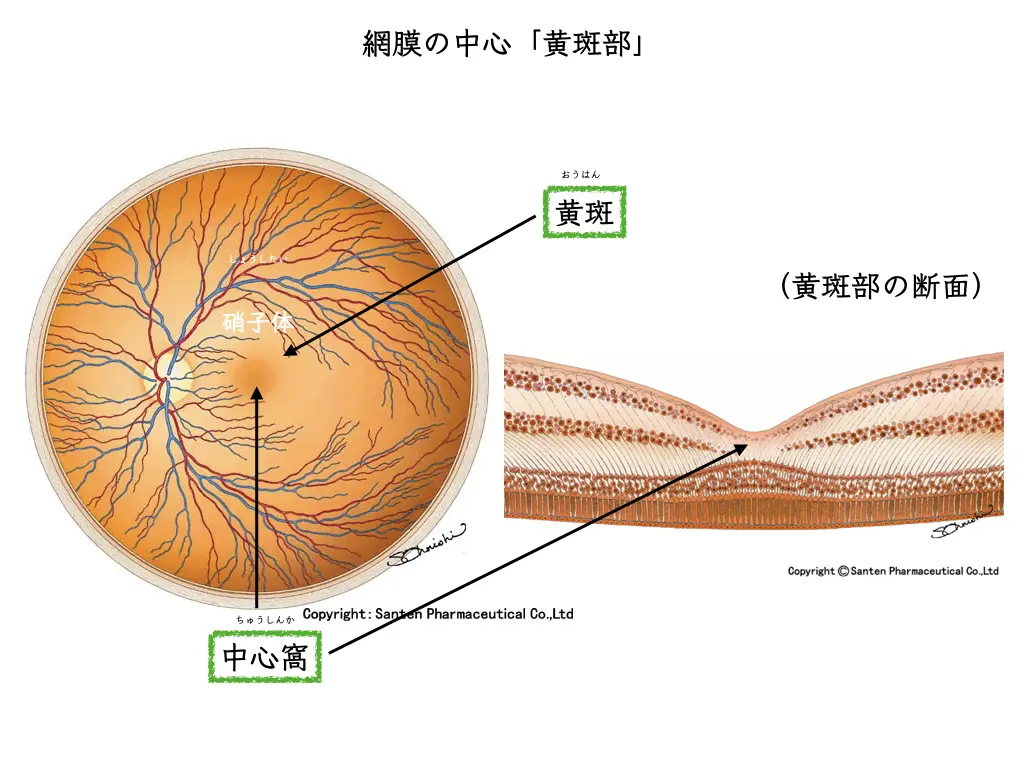
The retina is an extremely delicate layer of nerve tissue that plays the role of the film or image sensor in a camera. The most important part, the center of our vision, is called the “macula.” An epiretinal membrane (macular epiretinal membrane) is a condition in which a fibrocellular membrane forms on the front surface of this macula.
As this membrane contracts over time, it wrinkles the retina beneath it and physically distorts the retina. This mechanical distortion causes the characteristic symptom mentioned at the beginning: “objects look distorted (metamorphopsia).”
Causes of Epiretinal Membrane (Macular Epiretinal Membrane)
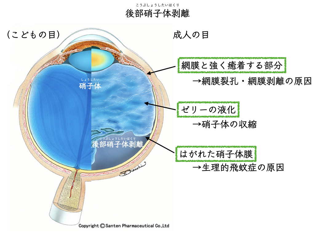
The most common cause of an epiretinal membrane (macular epiretinal membrane) is a naturally occurring age-related phenomenon called “posterior vitreous detachment.”
The inside of the eyeball is filled with a gel-like substance, similar to egg white, called the vitreous.
In youth, the vitreous is in contact with the retina, but with age it gradually liquefies and shrinks. At some point, it separates naturally from the surface of the retina. This is called posterior vitreous detachment.
In most cases, posterior vitreous detachment completes without problems. However, during the process of detachment, part of the vitreous may remain on the surface of the retina, or very minute injuries may occur. The eye then tries to repair the injury, and retinal cells and others migrate to the surface and proliferate, forming a membrane.
When aging or posterior vitreous detachment is the main cause, it is called “idiopathic,” and this accounts for the majority of cases1. On the other hand, when it is caused by diabetic retinopathy, retinal detachment, eye injury, or surgery, it is called “secondary.”

Epiretinal Membrane (Macular Epiretinal Membrane): Main Symptoms
Not every epiretinal membrane (macular epiretinal membrane) causes symptoms. In fact, many people have no symptoms, and the condition is found by chance during an eye checkup.
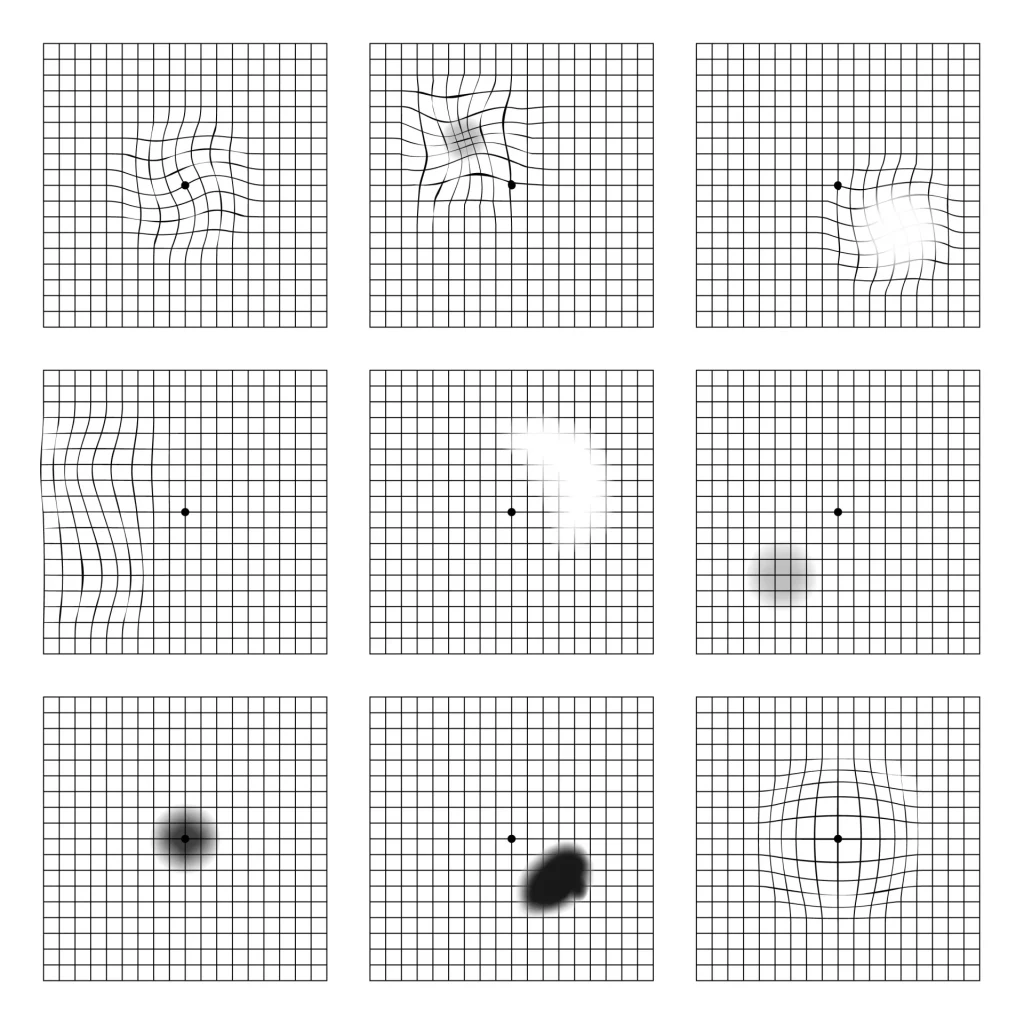
When symptoms do appear, the most typical is “metamorphopsia.” This is a symptom in which straight lines look wavy or bent, and it is a complaint that greatly affects the quality of life of many patients.
Other symptoms that may be seen include the following.
- Blurred central vision
- Objects look smaller than they really are (micropsia)
- Letters and other text become hard to read
- Objects may look double when viewed with only one eye (monocular diplopia)
An epiretinal membrane (macular epiretinal membrane) is a disease that mainly affects central vision; it does not cause loss of the visual field or lead to blindness.
Does an Epiretinal Membrane (Macular Epiretinal Membrane) Heal on Its Own? The Outlook
Many people worry, “If I leave it like this, won’t I lose my sight?”
However, it is known that many epiretinal membranes (macular epiretinal membranes) stop progressing and become stable after a certain period of time.
One study that followed patients with an epiretinal membrane and relatively good visual acuity (20/40; decimal 0.5 or better) reported that about 21% needed surgery after 4 years3.
This shows that many people can be managed with observation, without surgery. In addition, some early epiretinal membranes may progress slowly over several years.
Very rarely, the membrane may peel away from the retina on its own and the symptoms may improve.
Epiretinal Membrane (Macular Epiretinal Membrane): How It Is Diagnosed
For diagnosing and evaluating an epiretinal membrane (macular epiretinal membrane), the most important test today is “OCT (optical coherence tomography).”
OCT is a device that can capture cross-sections of the retina in very high definition. It can accurately determine whether a membrane is present on the surface of the retina, and can also evaluate in detail the thickness of the membrane, retinal wrinkling, the degree of swelling, and even the condition of the photoreceptors.
This information is essential for forming an outlook and deciding on a treatment plan.
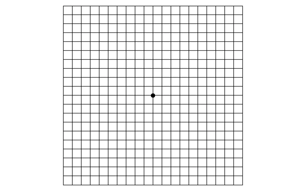
For checking distortion at home, a grid-like chart called the “Amsler grid” is useful.
Choosing Treatment: Observation and Surgery
There are no eye drops, oral medications, or supplements that are effective for an epiretinal membrane (macular epiretinal membrane).
When treatment becomes necessary, the only method is surgery.
Observation
If symptoms are mild or absent and there is no major impact on daily life, it is common to follow the condition with regular examinations.
In many cases the membrane is stable, so observation is a reasonable choice, also in the sense of avoiding unnecessary surgery.
Surgery (Vitrectomy)
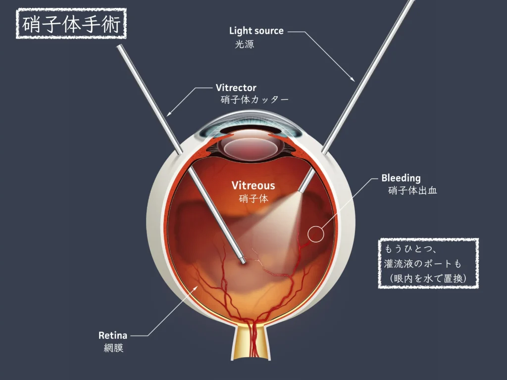
Surgery is considered when metamorphopsia or reduced vision clearly interferes with daily life, such as reading, driving, or work.
Timing of Surgery
There is no clear criterion for surgery, but it is often considered when visual acuity falls to about 0.5 (20/40) or when distortion greatly reduces quality of life.
If surgery is done too early, it may involve unnecessary risks.
On the other hand, if it is too late, the retinal tissue will have been distorted for a long time, and recovery after surgery may be limited.
Therefore, it is important to decide the best timing by comprehensively judging the condition of the retina on OCT and how much inconvenience the patient actually feels.
How the Surgery Is Done
The surgery is called “vitrectomy,” and in most cases it is performed as day surgery under local anesthesia.
Three small openings are made in the eyeball, and thin instruments are inserted through them. First, the vitreous that is pulling on the retina is removed.
Next, a special dye is used to stain the thin membrane on the retina so that it is easier to see, and then the membrane is carefully peeled off with extremely small tweezers called micro forceps.
Surgery for Epiretinal Membrane (Macular Epiretinal Membrane): Success Rate and Visual Recovery
The Course of Recovery
Visual recovery after surgery is characteristically slow. Immediately after surgery, vision may actually remain worse than before, but it gradually improves over several weeks to several months. Most of the improvement occurs in the first 3 to 6 months, but it can take more than a year to reach the final quality of vision.
Effects of Surgery
The success rate of surgery is high, and improvement in visual acuity has been reported in many patients8. Also, many patients feel a reduction in distortion more than the visual acuity numbers would suggest. However, it is important to understand that if the retina has been distorted for a long time, visual function will not return completely to its original state even after surgery.
Risks and Complications
Vitrectomy is a well-established procedure, but it is not entirely without risk.
The most frequent complication is “progression of cataract.”
In many cases, a cataract progresses within a few years, so cataract surgery may eventually be needed.
Other complications, though less frequent, that have been reported include retinal tears, retinal detachment, infection, and recurrence of the membrane8.
Summary
An epiretinal membrane is an eye condition that can occur in anyone with aging. Many cases have no symptoms or only mild symptoms and can be followed by observation. However, if symptoms such as “things look distorted” or “vision looks hazy” are lowering your quality of life, surgery can be expected to improve visual function.
What matters is to first accurately understand the condition of your own retina with tests such as OCT. Then, compare the inconvenience caused by how you see with the benefits and risks of treatment, and carefully decide on the best option for you.
References
- Ożóg, M. K., Nowak-Wąs, M., & Rokicki, W. (2023). Pathophysiology and clinical aspects of epiretinal membrane – review. Frontiers in Medicine, 10, 1121270. https://pubmed.ncbi.nlm.nih.gov/37636571/
- Stevenson, W., Prospero Ponce, C. M., Agarwal, D. R., Gelman, R., & Christoforidis, J. B. (2016). Epiretinal membrane: optical coherence tomography-based diagnosis and classification. Clinical Ophthalmology, 10, 527–534. https://pubmed.ncbi.nlm.nih.gov/27099458/
- Luu, K.-Y., Koenigsaecker, T., Yazdanyar, A., Mukkamala, L., Durbin-Johnson, B. P., Morse, L. S., Moshiri, A., Park, S. S., & Yiu, G. (2019). Long-term natural history of idiopathic epiretinal membranes with good visual acuity. Eye, 33(5), 714–723. https://pubmed.ncbi.nlm.nih.gov/31000833/
- Lee, M.-W., Jung, I., Song, Y.-Y., Baek, S.-K., & Lee, Y.-H. (2020). Long-term outcome of epiretinal membrane surgery in patients with internal limiting membrane dehiscence. Journal of Clinical Medicine, 9(8), 2470. https://pubmed.ncbi.nlm.nih.gov/32752171/
- Cacciamani, A., Cosimi, P., Ripandelli, G., Di Nicola, M., & Scarinci, F. (2020). Epiretinal membrane surgery: structural retinal changes correlate with the improvement of visual function. Journal of Clinical Medicine, 10(1), 90. https://pubmed.ncbi.nlm.nih.gov/33383922/
- Bailey, S. T., Vemulakonda, G. A., Kim, S. J., Kovach, J. L., Lim, J. I., Ying, G.-S., & Flaxel, C. J.; American Academy of Ophthalmology PPP Retina/Vitreous Committee. (2025). Idiopathic epiretinal membrane and vitreomacular traction Preferred Practice Pattern®. Ophthalmology, 132(4), P197–P233. https://pubmed.ncbi.nlm.nih.gov/39918520/
- Al-Khersan, H., Shaheen, A. R., Flynn, H. W., Jr., & Smiddy, W. E. (2022). Natural history and surgical timing for idiopathic epiretinal membrane. Ophthalmology Retina, 6(11), 978–984. https://pubmed.ncbi.nlm.nih.gov/35272083/
- Iuliano, L., Fogliato, G., Gorgoni, F., Corbelli, E., Bandello, F., & Codenotti, M. (2019). Idiopathic epiretinal membrane surgery: safety, efficacy and patient related outcome measures. Clinical Ophthalmology, 13, 1253–1265. https://pubmed.ncbi.nlm.nih.gov/31409964/
- Hartmann, K. I., Schuster, A. K., Bartsch, D. U., Kim, J. S., Chhablani, J., & Freeman, W. R. (2014). Restoration of retinal layers after epiretinal membrane peeling. Retina, 34(4), 647–654. https://pubmed.ncbi.nlm.nih.gov/24077089/
- Steinkerchner, M. S., Russell, M. W., Sharma, N., Kumar, M., Talcott, K. E., & Singh, R. P. (2024). Long-term visual outcomes in patients with idiopathic epiretinal membrane surgery. Ophthalmic Surgery, Lasers and Imaging Retina, 55(2), 71–77. https://pubmed.ncbi.nlm.nih.gov/38198614/
- Park, S. H., Moon, S., Kim, S.-J., Lee, J.-E., Park, S. W., Byon, I., & Lee, S. M. (2025). Idiopathic epiretinal membrane area changes in eyes with good vision and the association with visual function. PLOS ONE, 20(9), e0331437. https://pubmed.ncbi.nlm.nih.gov/40892798/