![Takeru Eye Clinic | Takatori Shopping Street, Sawara-ku, Fukuoka City [Nishijin Station / Fujisaki Station]](https://takeru-eye.com/wp-content/uploads/2022/10/takeru_logo_for-WP-header.png)

Your eyes feel gritty, the redness persists, light is dazzling, and your vision is a little blurred.
When symptoms like these continue, many people feel anxious.
What you thought was a stye or conjunctivitis may in fact be a viral infection called “ocular herpes.”
The most important thing in treating ocular herpes is to determine accurately whether “the virus itself is actively causing damage” or “the body’s immune response has become excessive.” The drugs to be used are completely different depending on this judgment.
In this article I explain step by step what ocular herpes is, why it occurs, and what types and treatments there are.

The “Latent Virus” That Many People Carry Without Knowing

Ocular herpes is caused by herpes simplex virus (HSV) or by varicella-zoster virus (VZV), the virus that causes chickenpox and shingles [2].
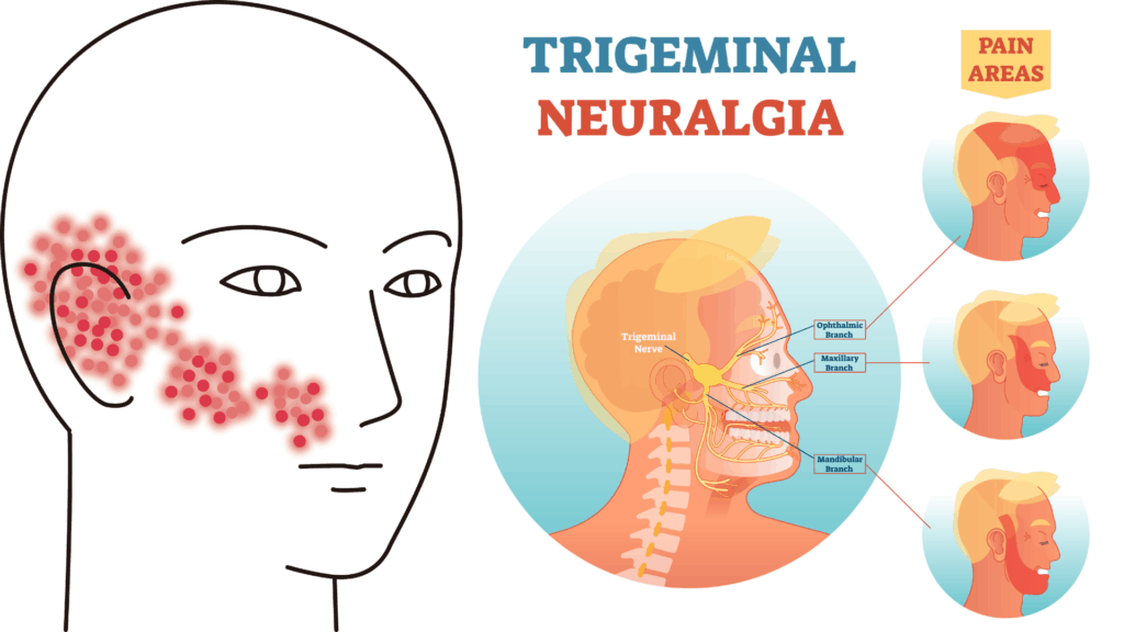
Once infected, these viruses do not completely leave the body. They lurk quietly in a ganglion at the root of the trigeminal nerve, the nerve that carries sensation from the face to the brain (latent infection) [2]. It is like a hibernating animal.
Usually they cause no symptoms, but triggered by fever, physical or mental stress, ultraviolet light, or reduced immunity, the virus becomes active again (reactivation) [5]. The awakened virus travels along the nerve to the surface or the inside of the eye and causes a variety of symptoms.
In developed countries, herpes simplex keratitis is one of the leading infectious causes of blindness [2].
Eye Diseases Caused by Herpes Simplex Virus (HSV)
Herpes simplex virus can cause disease in many parts of the eye, including the eyelid, conjunctiva, cornea (the black of the eye), iris (the brown part that determines eye color), and retina. Corneal disease is the most typical.
When inflammation occurs in the iris, it is called herpetic iritis (uveitis).

Herpetic Keratitis: Two Types That Require Different Treatment
Herpetic keratitis is broadly divided into two types according to how the disease arises, and the treatments are completely different.
1. The type in which the virus directly affects the cornea (epithelial keratitis)
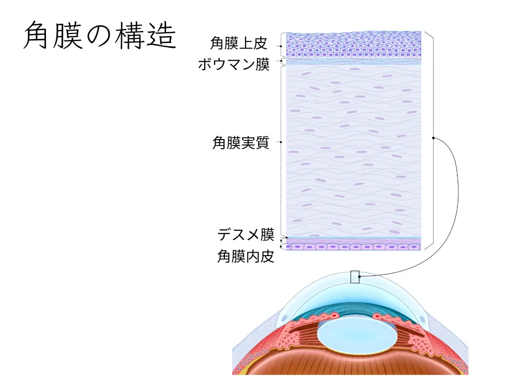
This is a state in which the virus is actively multiplying in the “epithelium,” the outermost layer of the cornea [8].
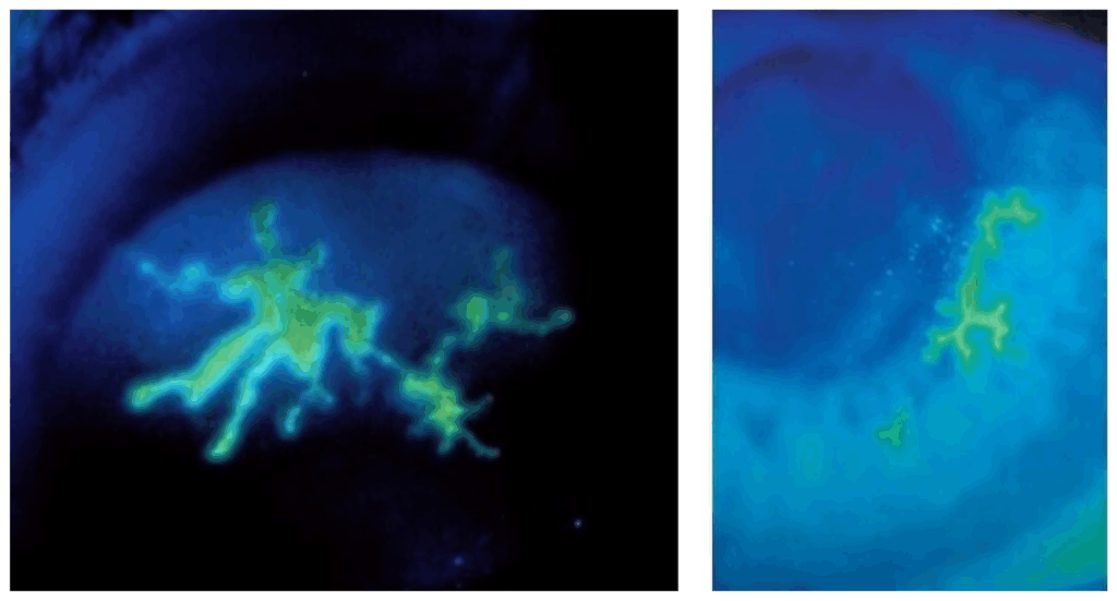
A characteristic finding is a lesion called a “dendritic ulcer.” It has a shape like a growing tree branch or a bolt of lightning, and an ophthalmologist can diagnose it by checking it with a special dye and a microscope [8]. This lesion is formed as the virus multiplies and destroys cells. Pain, a foreign-body sensation, redness, tearing, and light sensitivity tend to occur.
The mainstay of treatment for this type is antiviral medication that directly suppresses viral replication. Eye drops and eye ointment are mainly used [8]. However, caution is needed: if steroids are mistakenly used at this stage to suppress inflammation, they can promote viral replication and make the condition worse.
2. The type caused by the body’s immune response (stromal keratitis)
This, on the other hand, is inflammation in the “stroma,” a slightly deeper layer of the cornea.
It occurs when the body’s protective immune system overreacts to the viral fragments remaining in the cornea, rather than to viral replication itself [2].
It is as if the body’s well-meant defense “mistakes friend for foe and attacks,” injuring its own corneal tissue.
The main symptoms are blurring and decreased vision, and pain may be relatively mild.
The mainstay of treatment here is steroid drugs to suppress the excessive immune response.
However, using steroids alone can allow the lurking virus to become active again, so antiviral drugs are also used together to prevent this [6].
This treatment policy was established by a large clinical study called the Herpetic Eye Disease Study (HEDS) [6, 7].
In this way, even for the same herpetic keratitis, the treatment policy differs greatly depending on whether the main cause of the disease is “viral activity” or “the immune response.”
Herpes Simplex Spreading Beyond the Cornea
Herpes simplex can also cause inflammation outside the cornea.
Corneal endotheliitis
The endothelial cells on the innermost side of the cornea are damaged, the whole cornea swells, and vision decreases [2].
Iridocyclitis (uveitis)
Inflammation occurs in the iris (the brown part of the eye that determines eye color) and in the ciliary body, which lies behind it and is responsible for focusing, causing blurred vision, eye pain, and redness. Eye pressure may also rise sharply [2].
In these conditions as well, the immune response is the main cause, so combining steroids and antiviral drugs is the basis of treatment.
Eye Disease Caused by Varicella-Zoster Virus (VZV)
When shingles occurs on the face and is accompanied by eye symptoms, it is called herpes zoster ophthalmicus (HZO).
It develops when the same virus that causes chickenpox is reactivated in the first branch of the trigeminal nerve, the area from the forehead to the eye and nose [4].
A Rash on the Nose Is a Warning Sign: Hutchinson’s Sign
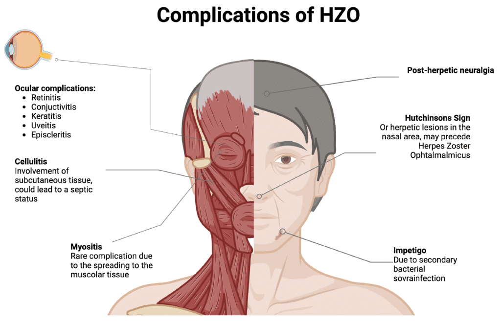
If, along with a rash on the forehead, a rash appears on the tip or side of the nose, particular caution is needed.
This is called “Hutchinson’s sign,” a sign indicating a very high risk of complications inside the eye [4].
This is because the tip of the nose and the eye are supplied by the same nerve (the nasociliary nerve).
If this sign is seen, please see an ophthalmologist immediately.
Diverse Eye Complications and Aftereffects
Herpes zoster ophthalmicus can cause conjunctivitis, keratitis, uveitis, and elevated eye pressure [4].
In addition, because the nerves are damaged by the virus, long-lasting nerve pain even after treatment (postherpetic neuralgia, PHN) can become a problem [4].
The most important thing in treatment is to start high-dose oral antiviral medication within 72 hours of the appearance of the rash. It is known that early treatment suppresses viral replication and greatly reduces the risk of eye complications and aftereffects [1].
A Warning Sign Not to Miss: Herpes of the Retina
Although very rare, herpesvirus can infect the retina and cause a serious disease called acute retinal necrosis (ARN) [5].
It progresses rapidly and destroys the retina over a wide area, so it is an ophthalmic emergency that carries a risk of blindness [5].
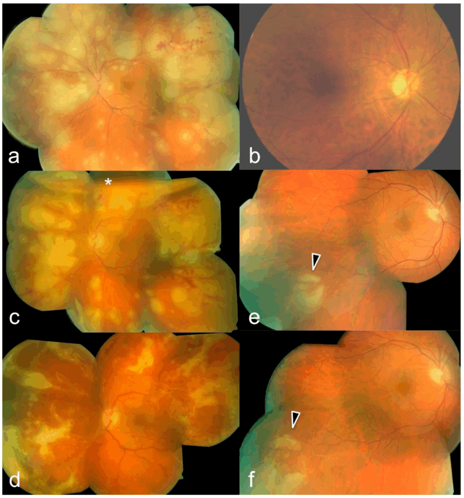
If you notice a sudden decrease in vision or a loss of visual field, you must see a specialized medical institution immediately.
Treatment is intensive, with intravenous antiviral drugs and injections into the eye [5].
Even if the inflammation is mild at first, lesions often arise from the periphery of the retina.
For that reason, it is important for ophthalmologists to observe the retinal periphery as well.
Today, with ultra-wide-field fundus imaging, it has become possible to screen the periphery too.
Preventing Recurrence
Ocular herpes tends to recur even after it has once completely healed. Preventive treatment is especially important for patients who repeatedly have stromal keratitis, which has a large effect on vision.
Long-Term Preventive Oral Medication
The HEDS study has proven that taking a low-dose antiviral drug every day for at least one year reduces the recurrence rate of all types of ocular herpes by about 40% and that of stromal keratitis by about 50% [7].
Prevention Before Eye Surgery
Eye surgeries such as cataract surgery and corneal transplantation can trigger reactivation of the lurking herpesvirus [2]. When a patient with a history of ocular herpes undergoes surgery, taking antiviral drugs preventively before and after surgery is very effective in preventing recurrence.
Herpes Zoster Vaccine
For people aged 50 and over, there is a highly effective vaccine (Shingrix®) that prevents the onset of shingles [3]. This vaccine helps prevent not only shingles itself but also complications such as herpes zoster ophthalmicus and postherpetic neuralgia [3].
Aqueous-humor PCR testing when herpes is suspected, and why treatment changes depending on whether it is infectious, are explained in the next article.
Summary
Ocular herpes is a single disease, yet its symptoms are extremely varied. The conditions range from those in which actively multiplying virus is the problem to those caused by the body’s immune response.
- In epithelial keratitis, antiviral drugs are the mainstay of treatment.
- In stromal keratitis and uveitis, the key to treatment is to use steroids carefully while using antiviral drugs as an adjunct.
- In herpes zoster ophthalmicus, it is especially important to start oral antiviral medication as soon as possible.
In any type, using over-the-counter eye drops on your own judgment or interrupting treatment is dangerous.
If eye symptoms continue, please do not put it off with “let’s wait and see”; see an ophthalmology specialist early. Early detection and early treatment are the best ways to protect your precious vision.
References
- Balfour, H. H., Jr, Bean, B., Laskin, O. L., Ambinder, R. F., Meyers, J. D., Wade, J. C., Zaia, J. A., Aeppli, D., Kirk, L. E., Segreti, A. C., & Keeney, R. E. (1983). Acyclovir halts progression of herpes zoster in immunocompromised patients. The New England journal of medicine, 308(24), 1448–1453. https://doi.org/10.1056/NEJM198306163082404
- Farooq, A. V., & Shukla, D. (2012). Herpes simplex epithelial and stromal keratitis: an epidemiologic update. Survey of ophthalmology, 57(5), 448–462. https://doi.org/10.1016/j.survophthal.2012.01.005
- Lal, H., Cunningham, A. L., Godeaux, O., Chlibek, R., Diez-Domingo, J., Hwang, S. J., Levin, M. J., McElhaney, J. E., Poder, A., Puig-Barberà, J., Vesikari, T., Watanabe, D., Weckx, L., Zahaf, T., Heineman, T. C., & ZOE-50 Study Group (2015). Efficacy of an adjuvanted herpes zoster subunit vaccine in older adults. The New England journal of medicine, 372(22), 2087–2096. https://doi.org/10.1056/NEJMoa1501184
- Sanjay, S., Huang, P., & Lavanya, R. (2011). Herpes zoster ophthalmicus. Current treatment options in neurology, 13(1), 79–91. https://doi.org/10.1007/s11940-010-0098-1
- Schoenberger, S. D., Kim, S. J., Thorne, J. E., Mruthyunjaya, P., Yeh, S., Bakri, S. J., & Ehlers, J. P. (2017). Diagnosis and Treatment of Acute Retinal Necrosis: A Report by the American Academy of Ophthalmology. Ophthalmology, 124(3), 382–392. https://doi.org/10.1016/j.ophtha.2016.11.007
- Barron, B. A., Gee, L., Hauck, W. W., Kurinij, N., Dawson, C. R., Jones, D. B., Wilhelmus, K. R., Kaufman, H. E., Sugar, J., & Hyndiuk, R. A. (1994). Herpetic Eye Disease Study. A controlled trial of oral acyclovir for herpes simplex stromal keratitis. Ophthalmology, 101(12), 1871–1882. https://doi.org/10.1016/s0161-6420(13)31155-5
- Acyclovir for the prevention of recurrent herpes simplex virus eye disease. Herpetic Eye Disease Study Group. (1998). The New England journal of medicine, 339(5), 300–306. https://doi.org/10.1056/NEJM199807303390503
- Wilhelmus K. R. (2015). Antiviral treatment and other therapeutic interventions for herpes simplex virus epithelial keratitis. The Cochrane database of systematic reviews, 1(1), CD002898. https://doi.org/10.1002/14651858.CD002898.pub5