![Takeru Eye Clinic | Takatori Shopping Street, Sawara-ku, Fukuoka City [Nishijin Station / Fujisaki Station]](https://takeru-eye.com/wp-content/uploads/2022/10/takeru_logo_for-WP-header.png)

During pregnancy,
・things may look harder to see, or your vision may drop
・your contact lenses or glasses may no longer suit you
・your vision may seem strange
— any of these may happen.
The tissues responsible for refracting the light entering the eye are
the cornea (the black of the eye) and the crystalline lens.
Both can temporarily change in thickness during pregnancy.
Pregnancy is also a time when the body’s immune system changes greatly.
Temporary changes in the front half of the eyeball (the anterior segment): the cornea and lens
may be the cause of reduced vision during pregnancy.1。
Most eye complications during pregnancy are mild and temporary and need no treatment.
Sometimes they are serious and permanent, and prompt judgment by an ophthalmologist may be needed.
However, we also need to watch for pathological changes inside the eye.
Reduced Vision During Pregnancy (Changes in Eye Prescription)
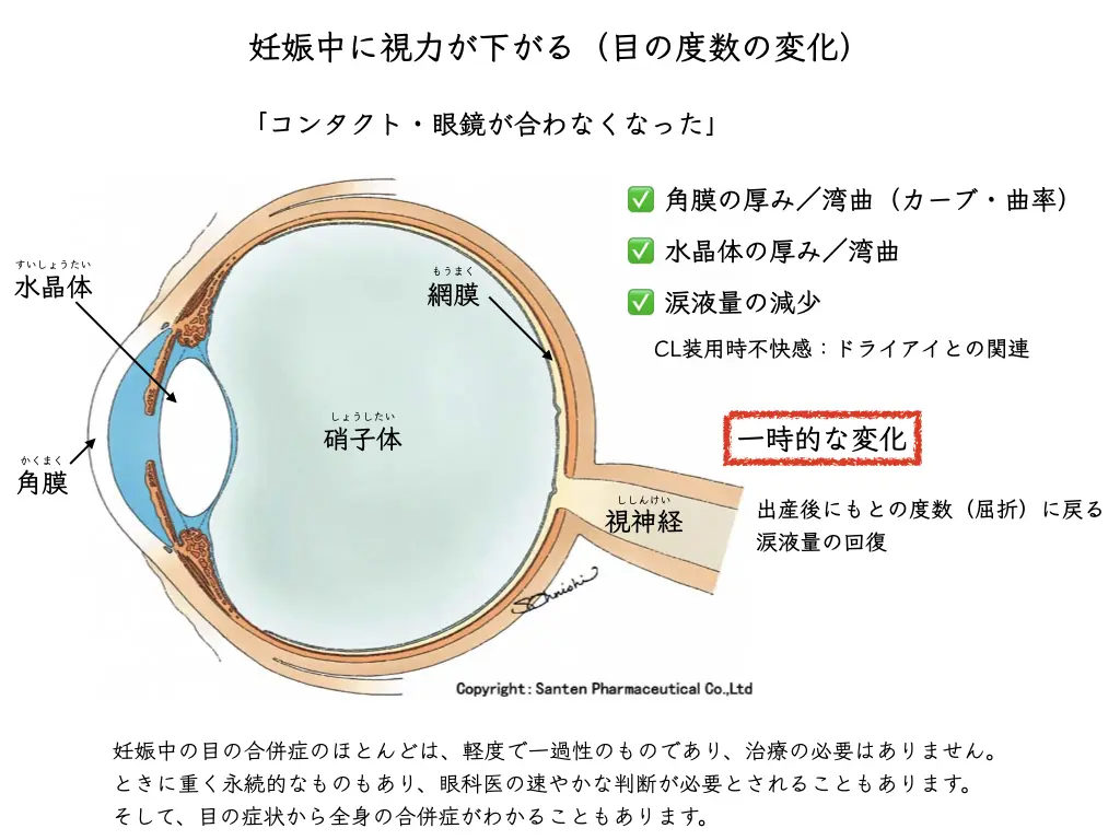
Here we mainly describe changes in the anterior segment (the cornea and lens).
Temporary changes in refraction (eye prescription): the cornea and lens during pregnancy
・the cornea becomes thicker
・the curvature of the cornea and lens increases
Either can cause a temporary change in refraction.
→ The eyes “get worse,” shifting toward myopia.

This shows up as reduced uncorrected vision / a change in the prescription of glasses and contact lenses
.
It is known to return to the original state after delivery.
Corneal thickness
: an increase of about 1–16 microns in corneal thickness, thought to be due to temporary corneal edema2。
Corneal curvature
: it is known to increase by an average of 1 D in late pregnancy3。
It resolves after childbirth or after breastfeeding ends.
Curvature of the lens
: during pregnancy and breastfeeding, the curvature of the lens also increases,and a temporary decrease in accommodation occurs4。
What is a micron?
It refers to a micrometer (μm).
There was an easy-to-understand picture comparing small things.
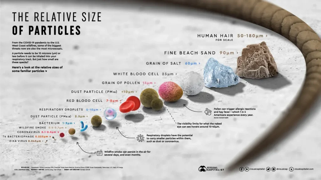
Hair: 50–180 microns
One grain of beach sand: 90 microns
One grain of salt: 60 microns
White blood cell: 25 microns
Pollen: 15 microns
Dust: 10 microns or less
Red blood cell: 7–8 microns
Droplets: 5–10 microns
Dust (PM2.5): 2.5 microns
Bacteria: 1–3 microns
Wildfire smoke: 0.4–0.7 microns
Coronavirus: 0.1–0.5 microns
Enterobacteria virus T4: 0.225 microns
Zika virus: 0.045 microns
The change in corneal thickness during pregnancy can be understood as
a change on the scale of bacteria to droplets to red blood cells to pollen
(a change far finer than the thickness of a hair).
Because refraction (the prescription) is unstable,
refractive surgery such as LASIK or ICL (implantable contact lens) is recommended to be postponed.
Keratoconus: changes during pregnancy
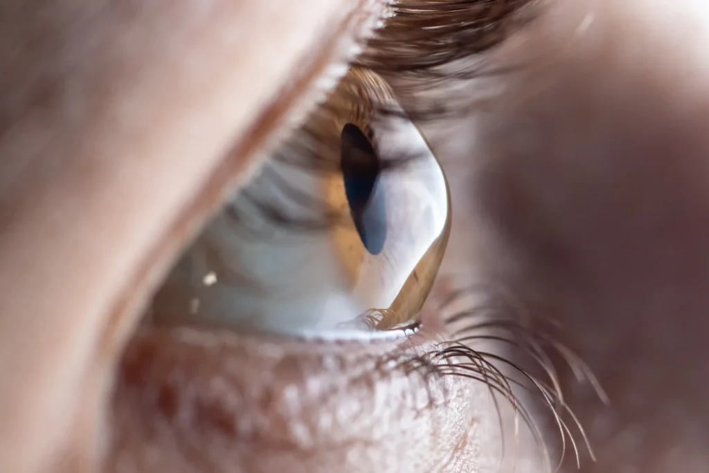
There are reports that during pregnancykeratoconusmay progress.5,6。
The cause is explained as softening of the cornea by estrogen and relaxin (a uterine relaxing factor).
Careful follow-up using corneal topography is recommended.
Other changes
Diseases that cause serious vision loss, such as retinal disease and glaucoma, may also be hidden.
If you notice anything unusual, it is important to consult an ophthalmologist.
(References)
- Sharma, S., Rekha, W., Sharma, T., Downey, G., 2006. Refractive issues in pregnancy. Aust N Z J Obstet Gynaecol 46, 186–188. https://doi.org/10.1111/j.1479-828X.2006.00569.x
- Weinreb, R.N., Lu, A., Beeson, C., 1988. Maternal Corneal Thickness During Pregnancy. American Journal of Ophthalmology 105, 258–260. https://doi.org/10.1016/0002-9394(88)90006-2
- Park SB, Lindahl KJ, Temnycky GO, Aquavella JV. The effect of pregnancy on corneal curvature. CLAO J 1992; 18: 256– 259.
- Duane’s Ophthalmology: Pregnancy and the Mother’s Eye (CD- ROM). Clinical volume 5, Chapter 32. Hagerstown, MD: Lippincott Williams & Wilkins, 2001.
- Naderan, M., Jahanrad, A., 2017. Topographic, tomographic and biomechanical corneal changes during pregnancy in patients with keratoconus: a cohort study. Acta Ophthalmologica 95, e291–e296. https://doi.org/10.1111/aos.13296
- Bilgihan, K., Hondur, A., Sul, S., Ozturk, S., 2011. Pregnancy-induced Progression of Keratoconus. Cornea 30, 991–994. https://doi.org/10.1097/ICO.0b013e3182068adc


 ポチップ
ポチップ