![Takeru Eye Clinic | Takatori Shopping Street, Sawara-ku, Fukuoka City [Nishijin Station / Fujisaki Station]](https://takeru-eye.com/wp-content/uploads/2022/10/takeru_logo_for-WP-header.png)

As blood glucose rises, blood vessels are gradually damaged from the inside.
The thinnest blood vessels in the body are known to be the most vulnerable.
One of the thinnest vessels is found in the retina:
Diabetes affects the blood vessels in the eye, the “retinal vessels,” and can impair vision without you noticing.
For people with diabetes, taking care of eye health is very important.


In eye examinations, particular attention is paid to a part called the “FAZ.”
This is a special area at the center of the retina that we use to see things clearly.
It has become clear that examining the condition of the FAZ can help detect eye abnormalities at an early stage.
This is an examination using OCT angiography.
Diabetes and the Eyes: The Importance of the “FAZ” in Detecting Retinopathy Early
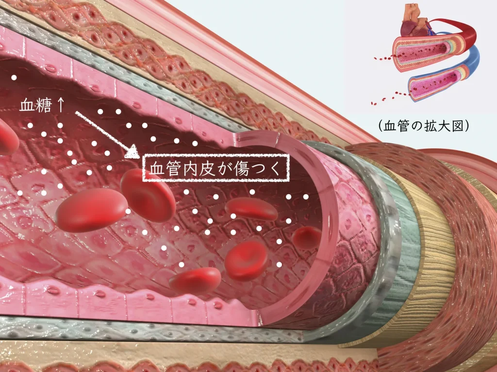
Elevated blood glucose affects blood vessels, and the fine vessels of the retina in particular are known to be damaged early¹. In recent years, evaluation of the foveal avascular zone (FAZ) has attracted attention as a way to detect minute changes in the retina.
What Is the FAZ?
The FAZ is a special avascular area located in the macula, at the center of the retina.
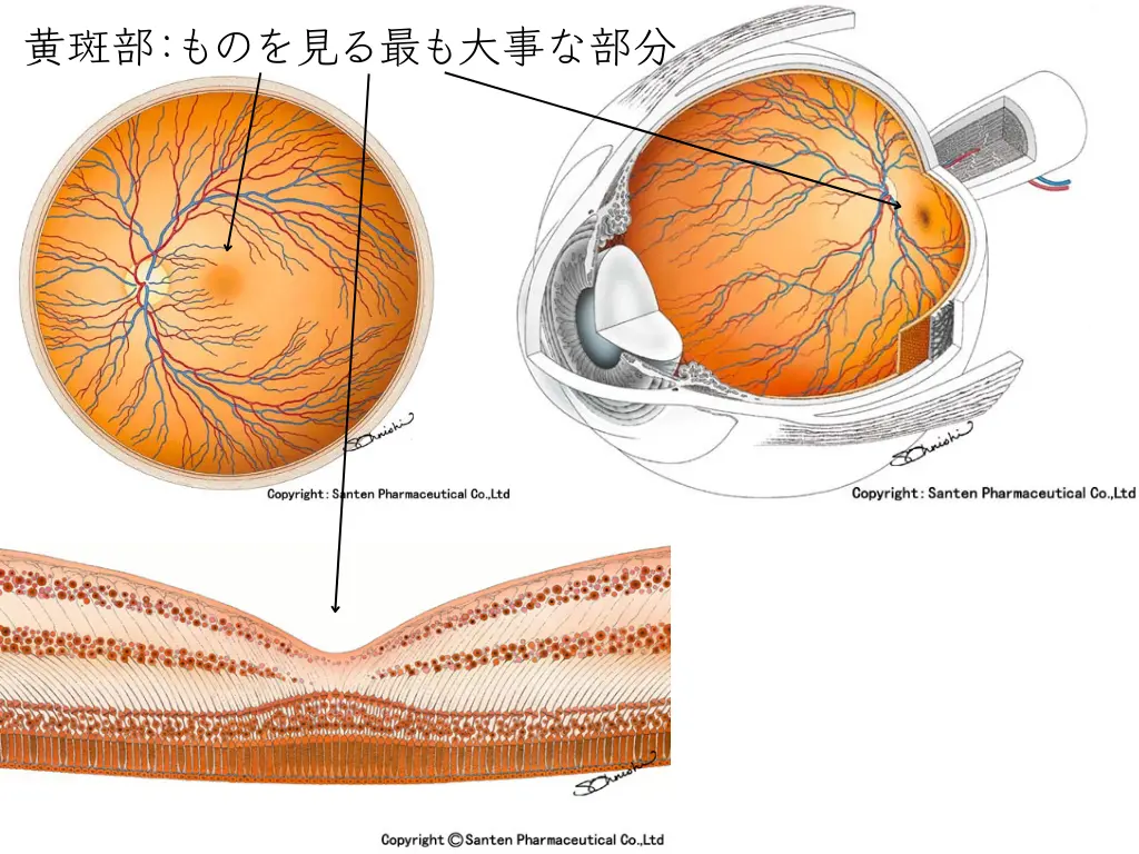
This area plays an important role in letting us see things clearly.
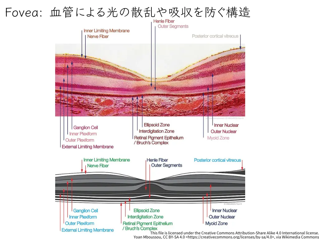
Its structure prevents light from being scattered or absorbed by blood vessels, so that light can reach the photoreceptors efficiently².
Development and Structure
The formation of the FAZ is a precise process that begins before birth. Formation of the FAZ begins at around 24–26 weeks of gestation and triggers the start of foveal pit formation³. The FAZ takes its complete form by birth and continues to develop afterward.
The basic structure of the FAZ has the following features:
- The inner retinal layers slope gently to form a depression
- Only cone photoreceptors and Müller cells are present at the center
- Photoreceptor axons run radially to form the Henle fiber layer
Retinal Changes in Diabetes
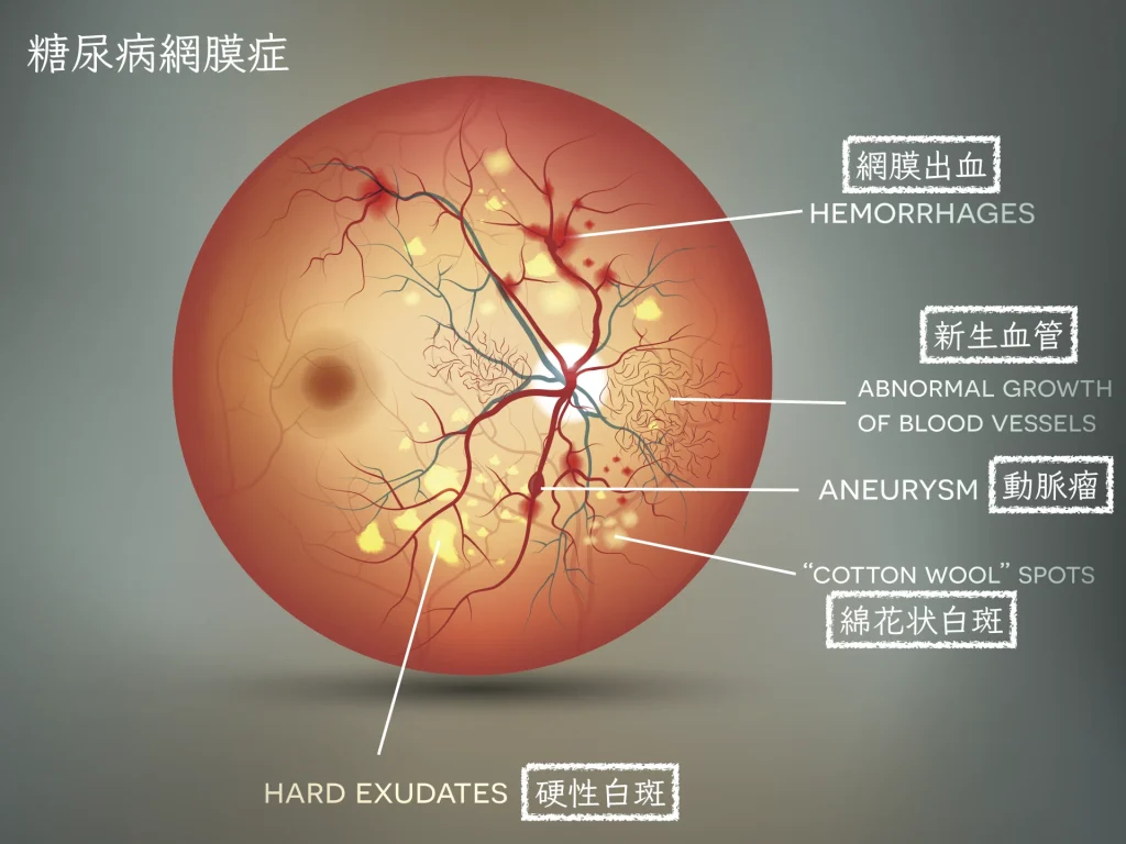
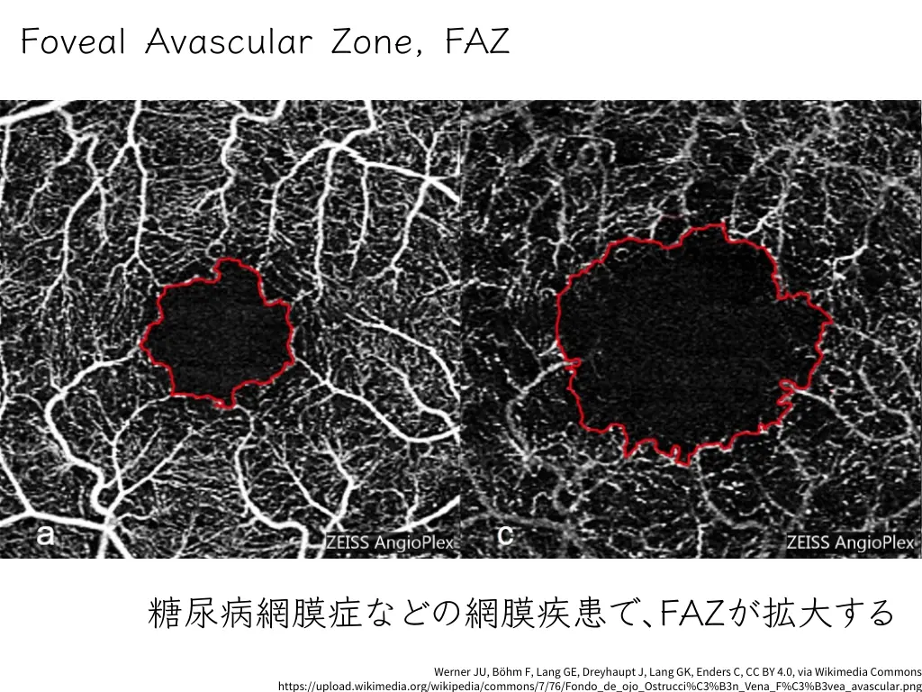
In diabetic retinopathy, characteristic changes are observed in the FAZ⁴. In particular, recent studies have shown that a decrease in retinal flow density is an important indicator⁶. The main changes are:
- Enlargement of the FAZ area
- Decreased vessel density
- Changes in the capillary network
- Irregularity of the vascular structure
It has been reported that these changes can be detected even before clinically apparent signs of retinopathy appear.
The Latest Imaging Technology
Instead of conventional fluorescein angiography (FA), optical coherence tomography angiography (OCTA) is now the mainstream⁵.
The features of OCTA are:
- Non-invasive, requiring no contrast dye
- Quick to perform
- Allows individual evaluation of the retinal vascular layers
- Allows quantitative evaluation
In OCTA evaluation, we focus in particular on the following:
- Measurement of the FAZ area
- Quantitative evaluation of vessel density
- Evaluation of shape irregularity
- Detection of changes over time
Notably, it has been shown that flow density measurement by OCTA is a useful biomarker in the diagnosis of diabetic retinopathy⁶.
This approach opens up the possibility of detecting lesions earlier than before.
Summary
Evaluation of the FAZ plays an important role in the early detection and follow-up of diabetic retinopathy. The latest imaging technologies are making more accurate diagnosis and more effective treatment possible.
Early detection through regular eye examinations is essential for maintaining visual function.
References
- Antonetti DA, Klein R, Gardner TW (2012) ‘Diabetic retinopathy’, New England Journal of Medicine, 366(13), pp. 1227-1239.
- Provis JM, Dubis AM, Maddess T, Carroll J (2013) ‘Adaptation of the central retina for high acuity vision: Cones, the fovea and the avascular zone’, Progress in Retinal and Eye Research, 35, pp. 63-81.
- Hendrickson AE, Yuodelis C (1984) ‘The morphological development of the human fovea’, Ophthalmology, 91(6), pp. 603-612.
- Durbin MK, An L, Shemonski ND, Soares M, Santos T, Lopes M, Neves C, Cunha-Vaz J (2017) ‘Quantification of Retinal Microvascular Density in Optical Coherence Tomographic Angiography Images in Diabetic Retinopathy’, JAMA Ophthalmology, 135(4), pp. 370-376.
- Spaide RF, Fujimoto JG, Waheed NK, Sadda SR, Staurenghi G (2018) ‘Optical coherence tomography angiography’, Progress in Retinal and Eye Research, 64, pp. 1-55.
- Kaizu Y, Nakao S, Arima M, et al. (2019) ‘Flow Density in Optical Coherence Tomography Angiography is Useful for Retinopathy Diagnosis in Diabetic Patients’, Scientific Reports, 9, 8668.