![Takeru Eye Clinic | Takatori Shopping Street, Sawara-ku, Fukuoka City [Nishijin Station / Fujisaki Station]](https://takeru-eye.com/wp-content/uploads/2022/10/takeru_logo_for-WP-header.png)

“Hayari-me” (epidemic keratoconjunctivitis, EKC) is inflammation caused by adenovirus.
Unlike allergic conjunctivitis, it heals on its own in about 2 weeks (a self-limiting disease).

Hayari-me (epidemic keratoconjunctivitis) causes symptoms such as
marked redness, strong pain, copious discharge, and being unable to open the eye.
Based on the ophthalmologist’s findings under the slit-lamp microscope, a test kit that detects the antigen may also be used if necessary.
Even a negative result does not rule out adenovirus infection (false negative).
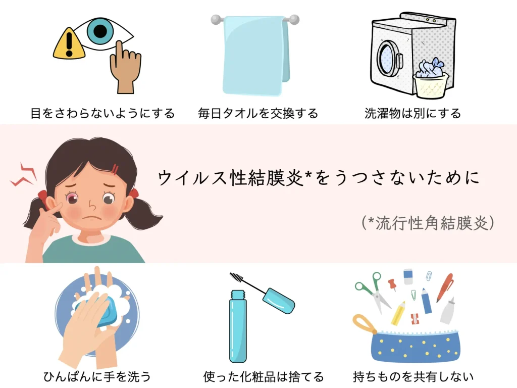
Because it is highly contagious, an accurate diagnosis and attention to daily life are important.
In addition, there is no drug that acts directly on adenovirus.

Steroid eye drops are prescribed to reduce inflammation.
If a bacterial component to the inflammation is also judged to be present, antibiotics may be used as well.
After the infection has healed, the black of the eye may become cloudy (nummular keratitis).
In that case, even after the redness has subsided, symptoms such as glare, blurred vision, and difficulty seeing continue.
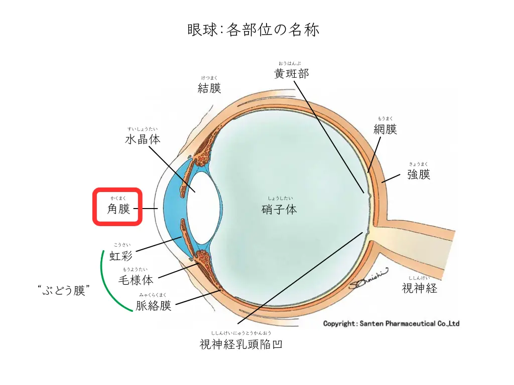
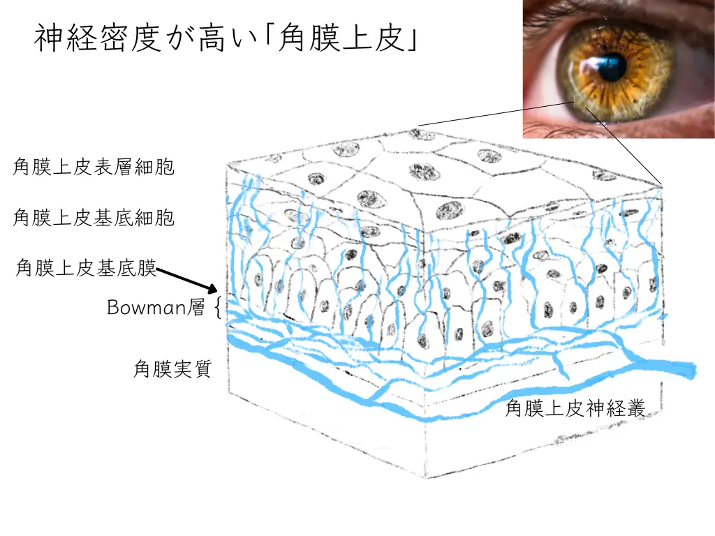
The cornea is known to have about 400 times as many sensory nerves as the skin1.
Many neurotransmitters and peptides are produced from the nerve endings, and they are thought to work to keep the cornea transparent2.
The immune reaction to adenovirus lurking in the corneal epithelium is the essence of corneal clouding (corneal opacity).
Adenovirus induces inflammatory cells within the corneal stroma.
Analysis with in vivo confocal microscopy has shown that the inflammation caused by adenovirus damages the corneal nerve plexus and also contributes to the clouding3.
Once corneal clouding has developed, it remains for several months to several years.
After the inflammation subsides it becomes a scar, so an early response is important:
When the cornea’s inflammation heals, it is very important that its transparency returns.
Nerves are known to have the ability to remodel (reconstruct)4.
Over a long period, the corneal nerve plexus may recover and transparency may return.
Our treatment policy is as follows.
We calm the inflammation appropriately, watch for recurrence, and aim to prevent aftereffects (corneal opacity) from persisting.
○ Suppress inflammation with steroid eye drops even before the hayari-me has healed
○ Even after the clouding has cleared, taper (reduce) the steroid eye drops carefully
: Keratitis often recurs when the dose is reduced too quickly
○ Even after clouding remains, reduce the eye drops as much as possible while waiting for the corneal nerve plexus to recover.
○ For changes in corneal shape caused by keratitis (irregular astigmatism), follow-up is carried out using a corneal topographer.
* If irregular astigmatism is strong and vision is hard to achieve, correction with hard contact lenses is also considered.
Even after the redness, eye discharge, and pain of hayari-me have disappeared, we keep a close watch on
whether any clouding (corneal opacity) remains
.
References
1. Rózsa, A.J., Beuerman, R.W., 1982. Density and organization of free nerve endings in the corneal epithelium of the rabbit. Pain 14, 105–120. https://doi.org/10.1016/0304-3959(82)90092-6
2. Muller LJ, Marfurt CF, KruseF, Tervo TM. Corneal nerves: structure, contents and function. Exp Eye Res 76: 521-542, 2003.
3. Dosso AA, Rungger-Brändle E. Clinical course of epidemic keratoconjunctivitis evaluation by in vivo confocal microscopy. Cornea 27: 263-268, 2008.
4. Patel DV, McGhee CNJ. In vivo laser scanning confocal microscopy confirms that the human corneal sub-basal nerve plexus is dynamic structure. Invest Ophthalmol Vis Sci 49: 3409-3412, 2008.