![Takeru Eye Clinic | Takatori Shopping Street, Sawara-ku, Fukuoka City [Nishijin Station / Fujisaki Station]](https://takeru-eye.com/wp-content/uploads/2022/10/takeru_logo_for-WP-header.png)

Color vision deficiency: we offer two tests.
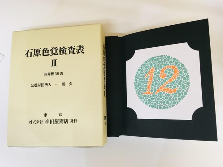
○ Ishihara color test plates
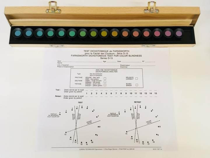
○ Panel D-15
“Color vision deficiency cannot be cured” — many people may feel this way.
People with it live in a world where colors look different from how most other people see them.
The word “deficiency” may not even be the right one.
Rather than something without a treatment, it may be better to see it as
a difference in how each person sees color.For everyone else:
It helps to know how colors look to people with color blindness or color weakness, and to be considerate about
colors that are easy to confuse,
- colors that are hard to see,
- and colors that are easy to see.
- and so on.
This is an explanation with photographs.
The World of Color Vision Deficiency
Colors that look different with color vision deficiency
We show various examples of how things look different to people with color blindness or color weakness, using photographs.
Writing on a green chalkboard with red chalk can be hard to read
Barbecue and grilled meat: how well the meat is cooked
Freshness of apples: light red and dark red
Pink and white are hard to tell apart
Colors of red and green bell peppers
Most color vision deficiency is red-green deficiency (described below).
In situations like the following,
it can bedifficultto tell colors apart.

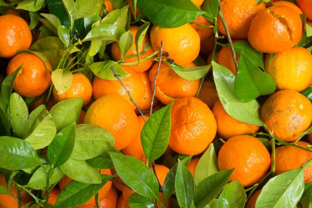











Confusion colors in congenital red-green color vision deficiency
- Red and green
- Orange and yellow-green
- Brown and green
- Blue and purple
- Pink and white or gray
- Green and gray or black
- Red and black
- Pink and light blue
People with protan deficiency tend to confuse 1 through 8 with each other, and those with deutan deficiency tend to confuse 1 through 6.
Red LED lamps, which are often used for warning displays, can be hard to recognize even for a person with protan anomalous trichromacy who passes Panel D-15.
Kaoru Nakamura, Nippon no Ganka 91(11) (2020): 1515-1519
.jpg)
How things look: simulation of the experience
You can simulate how things look to people with color blindness or color weakness.
What does it actually mean for the way colors look to be different? Using several examples, you can experience how things look to people with a color vision disability.
(The images are simulated based on a specific theory and formula. They do not show how every person with a color vision disability sees, so please keep this in mind.)
From the Toyo Ink website
»“Color vision disability” simulated experience
An introduction to an app that shows how colors look to people with color vision deficiency.
Check contrast ratios: use an app
When designing, you can check the text color against its background.
It checks the contrast ratio and gives a Pass/Fail result.
It is a handy tool.
Color vision tests
○ Ishihara color test plates (international edition, 38 plates)
Distinguishes normal color vision from suspected color vision deficiency.
○ Panel D-15
Classifies color vision deficiency as severe or moderate-or-milder.
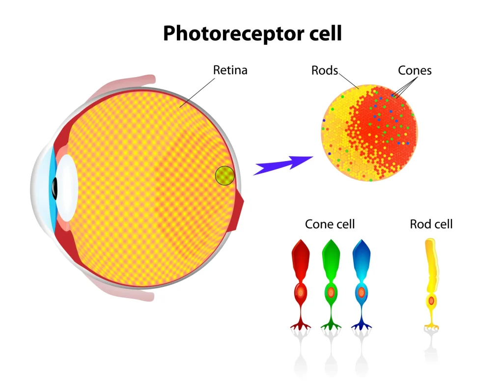
Retinal photoreceptors: “rods” that detect brightness, and “cones” that detect RGB (red, green, blue)
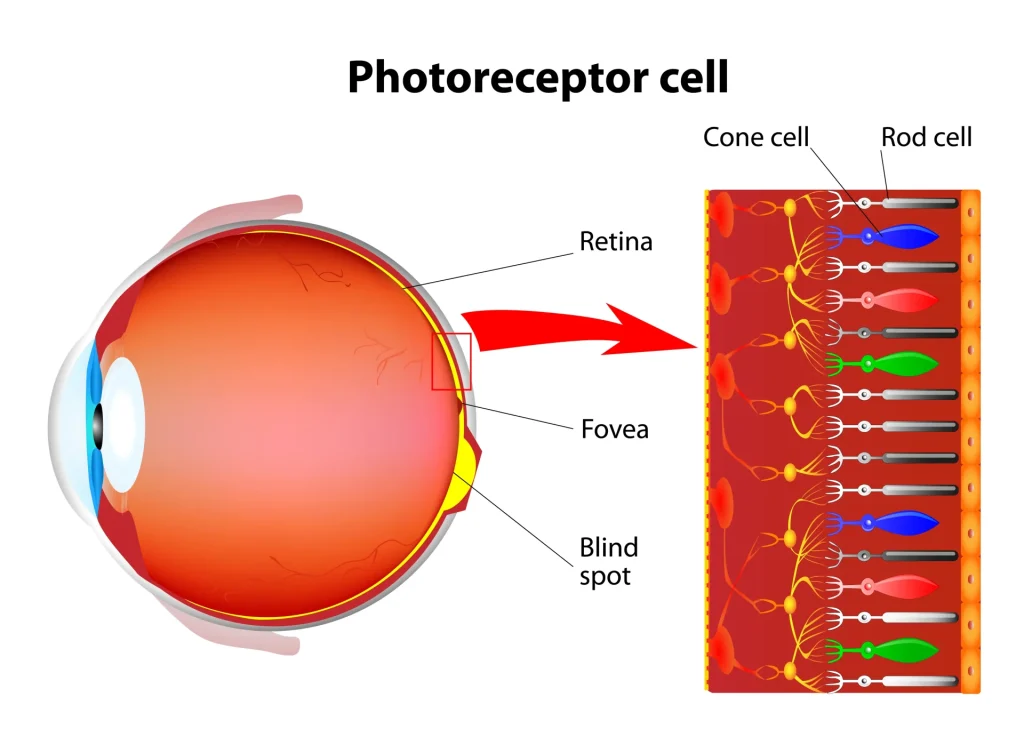
The retina’s photoreceptor cells come in two types.
○ RodsRod cell
Distinguish brightness and darkness:
They are especially useful for seeing in dim places.
○ ConesCone cell
Distinguish red, green, and blue (Red/Green/Blue)
When looking at the central area
Red cone L cone long wavelength R
Green cone M cone medium wavelength G
Blue cone S cone short wavelength B
The brain tells colors apart from the combination of each cone’s response
→ and recognizes it as “color.”



Causes and classification of color vision deficiency: “type __” “__ chromacy”
So-called “color blindness” is dichromacy, and “color weakness” is trichromacy (anomalous).
Severity runs in the order monochromacy > dichromacy > anomalous trichromacy.
They are also classified by RGB as type 1 (R), type 2 (G), and type 3 (B).
Type 1: abnormality of the red cone (L cone)
Type 2: abnormality of the green cone (M cone)
Type 3: abnormality of the blue cone (S cone)
Monochromacy (total color blindness): affects visual acuity
Rod monochromacy: only rods (no cones)
Cone monochromacy
Dichromacy: so-called “color blindness”; absence of just one of the RGBcone types Dichromacy
Protanopia (type 1 dichromacy): 1% of men
Deuteranopia (type 2 dichromacy): 1% of men
Tritanopia (type 3 dichromacy): below 1% in both men and women
Anomalous trichromacy: so-called “color weakness”; dysfunction or reduced sensitivity ofjust one of the RGB cone types Anomalous trichromacy
Protanomaly (type 1 anomalous trichromacy): 1% of men, 0.01% of women
Deuteranomaly (type 2 anomalous trichromacy): 6% of men, 0.4% of women
Tritanomaly (type 3 anomalous trichromacy): 0.01% in both men and women
Congenital cone color vision deficiency means
that the sensitivity of one of red, green, or blue (Red/Green/Blue) is low,
so that colors are perceived differently.
Change in the red cone: type 1 color vision
1 in 20 men (5%)
1 in 500 women (0.2%)
Change in the green cone: type 2 color vision
1 in 20 men (5%)
1 in 500 women (0.2%)
Color vision deficiency: difficult or restricted occupations
Work that tends to be difficult even with anomalous trichromacy
Train driver, color adjustment of video equipment, ink adjustment and color proofing in printing, dyeing, painting, titration experiments
Work thought to be difficult with dichromacy
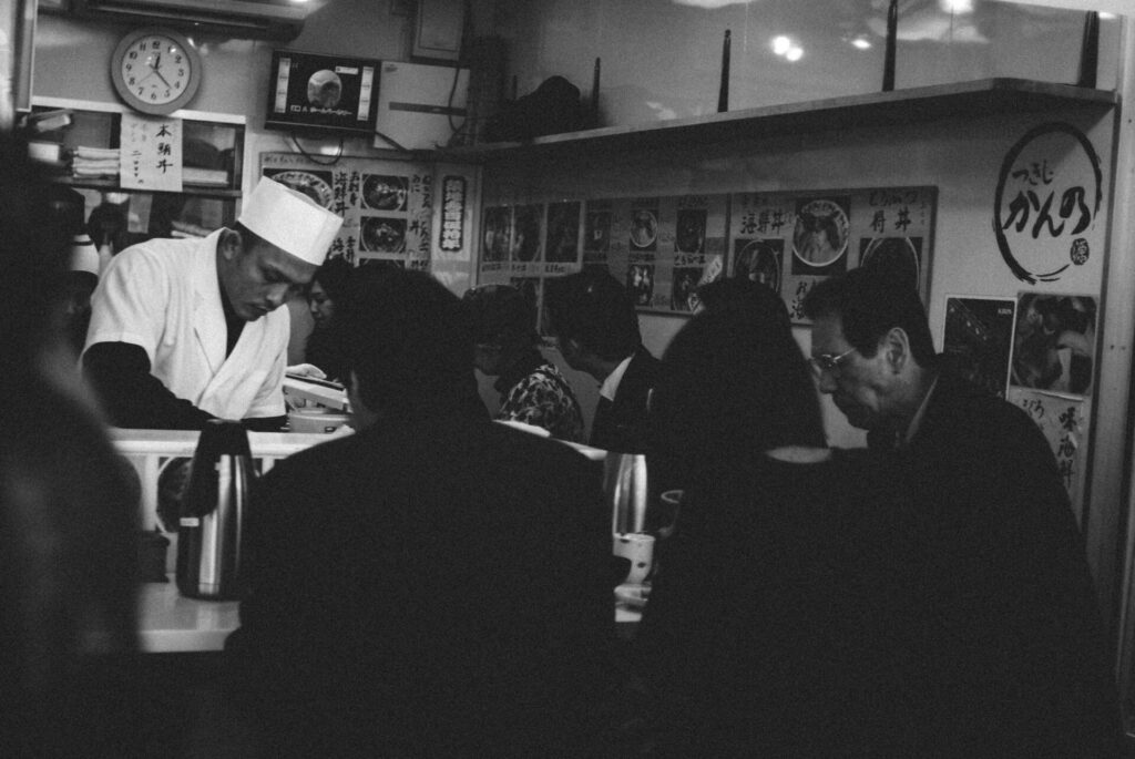
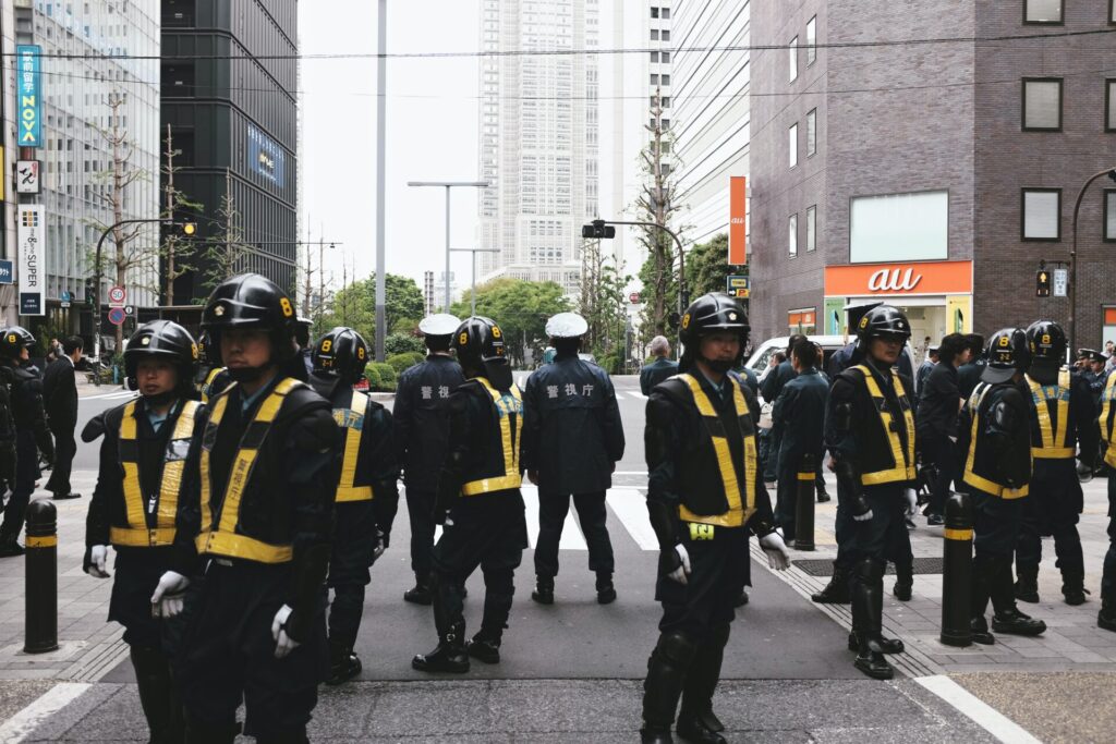
Ship navigator, airline pilot, aircraft and railway maintenance, police officer, commercial designer, photographer, paramedic, nurse, dental technician, veterinarian, hairdresser, clothing sales, server monitoring, kaiseki chef, work that judges food freshness
Work that can be done with little extra effort even with dichromacy
Physician, dentist, pharmacist, teacher, cook, barber, artist, architect, electrician, general office work involving a computer terminal
Work with no problem at all even with dichromacy
General office work with monochrome documents,
and other work that does not require color discrimination (including work in which all information other than color is also provided)
Kaoru Nakamura. Occupational problems of congenital color vision deficiency. Journal of Tokyo Women’s Medical University, vol. 82, 59-62, Extra, 2012










Diseases that cause color vision deficiency in adulthood (acquired color vision deficiency)
Color vision deficiency can also occur when the retina is damaged later in life.
The difference from congenital color vision deficiency is that the symptoms change.
(They can worsen or improve.)
Chronic diseases:
Diabetic retinopathy, glaucoma, leukemia, liver disease, Alzheimer’s disease, chronic alcoholism, macular degeneration, multiple sclerosis, Parkinson’s disease, sickle cell disease, retinitis pigmentosa
Accidents, brain disorders, and retinal vascular disorders:
Cerebral hemorrhage, cerebral thrombosis, retinal vein occlusion, retinal artery occlusion
Oral medications:
Antibiotics, tranquilizers, antituberculosis drugs, antihypertensives, and others
Aging:
Reduced retinal sensitivity with age
Famous people with color vision deficiency
Bill Clinton
Bill Clinton
ビル・クリントン – 停滞するアメリカをいかに建て直したか
Keanu Reeves
Keanu Reeves

Mark Zuckerberg
Mark Zuckerberg

Red-green color vision deficiency.
He started Facebook in a Harvard University dormitory in 2004.
In 2012 he brought Instagram under the company.
Facebook’s blue corporate color is reportedly the color that is easiest for Zuckerberg to see.
ソーシャル・ネットワーク (字幕版)
Facebookを創った男: ザッカーバーグ・ストーリー The Mark Zuckerberg Story
Mark Twain
Mark Twain
トム・ソーヤーの冒険 (新潮文庫) | マーク トウェイン
Bing Crosby
Bing Crosby
One of the best-known American pop musicians, with songs such as “White Christmas.”
Bing Crosby – White Christmas
John Dalton
John Dalton
British chemist, physicist, and meteorologist.
He published research on his own congenital color vision deficiency.
As a result, congenital color vision deficiency came to be called Daltonism.
Sim Daltonism (Mac App Store)
Paul Newman
Paul Newman
メッセージ・イン・ア・ボトル
Prince William
Prince William, Duke of Cambridge
Christopher Nolan
Christopher Nolan
プレステージ
About color vision deficiency: introducing books that explain it clearly
「家庭・保育園・学校でできるサポート術」
» コミックQ&A 色弱の子どもがわかる本