![Takeru Eye Clinic | Takatori Shopping Street, Sawara-ku, Fukuoka City [Nishijin Station / Fujisaki Station]](https://takeru-eye.com/wp-content/uploads/2022/10/takeru_logo_for-WP-header.png)

After a hyaluronic acid injection for cosmetic purposes, the blood vessels of the retina at the back of the eye can become blocked, and blindness can occur instantly.

Hyaluronic acid injection is called “dermal filler” in English, and in Japanese it is also called “dāmaru firā” or “hyaluronic acid filler.”
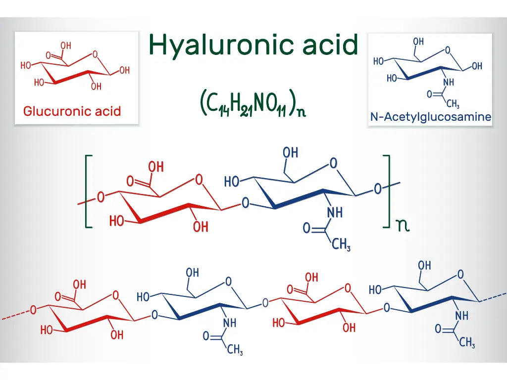
This is the general name for a type of aesthetic medicine in which hyaluronic acid, a moisturizing component found in the skin, is injected to fill wrinkle lines or to add volume to the lips and under-eye “tear bags.”

On the other hand, general risks include side effects such as swelling, bruising, and allergic reactions.
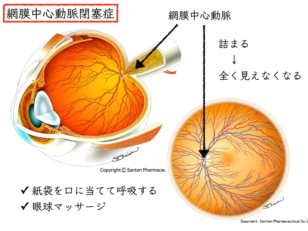
The cause of blindness from hyaluronic acid (filler) injection is that particles of hyaluronic acid block a blood vessel and cause “central retinal artery occlusion.”
Journal of the Japanese Ophthalmological Society, Vol. 118, No. 9 (2014)
A case of ophthalmic artery occlusion after hyaluronic acid injection into the nasal dorsum for cosmetic purposes
http://journal.nichigan.or.jp/Disp?style=abst&vol=118&year=2014&mag=0&number=9&start=783
There are almost no ways to rescue vision and almost no effective treatments.
Hyaluronic acid injections, which have drawn attention in cosmetic surgery, are expected to bring many cosmetic benefits such as improving wrinkles and sagging. Recently, however, it has also become clear that their use carries serious risks.
Among the most striking examples, cases of instant blindness after hyaluronic acid injection have been reported.
Careful choices are required in the pursuit of beauty.
Here I describe the details of blindness after hyaluronic acid injection, points to note to ensure safety, and what to do and the outlook once retinal vessel occlusion has occurred after hyaluronic acid injection.
I am sometimes asked about this by friends who are cosmetic surgeons, so I have summarized it below.
The Danger of Instant Blindness from Hyaluronic Acid Injection, and What to Do
Why Hyaluronic Acid Injections Can Enter Facial Arteries by Mistake: An Anatomical Perspective
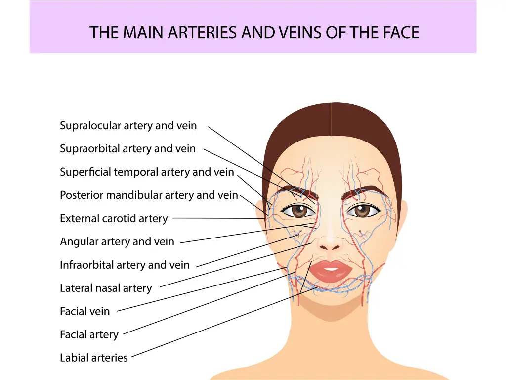
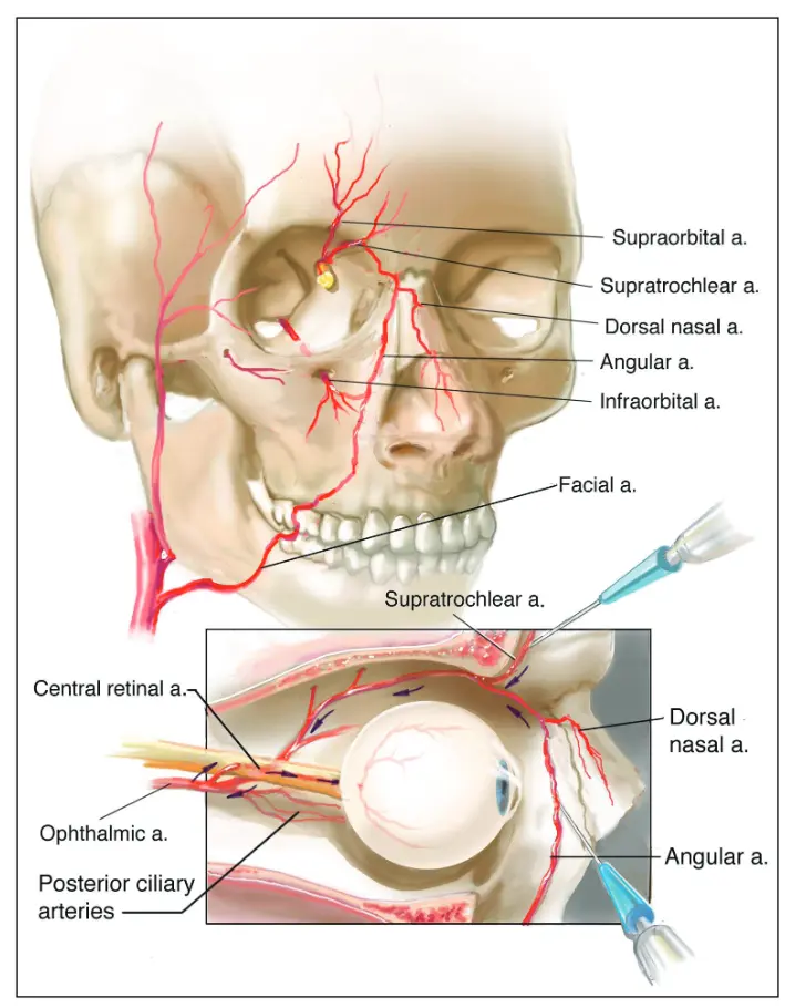
With facial filler injections, when injecting into the glabella*, medial midface, nasolabial fold, or nasal dorsum, the product can enter vessels via the superior orbital artery, supraorbital artery, artery of the oral commissure, or dorsal nasal artery.1。
*Glabella: the flat area between the eyebrows. It is an important region that affects emotional expression and facial expressions.
Beleznay et al. published a review of blindness after facial filler injection involving 98 cases.2。
The injection sites at highest risk of causing eye complications, in descending order of frequency, were:
Glabella (38.8%)
Nasal region (25.5%)
Nasolabial fold (13.3%)
Forehead (12.2%)
.
A new review in 2019 covers blindness from facial filler injection in 39 cases.3。
The injection sites with the highest risk of eye complications were:
Nasal region (56.3%)
Glabella (27.1%)
Forehead (18.8%)
Nasolabial fold (14.6%)
.
Why Hyaluronic Acid Injections Block the Arteries of the Eye: Particle Size and Artery Diameter

https://www.phdnest.com/morphology-of-bacteria-arrangement-shapes-sizes/

Hyaluronic acid particles: 200–1,000 microns (µm)
In contrast,
Diameter of the arteries of the eye: 500–1,000 microns (µm)
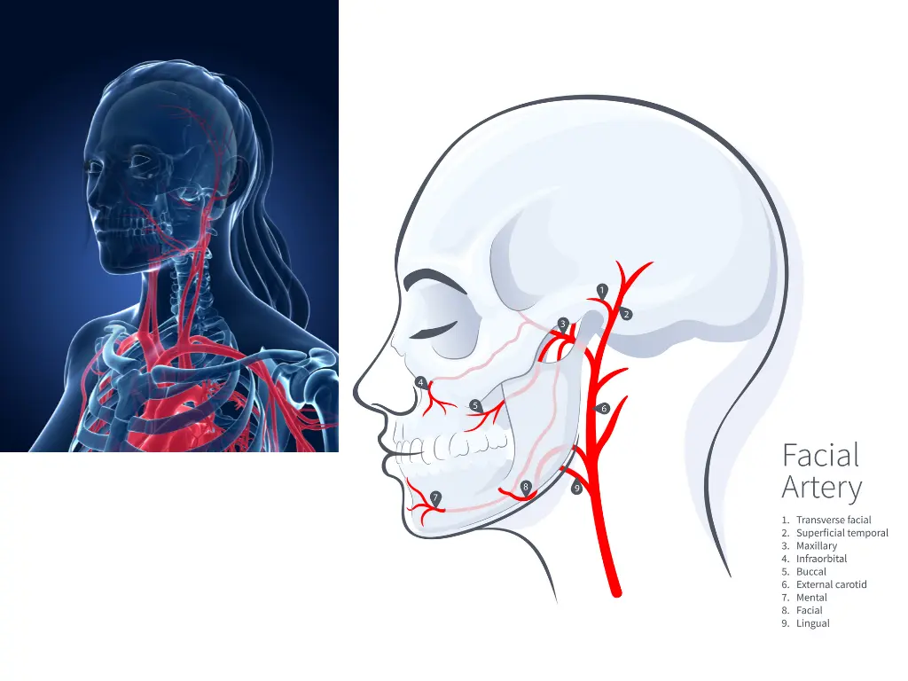
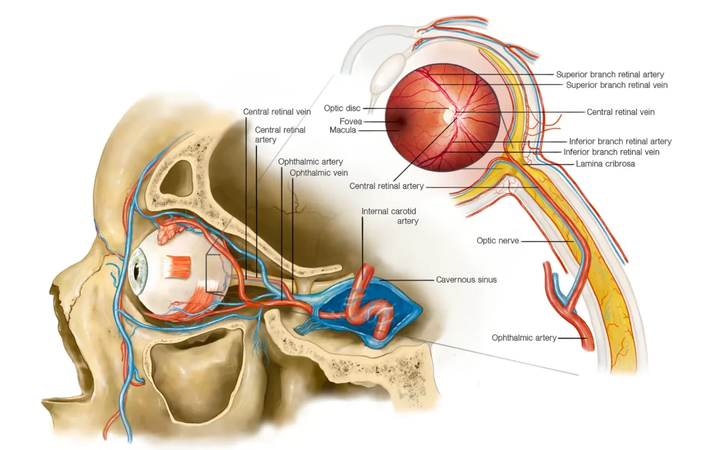
The ophthalmic artery is a major branch of the internal carotid artery and plays an important role in supplying blood to the structures of the orbit, including the eyeball and surrounding tissues.4。
If hyaluronic acid is injected into a blood vessel by mistake, particularly a small artery, the particles can block the vessel.
What Happens When Hyaluronic Acid Particles Block the Arteries of the Eye?
The retina, the part that corresponds to the film of a camera, is nourished by the retinal artery, which branches off the internal carotid artery.
If the central retinal artery becomes blocked, instant blindness can result.
Central retinal artery occlusion5:
occurs when blood flow stops reaching the whole retina within a short time and retinal function is impaired.
Central retinal artery occlusion, in which the main trunk of the artery that nourishes the retina is blocked, is among the most feared of ophthalmic emergencies.
Without pain, one eye suddenly becomes almost unable to see.
Cases of Instant Blindness After Hyaluronic Acid Injection
As the reviews above show, there are many case reports.
The prognosis is very poor in all of them.
As one example, I cite a case report published in 2022 in the American Journal of Ophthalmology Case Reports.6。
[Case report]
A 43-year-old woman received a hyaluronic acid injection in the eyelid and then experienced complete loss of vision in the left eye.
At presentation, she had
・Swelling of the left forehead and upper eyelid
・Ptosis (drooping eyelid)
・Complete ophthalmoplegia (paralysis of eye movement)
・Blindness
.
Eyeball massage was performed immediately, systemic therapy including acetylsalicylic acid (a nonsteroidal anti-inflammatory drug), tinzaparin sodium (a heparin-type anticoagulant), and cortisone (a steroid) was started, and hyaluronidase was injected at the original injection site.
In the course that followed, the patient developed necrotic and hemorrhagic skin and mucosal lesions, ptosis, anterior and posterior segment ischemia, and hypotony with successive deformation of the eyeball.
At 2.5 months of follow-up, the eyelid swelling and ptosis had resolved and the keratopathy had improved.
Blindness, skin lesions, and strabismus with reduced eye movement were still present, and loss of eyelashes (madarosis) and early enophthalmos were observed.
Central Retinal Artery Occlusion After Hyaluronic Acid Injection: What to Do When It Actually Happens
From multiple reviews, the following can be summarized.7, 8。
In every case, the visual prognosis is very poor.
Complications of cosmetic medicine (self-pay care) are not covered by health insurance.
Intra-arterial hyaluronidase
Hyaluronidase is an enzyme that dissolves hyaluronic acid.
This enzyme breaks down the particles in the hyaluronic acid filler and may restore blood flow in the blocked artery.
It is a method of injecting hyaluronidase directly into the artery.
However, its effect is limited when vision has been completely lost.
(It is not an easy procedure, and I believe almost no facilities perform it.)
Retrobulbar hyaluronidase (retrobulbar injection)
By injecting hyaluronidase behind the eyeball (retrobulbar), attempts have been made to dissolve the particles in the hyaluronic acid filler and restore blood flow.
However, whether blood flow can be successfully restored when vision has been completely lost is limited.
Retrobulbar injection:
It used to be performed routinely as a way of injecting anesthetic behind the eyeball during eye surgery.
Because it is a blind procedure, there is a possibility of globe perforation (the needle entering the eyeball).
In eye surgery, anesthesia methods with fewer complications have come into use.
As a result, it is becoming a procedure that is not common among ophthalmologists.
Subcutaneous or sub-Tenon hyaluronidase
Hyaluronidase is injected under the skin or into the space on the outside of the eyeball (sub-Tenon space).
It can be injected relatively more safely than a retrobulbar injection.
However, its effectiveness when vision has been completely lost is unknown.
Observation
Especially when the occlusion has led to complete loss of vision, observation may be chosen without active intervention.
Other measures
Anterior chamber paracentesis
This is expected to lower eye pressure and restart blood flow.
Intra-arterial thrombolysis / anticoagulation
Tissue plasminogen activator (tPA), urokinase, and others
Heparin, warfarin, and others
This is controversial because the risk of bleeding complications must be considered.
Corticosteroid therapy
It does not dissolve the clot directly.
By reducing inflammation and swelling in the affected area, it can improve blood flow and lower the risk of further clots forming.
It also helps reduce the risk of complications that can result from the occlusion, such as necrosis and bleeding.
Eye pressure–lowering drugs
They do not dissolve the clot directly.
By improving blood flow and lowering pressure in the blocked vessel, they help reduce the risk of further clots forming.
References
- Khan, T.T., Colon-Acevedo, B., Mettu, P., DeLorenzi, C., Woodward, J.A., 2017. An Anatomical Analysis of the Supratrochlear Artery: Considerations in Facial Filler Injections and Preventing Vision Loss. Aesthet Surg J 37, 203–208. https://doi.org/10.1093/asj/sjw132
- Beleznay, K., Carruthers, J.D.A., Humphrey, S., Jones, D., 2015. Avoiding and Treating Blindness From Fillers: A Review of the World Literature. Dermatol Surg 41, 1097–1117. https://doi.org/10.1097/DSS.0000000000000486
- Beleznay, K., Carruthers, J.D.A., Humphrey, S., Carruthers, A., Jones, D., 2019. Update on Avoiding and Treating Blindness From Fillers: A Recent Review of the World Literature. Aesthetic Surgery Journal 39, 662–674. https://doi.org/10.1093/asj/sjz053
- Li, X., Du, L., Lu, J., 2015. A Novel Hypothesis of Visual Loss Secondary to Cosmetic Facial Filler Injection. Annals of Plastic Surgery 75, 258. https://doi.org/10.1097/SAP.0000000000000572
- Scott, I.U., Campochiaro, P.A., Newman, N.J., Biousse, V., 2020. Retinal vascular occlusions. The Lancet 396, 1927–1940. https://doi.org/10.1016/S0140-6736(20)31559-2
- Davidova, P., Müller, M., Wenner, Y., König, C., Kenikstul, N., Kohnen, T., 2022. Ophthalmic artery occlusion after glabellar hyaluronic acid filler injection. Am J Ophthalmol Case Rep 26, 101407. https://doi.org/10.1016/j.ajoc.2022.101407
- Kapoor, K.M., Kapoor, P., Heydenrych, I., Bertossi, D., 2020. Vision Loss Associated with Hyaluronic Acid Fillers: A Systematic Review of Literature. Aesth Plast Surg 44, 929–944. https://doi.org/10.1007/s00266-019-01562-8
- Park, K.H., Kim, Y.-K., Woo, S.J., Kang, S.W., Lee, W.K., Choi, K.S., Kwak, H.W., Yoon, I.H., Huh, K., Kim, J.W., for the Korean Retina Society, 2014. Iatrogenic Occlusion of the Ophthalmic Artery After Cosmetic Facial Filler Injections: A National Survey by the Korean Retina Society. JAMA Ophthalmology 132, 714–723. https://doi.org/10.1001/jamaophthalmol.2013.8204