![Takeru Eye Clinic | Takatori Shopping Street, Sawara-ku, Fukuoka City [Nishijin Station / Fujisaki Station]](https://takeru-eye.com/wp-content/uploads/2022/10/takeru_logo_for-WP-header.png)

Blood vessels in the eye are the only vessels in the body that can be seen directly.
For this reason, doctors in specialties other than ophthalmology, such as neurosurgery, cardiology and neurology, sometimes use fundus findings to help with diagnosis.
In early 2022, findings were published on vascular changes after COVID-19 infection in
• the heart and blood vessels
• retinal blood vessels
.

Eyes after COVID-19: retinal vascular disorders
Cardiovascular disorders
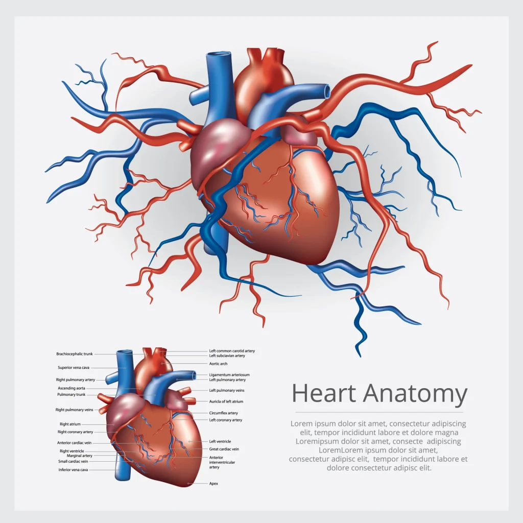
In February 2022, results of a study on long-term cardiovascular disorders after COVID-19 infection were reported.

Analysis of 153,760 people who had COVID-19 showed that
- Cerebrovascular disorders (stroke, transient ischemic attack)
- Arrhythmias (atrial fibrillation, sinus bradycardia, ventricular arrhythmias, atrial flutter)
- Inflammatory heart disease (pericarditis, myocarditis)
- Ischemic heart disease (acute coronary disease, myocardial infarction, ischemic cardiomyopathy, angina)
- Other heart disease (heart failure, non-ischemic cardiomyopathy, cardiac arrest, cardiogenic shock)
- Thromboembolic disease (pulmonary embolism, deep vein thrombosis, superficial vein thrombosis)
the risk of all of these increases after COVID-19 infection.1。
Eyes after COVID-19: are retinal hemorrhage and retinal ischemia seen?
It has also been described that vascular changes after COVID-19 infection can affect the eyes.
In the early period, there were many reports of conjunctivitis and eye pain with COVID-19 infection.
These are mainly lesions of the “anterior segment,” the surface and front part of the eyeball.
» “Eye pain” as an initial symptom of COVID-19

After that, research papers on the retina in the posterior segment (fundus) appeared one after another.
When COVID-19 damages the retina, it can lead to symptoms such as
• difficulty seeing
• blurred vision
• distorted vision
and the like.
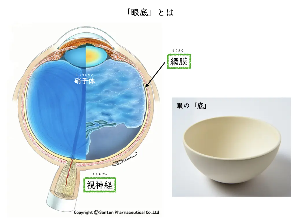

In May 2020, The Lancet carried the first description of retinal changes after COVID-19 infection2。
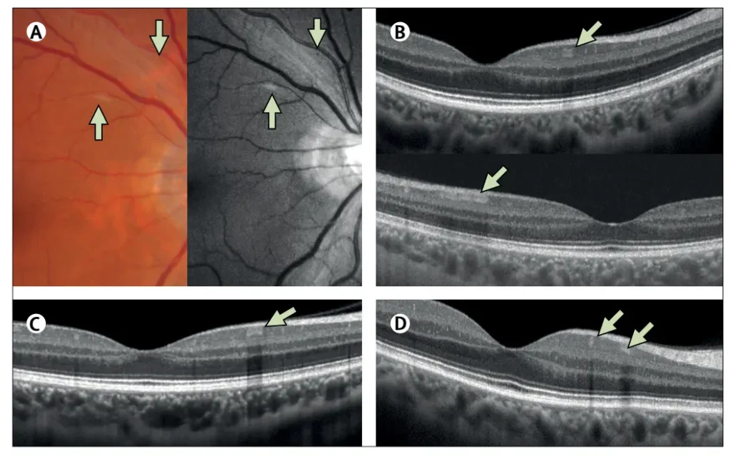
- Cotton-wool spots in the retina (a finding suggesting “ischemia,” poor blood flow in the retina)
- Retinal hemorrhage
Results of OCT (retinal cross-section) analysis were also described.
“Couldn’t the OCT findings above also be seen in normal eyes?”
Dr. Vavvas and colleagues at Harvard expressed a cautious opinion3(December 2020).
Eyes after COVID-19: decreased retinal vessel density
As cases accumulated with the spread of COVID-19, analyses of the “density” of retinal vessels were carried out.
These are studies using OCT angiography (OCTA).
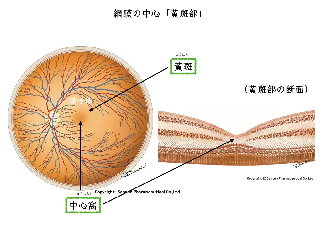
The most important part of the retina for seeing is called the “macula.”
The very center of it is called the “fovea,” which is known to be an area without blood vessels.
The depressed area seen on a retinal cross-section, the “foveal pit,” is an avascular zone.
Here we introduce an analysis (meta-analysis) based on 227 research papers on the effect of COVID-19 infection on retinal blood vessels.
From the 227 papers, after applying inclusion criteria, 31 were included in the meta-analysis.
With OCT angiography, researchers examined
- the density of blood vessels near the “fovea”
- and the size of the foveal avascular zone (FAZ)
.
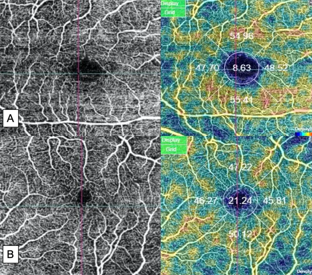
A representative photograph from the study with the largest number of subjects (Zapata, et al.4) is shown.
In case A (COVID-19 positive), compared with B (control: COVID-19 negative),
the foveal avascular zone (FAZ) is shown to be enlarged.
In this 2022 meta-analysis,
a total of 972 people with COVID-19 and 401 uninfected people (control group):
analysis using OCT angiography concluded that “retinal vessel density decreases after COVID-19 infection”5。
(a statistically significant difference was found).
It is known that the coronavirus enters the body through ACE-2 receptors on blood vessels.
ACE-2 receptors are also expressed in the vessels of the retina and choroid, as in other blood vessels,
so effects like those seen in other vascular systems are expected to be seen there as well.
The detailed molecular biological basis is not yet clear.
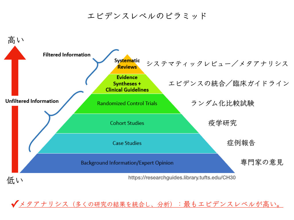
The scientific evidence from a meta-analysis, a paper that analyzes many papers, is high.
It seems reasonable to take it that “retinal blood vessels do change after COVID-19 infection.”
Long-term analyses are needed.
Retinal vessels as a “window” on the cardiovascular system
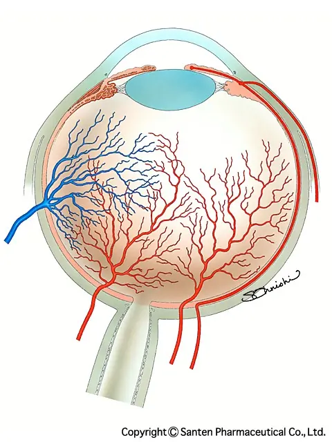
The retina at the back of the eye:
Using special lenses and a microscope, ophthalmologists observe the state of the retinal blood vessels.
“Vascular researchers” in fields far from ophthalmology also often use retinal vessels in animal experiments.
Retinal vessels are what the large vessels of the heart become after branching down to the finest caliber.
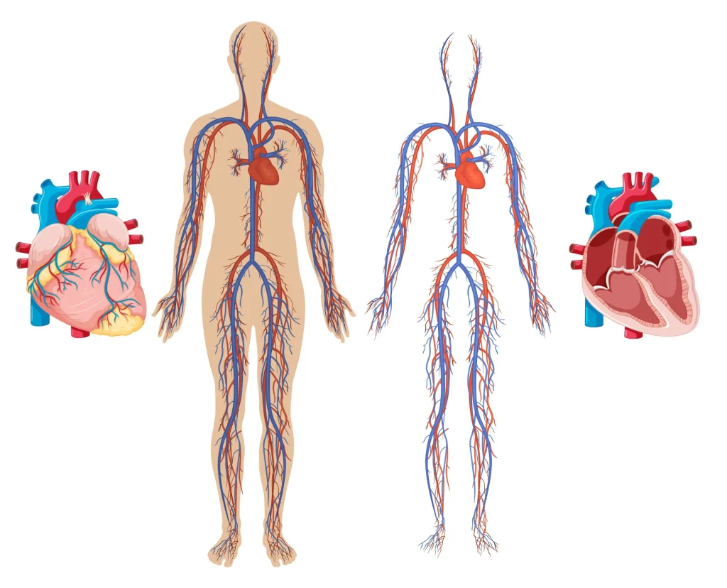
The heart and the eye, two organs at first sight not linked to each other, have more in common than one would expect. The vascu- lature of the eye, although some peculiarities do exist, shares many features with the vasculature of the heart and is often exposed to the same intrinsic and environmental influences. Thus, the eye, with its easily accessible vasculature, may indeed be a window to the heart, but knowledge about some unique vascular features is necessary.
The heart and the eyes seem to be unrelated organs, yet they have surprisingly much in common.
Although the vessels of the eye have some special features, they share many characteristics with the vascular system of the heart.
They are therefore often affected by intrinsic properties of the vessels and by the external environment.
Therefore, the eye, with its easily accessible vasculature, can truly become a “window to the heart,” but knowledge of several features of the vessels is also required.6。
Flammer, J., et al. The eye and the heart. Eur Heart J 34, 1270–1278.
Studies following vascular changes after COVID-19 infection have only just begun.
It may still be too early to call them “long COVID.”
There are broadly two ways to look at retinal vessels
• Direct viewing of retinal vessels with an examination microscope and lens
• Image analysis: OCT,OCT angiography,ultra-widefield fundus imaging
Using all of the above, ophthalmologists predict changes in the vascular system throughout the body.
References
- Xie, Y., Xu, E., Bowe, B., Al-Aly, Z., 2022. Long-term cardiovascular outcomes of COVID-19. Nat Med 1–8. https://doi.org/10.1038/s41591-022-01689-3
- Marinho, P.M., Marcos, A.A.A., Romano, A.C., Nascimento, H., Belfort, R., 2020. Retinal findings in patients with COVID-19. The Lancet 395, 1610. https://doi.org/10.1016/S0140-6736(20)31014-X
- Vavvas, D.G., Sarraf, D., Sadda, S.R., Eliott, D., Ehlers, J.P., Waheed, N.K., Morizane, Y., Sakamoto, T., Tsilimbaris, M., Miller, J.B., 2020. Concerns about the interpretation of OCT and fundus findings in COVID-19 patients in recent Lancet publication. Eye (Lond) 34, 2153–2154. https://doi.org/10.1038/s41433-020-1084-9
- Zapata, M.Á., Banderas García, S., Sánchez-Moltalvá, A., Falcó, A., Otero-Romero, S., Arcos, G., Velazquez-Villoria, D., García-Arumí, J., 2020. Retinal microvascular abnormalities in patients after COVID-19 depending on disease severity. Br J Ophthalmol bjophthalmol-2020-317953. https://doi.org/10.1136/bjophthalmol-2020-317953
- Teo, K.Y., Invernizzi, A., Staurenghi, G., Cheung, C.M.G., 2022. COVID-19-Related Retinal Micro-vasculopathy – A Review of Current Evidence. Am J Ophthalmol 235, 98–110. https://doi.org/10.1016/j.ajo.2021.09.019
- Flammer, J., Konieczka, K., Bruno, R.M., Virdis, A., Flammer, A.J., Taddei, S., 2013. The eye and the heart. Eur Heart J 34, 1270–1278. https://doi.org/10.1093/eurheartj/eht023