![Takeru Eye Clinic | Takatori Shopping Street, Sawara-ku, Fukuoka City [Nishijin Station / Fujisaki Station]](https://takeru-eye.com/wp-content/uploads/2022/10/takeru_logo_for-WP-header.png)

This article is a major update of “Eyes After COVID-19 Infection: Retinal Vascular Damage,” which we published in February 2022, based on research findings as of August 2025.
“Since I had COVID, my vision has felt different.” “I tire easily and find computer work difficult.”
Are you troubled by symptoms like these? It has become clear that COVID-19 can cause a variety of effects over a long period even after recovery. Research shows that the effects on the eyes in particular are more serious than expected and may last a long time.
In conclusion,after COVID-19 infection, even mild cases can show effects on the retinal blood vessels, and these effects have been confirmed to persist for at least one year after infection.

What the Blood Vessels in Your Eyes Tell Us
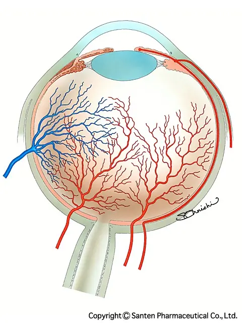
The blood vessels in the eye are the only blood vessels in the body that can be observed directly.
For this reason, they are also called a “window” that reflects the condition of the blood vessels throughout the body.
In fact, doctors outside ophthalmology, such as neurosurgeons, cardiologists, and neurologists, also use fundus findings as a reference for diagnosis.
In recent years, important discoveries about the state of blood vessels after COVID-19 infection have been made one after another through this “window.”
2020 to 2025: Progress in Research

Early Findings (2020)
In May 2020, retinal changes after COVID-19 infection were first reported in The Lancet1.
- Cotton-wool spots in the retina (changes suggesting retinal ischemia)
- Retinal hemorrhage
At first, attention focused mainly on symptoms at the front of the eye, such as conjunctivitis and eye pain, but it became clear that the retina, an important part at the back of the eye, is also affected.
Use of Detailed Imaging Technology (2021 Onward)
Research using a test technology called OCT angiography (OCTA) then began in earnest.
The following important findings have been reported2.
- Reduced retinal vessel density
- Other microvascular changes
This test has made it possible to observe the state of retinal blood vessels in more detail.
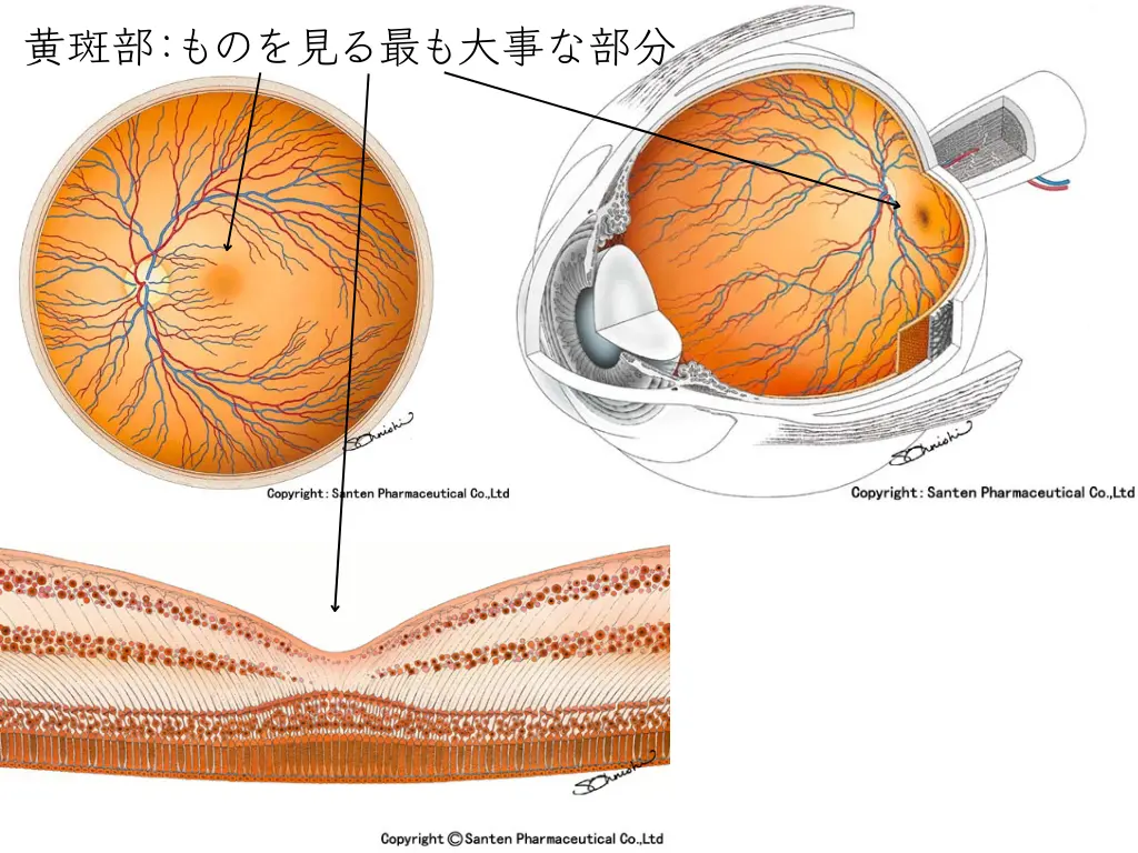
The most important part of the retina is the “macula.” The “fovea” at its center is originally an area without blood vessels (the foveal avascular zone), and changes in this area have a major effect on vision.
Accumulating Solid Evidence (2022)
A 2022 meta-analysis (a large-scale analysis combining multiple studies) performed OCTA analysis of a total of 972 people infected with COVID-19 and 401 uninfected people3.
As a result, it was established that “retinal vessel density is statistically significantly reduced after COVID-19 infection.”
Long-Term Effects Revealed (2023–2024)
An even more important finding followed. A study published in Frontiers in Medicine, with 12 months of long-term follow-up, found that the reduction in retinal vessel density persists for at least one year after infection4.
This study also showed that the reduction in retinal vessel density is associated with systemic inflammatory markers (CRP), aortic stiffness, and impaired kidney function.
Meanwhile, a 2024 study in Frontiers in Immunology reported changes in the retinal microvessels of patients with mild COVID-195.
In mild cases:
- Even when changes are seen in the retinal microvessels, they are usually temporary and often return to normal within 2 months.
In severe cases:
- Changes in vessel density and FAZ size may be permanent or last for a long time.
However, stricter studies that take into account the effects of high blood pressure, diabetes, and reinfection are needed in the future.
Points worth particular attention:
- Retinal vascular changes are seen not only in people who were severely ill but also in those whose illness was mild
- Some point out that similar changes have been reported in children as well as adults (though they are said to be milder in children).
- An association has been suggested between symptoms of long COVID (fatigue, brain fog, headache, and others) and retinal vascular changes
Current Understanding (as of 2025)

A 2025 prospective study (original research) has followed patients over the long term6.
This study examined in detail the long-term effects on the optic disc and the retinal microvasculature.
It showed that regular eye examinations are important for people who have recovered from COVID-19.
It provides new evidence that complications of the retina and optic nerve may persist for a long time after recovery.
The link between coronavirus disease and retinal artery occlusion has also been mentioned7.
The review recommends clinical vigilance for retinal vascular occlusion in people after COVID-19 infection or vaccination.
It also emphasizes that larger studies are needed to clarify the mechanisms by which retinal vascular occlusion develops.
At present, the following is known about changes in retinal blood vessels after COVID-19 infection.
Mechanisms of onset:
- Damage to vascular endothelial cells (the cells lining the inside of blood vessels)
- Inflammation caused by an overreaction of the immune system
- Formation of tiny blood clots
Effects on the whole body:
- Retinal vascular changes may serve as an indicator that reflects the condition of blood vessels throughout the body
- Correlate with elevated inflammatory markers (IL-6, D-dimer, and others)
- May be useful as a clue for predicting future risk of cardiovascular disease and cognitive decline
About Symptoms

If COVID-19 infection damages the retina, the following symptoms may appear:
- Difficulty seeing
- Blurred vision
- Distorted vision
- Increased eye strain
- Reduced concentration
However, these symptoms can also be caused by other diseases and factors.
An accurate diagnosis requires specialized tests.
Effects on the Cardiovascular System as a Whole

The effects of COVID-19 on blood vessels are not limited to the eyes.
A 2022 large-scale study showed increased risk of the following in the group that had COVID-198:
Cerebrovascular problems:
- Stroke
- Transient ischemic attack
Heart problems:
- Arrhythmia (atrial fibrillation, sinus bradycardia, ventricular arrhythmia, atrial flutter)
- Inflammatory heart disease (pericarditis, myocarditis)
- Ischemic heart disease (acute coronary syndrome, myocardial infarction, ischemic cardiomyopathy, angina pectoris)
- Other heart disease (heart failure, non-ischemic cardiomyopathy, cardiac arrest, cardiogenic shock)
Blood clot problems:
- Pulmonary embolism
- Deep vein thrombosis
- Superficial vein thrombosis
Why Retinal Blood Vessels Are an Important Indicator
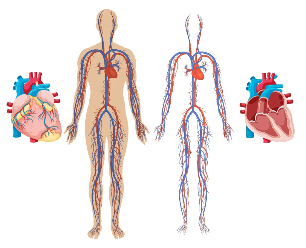
The retinal blood vessels of the fundus are the very thin peripheral vessels branching off from the large vessels leaving the heart. Because they are easy to observe, they are also called a “window to the heart.”
Blood vessels in the heart and in the eye share many characteristics and are influenced by the same constitution and living environment.
By examining the state of the retinal blood vessels, we can infer the health of blood vessels throughout the body.
Advances in Diagnostic Technology

Main tests currently used:
OCT (optical coherence tomography): Takes cross-sectional images of the retina and allows detailed observation of structural changes.
OCTA (OCT angiography): A technology that lets us see blood flow in the retinal vessels as an image without using a contrast agent. Changes in vessel density and blood flow can be examined in detail as numerical values.
These tests are painless and can be done in a short time.
Future Prospects and Challenges

Directions of research:
- Use as a predictive tool: Research is examining whether changes in retinal vessels could predict future risk of cardiovascular disease and cognitive decline.
- Development of treatments: At present, no method has been established to directly treat the retinal vascular changes themselves. For this reason, improving lifestyle habits is important for keeping blood vessels healthy throughout the body.
- Long-term follow-up: Further research is needed on changes several years after infection and on the possibility of recovery.
Use in clinical practice:
OCTA is currently recommended in the following cases:
- When there are visual symptoms after COVID-19 infection
- Patients after COVID-19 infection who have risk factors for cardiovascular disease
- Evaluation of long-lasting post-COVID symptoms (long COVID)
Points to Note in Daily Life

To minimize the effects on retinal blood vessels after COVID-19 infection:
(This is general information.)
Lifestyle habits that keep blood vessels healthy:
- Quit smoking
- Moderate exercise
- A balanced diet
- Maintaining an appropriate weight
- Managing blood pressure, blood sugar, and cholesterol levels
Eye health care:
- Regular eye examinations
- Working under appropriate lighting
- Avoiding prolonged use of computers and smartphones
- Getting enough sleep
Summary

COVID-19 can affect the retinal blood vessels over a long period even after recovery. It is known that these changes are also seen in people with mild illness and may persist for at least one year after infection.
What matters is that changes in the retinal blood vessels serve as an indicator reflecting the condition of blood vessels throughout the body9.
Eye examinations may make it possible to detect the risk of future cardiovascular disease early.
If visual symptoms or fatigue persist after COVID-19 infection, we recommend having specialized tests.
Early detection makes appropriate health management possible and may reduce future health risks.
Research is still ongoing, and the development of more effective treatments and preventive measures is expected.
It is important to receive appropriate care based on the latest medical knowledge.
References
- Marinho, P.M., Marcos, A.A.A., Romano, A.C., Nascimento, H., Belfort, R., 2020. Retinal findings in patients with COVID-19. The Lancet 395, 1610. https://doi.org/10.1016/S0140-6736(20)31014-X
- Hazar, L., Karahan, M., Vural, E., Ava, S., Erdem, S., Dursun, M.E., Keklikçi, U., 2021. Macular vessel density in patients recovered from COVID 19. Photodiagnosis and Photodynamic Therapy 34, 102267. https://doi.org/10.1016/j.pdpdt.2021.102267
- Teo, K.Y., Invernizzi, A., Staurenghi, G., Cheung, C.M.G., 2022. COVID-19-Related Retinal Micro-vasculopathy – A Review of Current Evidence. Am J Ophthalmol 235, 98–110. https://doi.org/10.1016/j.ajo.2021.09.019
- Castellino, N., Longo, A., Russo, A., Bonfiglio, V., Fallico, M., Toro, M.D., Cappellani, F., Grillo, M., Gaudio, A., Lo Cicero, L., Sessa, C., Colaci, M., Malatino, L., Castellino, P., Avitabile, T., Zanoli, L., 2024. COVID-19-related retinal microvasculopathy and systemic implications in patients with severe disease: results from the Methuselah study. Front. Med. 11. https://doi.org/10.3389/fmed.2024.1294432
- Zhang, C., Cheng, S., Chen, H., Yang, J., Chen, Y., 2024. New findings on retinal microvascular changes in patients with primary COVID-19 infection: a longitudinal study. Front. Immunol. 15. https://doi.org/10.3389/fimmu.2024.1404785
- Ozturk, M., Kumova Guler, D., Oskan, E.E., Onder, F., 2025. Long-Term Effects of COVID-19 on Optic Disc and Retinal Microvasculature Assessed by Optical Coherence Tomography Angiography. Diagnostics (Basel) 15, 114. https://doi.org/10.3390/diagnostics15010114
- Tzamalis, A., Foti, M., Georgiadou, M., Tsaftaridis, N., Ziakas, N., 2025. COVID-19 Related Retinal Vascular Occlusion: A Systematic Review. Journal of Clinical Medicine 14, 1183. https://doi.org/10.3390/jcm14041183
- Xie, Y., Xu, E., Bowe, B., Al-Aly, Z., 2022. Long-term cardiovascular outcomes of COVID-19. Nat Med 28, 583–590. https://doi.org/10.1038/s41591-022-01689-3
- Runsewe, O.I., Srivastava, S.K., Sharma, S., Chaudhury, P., Tang, W.H.W., 2024. Optical coherence tomography angiography in cardiovascular disease. Progress in Cardiovascular Diseases, Assorted Topics III 87, 60–72. https://doi.org/10.1016/j.pcad.2024.10.011