![Takeru Eye Clinic | Takatori Shopping Street, Sawara-ku, Fukuoka City [Nishijin Station / Fujisaki Station]](https://takeru-eye.com/wp-content/uploads/2022/10/takeru_logo_for-WP-header.png)

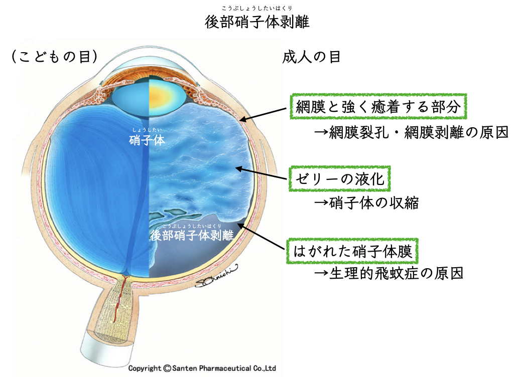
Posterior vitreous detachment, a normal change of the gel inside the eye (the vitreous), happens to everyone.
When posterior vitreous detachment occurs, floaters suddenly increase.
Posterior vitreous detachment (PVD)
When you see an ophthalmologist, we perform a comprehensive, whole-eye examination of the retina and vitreous.
(Since September 2021,a detailed fundus examination without dilating the pupilhas become possible.)
Sometimes a retinal detachment or retinal tear (a “hole in the retina”) is found at that visit.
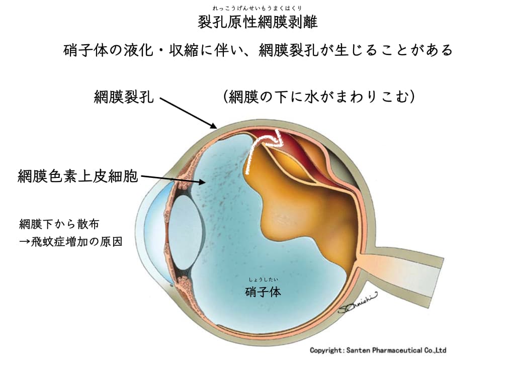
Posterior vitreous detachment: the probability (risk) of a retinal tear or retinal detachment

Symptoms of posterior vitreous detachment
Floaters
Also known as: “myodesopias”
Also known as: “entopsias”
Seeing streaks of light or flickering light (light flashes)
Also known as: “photopsias”
✔️If there is a part of your vision you cannot see (a central scotoma), a retinal detachment may be present.
Also known as: “scotoma”
Posterior vitreous detachment, retinal tears and retinal detachment: results of large clinical studies
Large studies were published in Ophthalmology last year and this year.
Ophthalmology, 20201:
From a medical institution in Pennsylvania (Wills Eye Hospital)
[Subjects] Patients who presented with acute symptoms of posterior vitreous detachment, such as floaters:7,999patients
[Purpose] To investigate the incidence of retinal tears and retinal detachment, and the factors behind complications
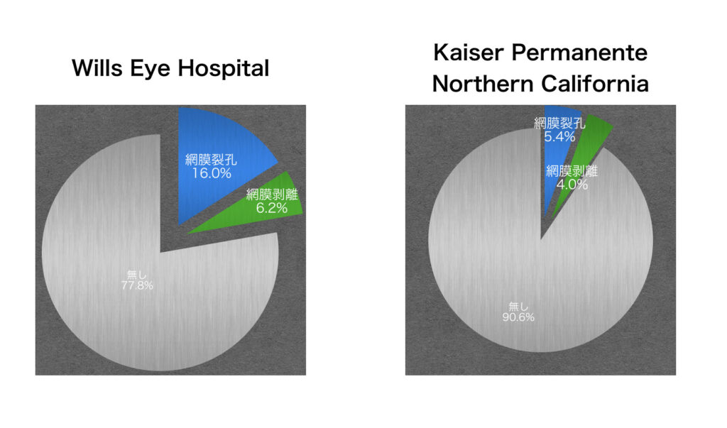
[Results] At the first visit, 1,280 patients (16.0%) were diagnosed with a retinal tear and 499 (6.2%) with a retinal detachment.
Later, 209 patients (2.6%) were diagnosed with a retinal tear and 80 (1.0%) with a retinal detachment, after a delay.
Of these,
within 6 weeks: retinal tear 116 patients (55.5%), retinal detachment 26 patients (32.5%)
after 6 weeks: retinal tear 93 patients (44.5%), retinal detachment 54 patients (67.5%)
2021, Ophthalmology2:
From the electronic medical records of a medical institution in California (Kaiser Permanente Northern California)
[Subjects] Patients who presented with acute symptoms of posterior vitreous detachment, such as floaters:8,305patients
[Purpose] To investigate the incidence of retinal tears and retinal detachment, and the factors behind complications
[Results] At the first visit, 448 patients (5.4%) were diagnosed with a retinal tear and 335 (4%) with a retinal detachment.
Retinal detachment and retinal tears with floaters: how high the risk is, judged from an urgent phone call
When it is hard to get to an eye clinic, a telephone interview can also help.2
[Risk of retinal detachment or retinal tear from posterior vitreous detachment]
– Reduced vision, or vision has become harder to see
– Being male
– A family member diagnosed with a retinal tear or retinal detachment
– Having had corneal refractive surgery (LASIK)
– Having had cataract surgery
People with these features are considered to be at higher risk.
After the visit, through various eye tests and the ophthalmologist’s own eyes (the slit-lamp microscope):
the risk of retinal detachment or retinal tear is sorted out further.2
Pigmented cells in the vitreous: a finding available before dilation
At every eye examination we check behind the crystalline lens (for cells in the anterior vitreous).
This is a finding that can be seen right away, before an exam that dilates the pupil.
If there are pigmented cells (retinal pigment epithelial cells) in the vitreous, the risk of an existing retinal tear or retinal detachment is judged to be high.2
Retinal tears and retinal detachment that are sometimes found later
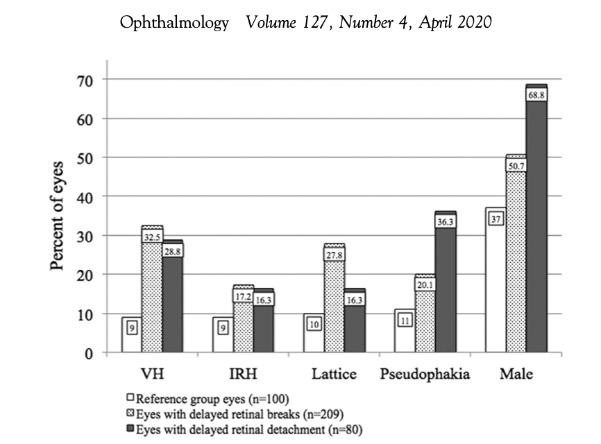
In the Pennsylvania paper above,1out of a total of 7,999 patients, some were diagnosed some time after the first visit (retinal tears were found a median of about 100 days later, and retinal detachment tends to be found even later):
Retinal tear: 209 patients (2.6%)
Retinal detachment: 80 patients (1.0%)
These were the diagnoses.
The following are listed as risk factors.
– Vitreous hemorrhage
– Intraretinal hemorrhage
– Lattice retinal degeneration
– Eyes with an intraocular lens, after cataract surgery (pseudophakia)
– Male sex (male gender)
If the items above are present at the visit,
the ophthalmologist keeps in mind a somewhat higher risk of retinal tear or retinal detachment and follows the eye over time.
What to watch for with floaters (checklist)
It is important to know whether your eye is at high risk.
- High myopia
- Male sex
- A family history of retinal tear or retinal detachment
- Having had eye surgery
Two papers in Ophthalmology in 2020 and 2021: why the numbers differ so much

In the United States, ophthalmology is divided into subspecialties,
retina specialists
cataract specialists
glaucoma specialists
cornea specialists
and so on, each with their own doctors.
For that reason,
it is usual for a retina specialist not to perform cataract surgery or glaucoma surgery.
is the norm.
After vitrectomy, if glaucoma develops,
you may need to book an appointment with a glaucoma specialist a few weeks later.
That was an everyday occurrence.
Wills Eye Hospital is one of the most famous eye research institutes and eye hospitals in the United States,
and people come from all over the country to see its retina specialists.
In the analysis of the California electronic medical records,
Kaiser Permanente Northern California: information on 4 million people held by an insurer
it includes overall, comprehensive eye care information.
The large difference is thought to come from the difference in the populations studied.
In Japan, ophthalmologists (in clinics, general hospitals and eye hospitals) see all eye diseases.
For that reason, the results of the California study are thought to be closer to the Japanese situation.
Treatment of floaters and its complications: a decision made by weighing risk
There are two main treatments for floaters.
(* Floaters caused by posterior vitreous detachment, when none of the retinal tears or retinal detachment described above are present)
- “Vitrectomy”: removes the clouded vitreous
- “Laser vitreolysis”: shatters thickened vitreous membranes with a shock-wave laser (YAG laser)
It requires a careful decision on whether you can accept the iatrogenic complications (those caused by performing the procedure).
References
- Uhr, J.H., Obeid, A., Wibbelsman, T.D., Wu, C.M., Levin, H.J., Garrigan, H., Spirn, M.J., Chiang, A., Sivalingam, A., Hsu, J., 2020. Delayed Retinal Breaks and Detachments after Acute Posterior Vitreous Detachment. Ophthalmology 127, 516–522. https://doi.org/10.1016/j.ophtha.2019.10.020
- Seider, M.I., Conell, C., Melles, R.B., 2021. Complications of Acute Posterior Vitreous Detachment. Ophthalmology 129, 67-72. https://doi.org/10.1016/j.ophtha.2021.07.020
» Advances in retinal imaging: ultra-widefield fundus imaging (in Japanese)