![Takeru Eye Clinic | Takatori Shopping Street, Sawara-ku, Fukuoka City [Nishijin Station / Fujisaki Station]](https://takeru-eye.com/wp-content/uploads/2022/10/takeru_logo_for-WP-header.png)

Glaucoma is a word that carries a particularly strong image, I think.

What Is the “Definition of Glaucoma”?
Glaucoma is
a disease with characteristic changes in the optic nerve and visual field,
in which, usually by sufficiently lowering intraocular pressure,
optic neuropathy can be improved or halted; it is characterized by functional and structural abnormalities of the eye.
I have quoted the definition word for word from the guidelines in the journal of the Japanese Ophthalmological Society1.
Guidelines for Glaucoma Practice (5th Edition). Journal of the Japanese Ophthalmological Society (Nippon Ganka Gakkai Zasshi), Vol. 126, No. 2 (in Japanese)
I think this wording is hard for patients to follow.
In this article, I explain glaucoma in plain terms, following the definition.
The Meaning of “Glaucoma,” a Word with a Gradient:
By understanding the state each person is in,
we talk at the eye clinic about whether there is cause for concern or not.
What Does “Glaucoma” Mean? Its Definition and Tests According to Disease Progression

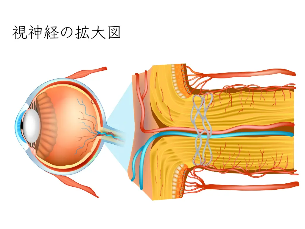
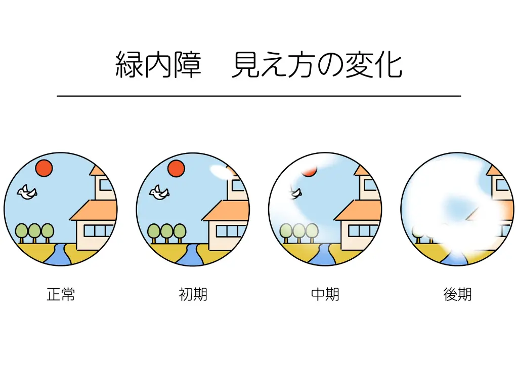
The Nerve at the Back of the Eye, the “Retina”: The Process from Structural Change to Functional Impairment
A paper published in 2004 by Dr. Weinreb of the University of California, San Diego (UCSD)2 contains an important figure.
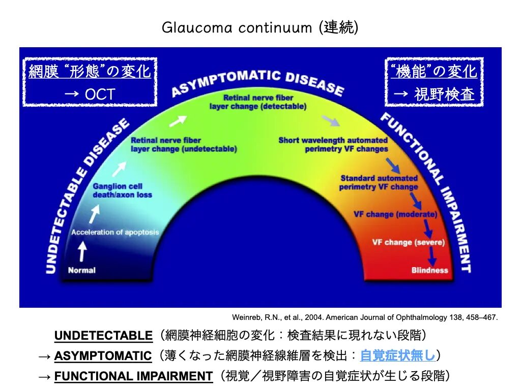
Changes in shape appear first, and after a while, changes in function appear.
“Glaucoma continuum”
This is a concept that is still supported today.
In the Guidelines for Glaucoma (5th edition) cited above, the term
functional and structural abnormalities of the eye corresponds to this.
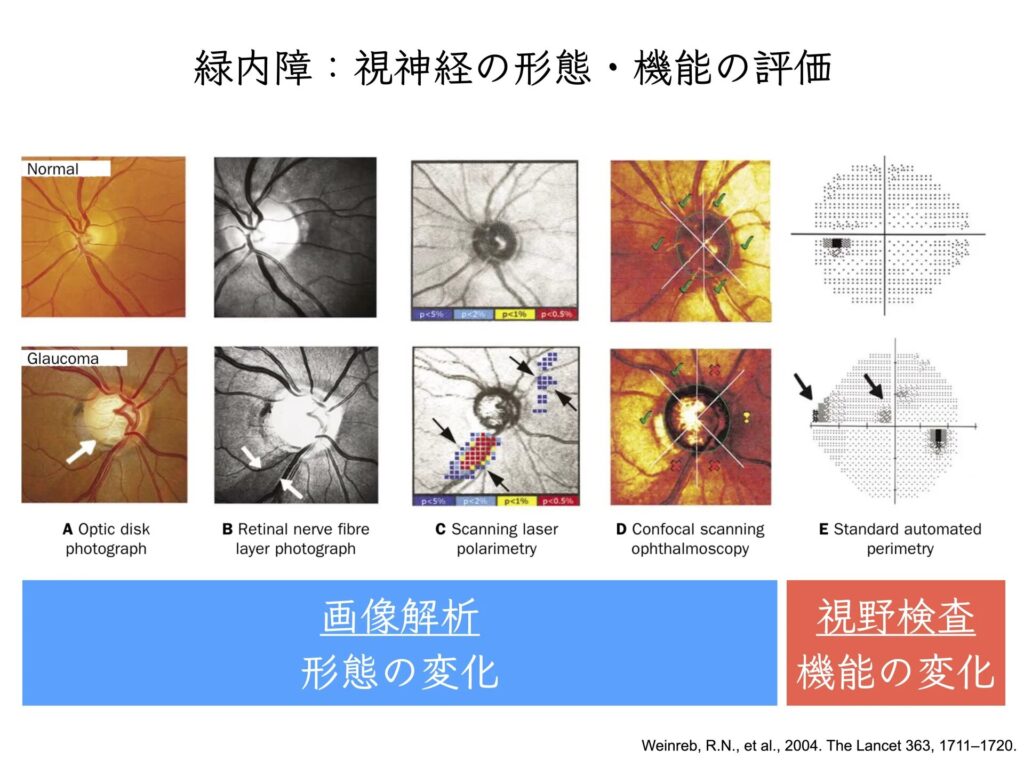
What Are Changes in Shape?
These are the areas where the retina has become thinner.
The retina can be compared to the film in a camera.
(If you have never seen film, imagine a retro camera📷)
What Are Changes in Function?
They are changes in the visual field.
When you hear the word “visual field,”
Is the visual field narrowing?
Is part of the visual field missing?
These questions come to mind instantly.
In eye care,
whether the sensitivity of the visual field is affected
is examined in detail.
Detection of visual field damage → “glaucoma is progressing”
is what this indicates.
If the visual field has actually narrowed or has missing areas,
it may be classified as “advanced glaucoma.”
“Early Detection of Glaucoma”
It is important to detect structural abnormalities early.
A fundus photograph can reveal them to some extent.
This is the stage at which they are found in workplace checkups and comprehensive health screenings (ningen dock).
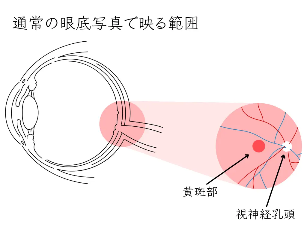
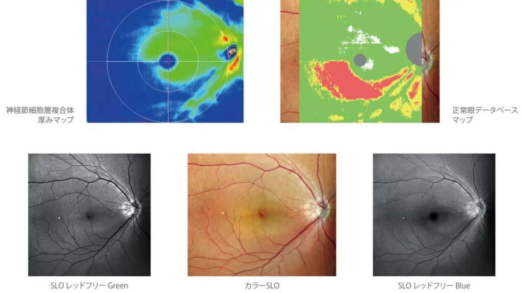
Detailed examination:
In the eye clinic, we use OCT (Optical Coherence Tomography; three-dimensional retinal image analysis).
Qualitative and Quantitative Assessment of Glaucoma: What Are They?
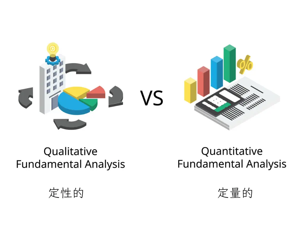
To understand each patient’s condition, we approach it from two directions.
• Qualitative assessment
• Quantitative assessment
Qualitative assessment is used to gain a deeper understanding of the background and reasons for a phenomenon, and collects data through words and descriptions.
On the other hand,
quantitative assessment measures the quantity or frequency of a phenomenon and analyzes it statistically using numerical data.
Combining the two allows a more comprehensive evaluation.
Each has a different purpose and method, and both are useful for diagnosing glaucoma and monitoring its progression.
Qualitative Assessment of Glaucoma
Optical coherence tomography (OCT)
- Qualitative assessment with OCT is a method of detecting glaucoma progression using wide-field scans.
By overlaying scan results and evaluating the changes, progression can be detected more often than with quantitative assessment.
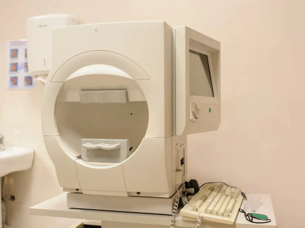
Visual field testing
- The 10-2 and 24-2 visual field tests and Goldmann perimetry (GP) are used to detect abnormalities in the central and peripheral visual field.
Assessment of patient knowledge
- A questionnaire to assess a patient’s knowledge about glaucoma is also part of qualitative assessment.
This lets us evaluate how effective our explanations to patients are and how well they are understood. - In practice, this leads into explanations given in conversation.
Quantitative Assessment of Glaucoma
Intraocular pressure measurement
- Measurement of intraocular pressure (IOP) is an important indicator in the diagnosis and management of glaucoma.
High intraocular pressure is the main risk factor for glaucoma and is measured quantitatively.
Measurement of retinal nerve fiber layer thickness by OCT
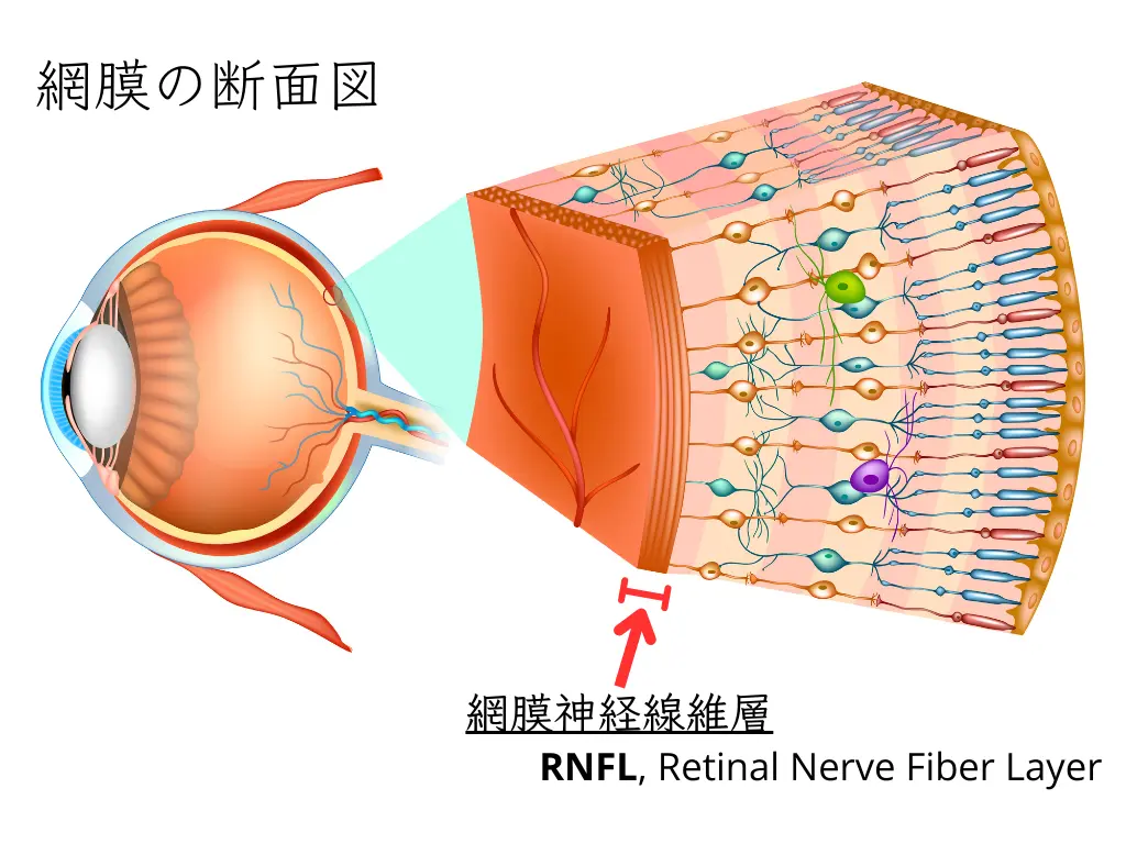
- Glaucoma progression is evaluated by quantitatively measuring the thickness of the retinal nerve fiber layer (RNFL). In particular, it is common to track changes in the thickness of the cpRNFL (circumpapillary retinal nerve fiber layer).
Quantitative analysis of visual field tests
- Visual field test results are converted into numerical values to evaluate the degree of visual field loss. This allows the degree of glaucoma progression to be assessed objectively.
OCT angiography (OCTA)
- OCT angiography is used to quantitatively evaluate blood flow inside the eye.
- It can reveal differences in blood flow between patients with glaucoma and healthy individuals.
Both qualitative and quantitative assessment are important in evaluating glaucoma.
Qualitative assessment evaluates visual changes and patient knowledge,
while quantitative assessment uses numerical data to grasp the condition objectively.
Eye examinations in general are handled by certified orthoptists.
Qualitative and quantitative assessment
Combining the two enables more accurate diagnosis and more effective treatment of glaucoma.

Types of Visual Field Tests and Their Differences
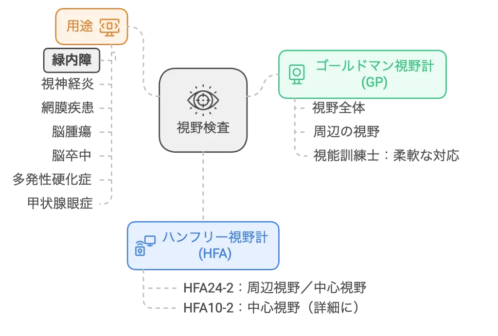
Visual field testing is very important for diagnosing glaucoma and monitoring its progression.
(Visual field testing is also important in optic neuritis, retinal diseases, brain tumors, stroke, multiple sclerosis, thyroid eye disease, and more.)
Tests fall broadly into the automated “Humphrey®” and the manual “GP.”
Characteristics, Differences, and Uses of the Humphrey® Field Analyzer (HFA 24-2 and HFA 10-2) and the Goldmann Perimeter (GP)
Humphrey® Field Analyzer (HFA) 24-2:
- Advantages: Highly accurate, standardized testing is possible. Both central and peripheral visual fields can be evaluated.
- Disadvantages: Peripheral field evaluation is not as extensive as with the GP.
Humphrey® Field Analyzer (HFA) 10-2:
- Advantages: Excellent for detailed evaluation of the central visual field. Can detect central visual field defects in early glaucoma at an early stage.
- Disadvantages: Not suited to evaluating the entire visual field.
Goldmann perimeter (GP):
- Advantages: Evaluates the entire visual field broadly and can capture peripheral field defects in detail. Flexible adaptation to the patient’s condition is possible.
- Disadvantages: The test takes time, and because results depend on the examiner’s skill, standardization can be difficult.
HFA 24-2 captures defects across the whole visual field, while HFA 10-2 performs a detailed evaluation of the central visual field.
The Goldmann perimeter (GP) evaluates the sensitivity and extent of the entire visual field broadly, and is therefore especially good at evaluating the peripheral visual field.
Here is a little more detail.
Humphrey® Field Analyzer (HFA)
The Humphrey® Field Analyzer is a type of static perimetry and is a computer-controlled automated perimeter.
HFA 24-2 Visual Field Test
- Range: Covers the visual field within 30 degrees of the center.
- Features: There are 54 measurement points at 6-degree intervals, evaluating both the central and peripheral visual field. Suitable for grasping the overall visual field loss in glaucoma.
- Use: Used to grasp the overall picture of glaucoma and to monitor the progression of visual field loss.
HFA 10-2 Visual Field Test
- Range: Examines in detail the visual field within 10 degrees of the center.
- Features: There are 68 measurement points at 2-degree intervals, allowing detailed evaluation of the central visual field. Effective for detecting central visual field defects early.
- Use: Especially useful in early glaucoma or when there are abnormalities in the central visual field. Used to grasp changes in the central visual field in detail.
Goldmann Perimeter (GP)
The Goldmann perimeter is a type of kinetic perimetry, a visual field test performed manually by a certified orthoptist.
- Range: Examines the entire visual field broadly. Especially good for evaluating the peripheral visual field.
- Features: Uses a moving light to examine the extent and sensitivity of the visual field. Because sensitivity is evaluated by varying the intensity and size of the light, it is also described as “kinetic quantitative perimetry.”
- Use: Because it evaluates the sensitivity and extent of the entire visual field, it is suited to evaluating advanced glaucoma. It is especially useful when grading of visual disability or detailed visual field evaluation is needed.
By combining the visual field tests above, glaucoma progression is monitored accurately.
The Roles of Central and Peripheral Vision and Their Importance in Daily Life
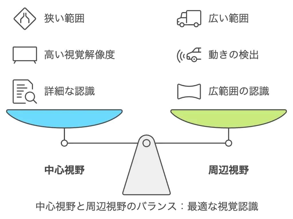
Vision consists of
“central vision,” which accurately recognizes the shape and details of objects,
and “peripheral vision,” which loosely perceives a wide area.
Central vision: needed to look at the part you want to see
Peripheral vision: needed to see what is around you
These two kinds of visual field have different roles, and both play important roles in daily life3, 4.
Central Vision
Role
- Detailed recognition: Central vision is important for accurately recognizing the shapes, colors, and details of objects. It is used, for example, in recognizing letters when reading, reading the text on signs, and identifying faces.
- High visual resolution: Central vision has high visual resolution and is suited to fine tasks and the processing of precise visual information.
Range
The range of central vision is very narrow, only about 1 to 2 degrees from the center of the visual field.
- Detailed recognition: Central vision is important for accurately recognizing the shapes, colors, and details of objects. It is used, for example, in recognizing letters when reading, reading the text on signs, and identifying faces.
- High visual resolution: Central vision has high visual resolution and is suited to fine tasks and the processing of precise visual information.
- Range: The range of central vision is very narrow, only about 1 to 2 degrees from the center of the visual field.5
Peripheral Vision
Role
- Wide-area recognition: Peripheral vision loosely captures a wide area of the visual field and helps us notice moving objects and changes in the environment. It is used, for example, in sensing the movements of pedestrians and other vehicles while driving, or in keeping track of surrounding players in sports.
- Motion detection: Peripheral vision is excellent at detecting moving objects and helps us sense danger early.
Range
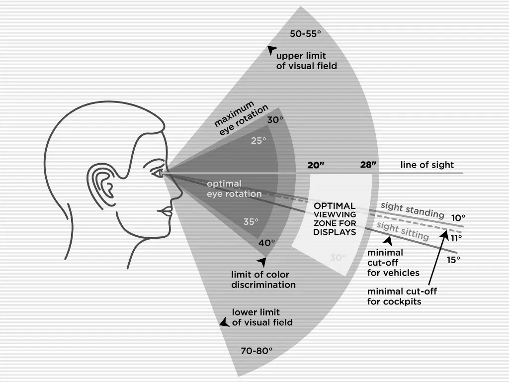
- Peripheral vision extends outside central vision and covers the whole visual field. With both eyes together, the visual field reaches 180 degrees or more6.
Which Is More Important, Central or Peripheral Vision? Importance in Real Life
In real life, central and peripheral vision each play a different role.
Importance of Central Vision
- Detailed tasks: Central vision is essential for activities that require detailed visual information, such as reading, checking documents, identifying faces, and fine work.
- Processing of precise visual information: Central vision has high visual resolution and is suited to processing precise visual information.
Importance of Peripheral Vision
- Ensuring safety: By sensing movement around you while driving or walking, it helps prevent accidents. A wide peripheral visual field lets you notice changes in the environment and moving objects sooner.
- Sports and daily activities: In sports, it is important to use peripheral vision to keep track of other players’ movements and make quick decisions. In daily life, too, we need peripheral vision to respond to changes in our surroundings.
In real life, both central and peripheral vision are important.
Central vision is essential for recognizing detailed visual information, while peripheral vision helps with recognizing the surroundings broadly and detecting motion.
Central and peripheral vision complement each other and work together to ensure quality of life and safety.
It is not that only one of them matters; what is important is that both visual fields function in good balance.
Evaluating Advanced Glaucoma: Central and Whole Visual Field Tests / What Is the “Floor Effect”?
In advanced glaucoma, the retinal nerve fiber layer (RNFL) is very thin7.
(If you compare the eye to a camera, it is as though the film has been damaged and thinned.)
Visual field testing and evaluation of intraocular blood flow are the main means of assessment.
Visual field testing:
Use the Goldmann perimeter (GP) to grasp the wide visual field,
and the Humphrey® Field Analyzer (HFA) 10-2 to grasp the central visual field.
In advanced glaucoma,
the Goldmann perimeter (GP), in which the orthoptist checks the patient’s responses as the test proceeds, becomes more important.
To evaluate the central visual field needed for daily life, we combine this with an evaluation of the central 10 degrees.
Central and Whole Visual Field Tests: Advantages of Combining Them
In evaluating advanced glaucoma, combining the GP and HFA 10-2 is effective in the following ways:
Broad visual field evaluation:
- Because the GP can evaluate the entire visual field broadly, it can capture peripheral field defects and reduced sensitivity in detail. This makes it possible to evaluate the overall state of the visual field comprehensively.
Detailed evaluation of the central visual field:
- HFA 10-2 excels at detailed evaluation of the central visual field and can accurately detect changes in the central visual field, which are especially important in advanced glaucoma. By grasping visual field defects within 10 degrees of the center in detail, the effect of treatment and the degree of progression can be evaluated.
Complementary information:
- Combining the GP and HFA 10-2 provides both broad information on the whole visual field and detailed information on the central visual field. This makes the evaluation of advanced glaucoma more accurate and comprehensive.
Evaluating Advanced Glaucoma: Dealing with the Floor Effect in Retinal Thickness
The floor effect refers to the phenomenon in which, once the thickness of the retinal nerve fiber layer (RNFL) falls below a certain value, further structural damage can no longer be measured.
This is because, although the RNFL continues to thin as glaucoma progresses, it never reaches zero and stops at a certain thickness.
In evaluating advanced glaucoma in particular, we determine the floor effect8.
In advanced glaucoma, the retinal nerve fiber layer (RNFL) is very thin, so evaluating intraocular blood flow is also important.
We use OCT angiography (OCTA)9.
- Features: A technology that evaluates intraocular blood flow non-invasively. It can evaluate blood flow in the optic nerve head and retina in particular in detail.
- Use: By detecting changes in blood flow, glaucoma progression can be found early. Even when the RNFL thickness has reached the “floor effect,” OCTA is useful for evaluating progression through changes in blood flow.
By using blood flow evaluation techniques such as OCT angiography, progression can be monitored even when the thickness of the retinal nerve fiber layer has reached its limit.
References
- Japan Glaucoma Society Guidelines Revision Committee. Guidelines for Glaucoma Practice (5th Edition). Journal of the Japanese Ophthalmological Society (Nippon Ganka Gakkai Zasshi), Vol. 126, No. 2, February 2022 (in Japanese).
- Weinreb, R.N., Friedman, D.S., Fechtner, R.D., Cioffi, G.A., Coleman, A.L., Girkin, C.A., Liebmann, J.M., Singh, K., Wilson, M.R., Wilson, R., Kannel, W.B., 2004. Risk assessment in the management of patients with ocular hypertension. American Journal of Ophthalmology 138, 458–467. https://doi.org/10.1016/j.ajo.2004.04.054
- Rehman, I., Mahabadi, N., Motlagh, M., Ali, T., 2024. Anatomy, Head and Neck, Eye Fovea, in: StatPearls. StatPearls Publishing, Treasure Island (FL).
- Bernard, J.-B., Chung, S.T.L., 2016. The Role of External Features in Face Recognition with Central Vision Loss. Optom Vis Sci 93, 510–520. https://doi.org/10.1097/OPX.0000000000000819
- Loschky, L.C., Nuthmann, A., Fortenbaugh, F.C., Levi, D.M., 2017. Scene perception from central to peripheral vision. Journal of Vision 17, 6. https://doi.org/10.1167/17.1.6
- Vater, C., Wolfe, B., Rosenholtz, R., 2022. Peripheral vision in real-world tasks: A systematic review. Psychon Bull Rev 29, 1531–1557. https://doi.org/10.3758/s13423-022-02117-w
- Leung, C.K., Cheung, C.Y.L., Weinreb, R.N., Qiu, K., Liu, S., Li, H., Xu, G., Fan, N., Pang, C.P., Tse, K.K., Lam, D.S.C., 2010. Evaluation of Retinal Nerve Fiber Layer Progression in Glaucoma: A Study on Optical Coherence Tomography Guided Progression Analysis. Invest. Ophthalmol. Vis. Sci. 51, 217. https://doi.org/10.1167/iovs.09-3468
- Tomita, R., Rawlyk, B., Sharpe, G.P., Hutchison, D.M., Shuba, L.M., Nicolela, M.T., Chauhan, B.C., 2024. Progressive Changes in the Neuroretinal Rim and Retinal Nerve Fiber Layer in Glaucoma: Impact of Baseline Values and Floor Effects. Ophthalmology 131, 700–707. https://doi.org/10.1016/j.ophtha.2023.12.032
- Werner, A.C., Shen, L.Q., 2019. A Review of OCT Angiography in Glaucoma. Seminars in Ophthalmology 34, 279–286. https://doi.org/10.1080/08820538.2019.1620807