![Takeru Eye Clinic | Takatori Shopping Street, Sawara-ku, Fukuoka City [Nishijin Station / Fujisaki Station]](https://takeru-eye.com/wp-content/uploads/2022/10/takeru_logo_for-WP-header.png)

As a cataract progresses, the crystalline lens becomes larger.
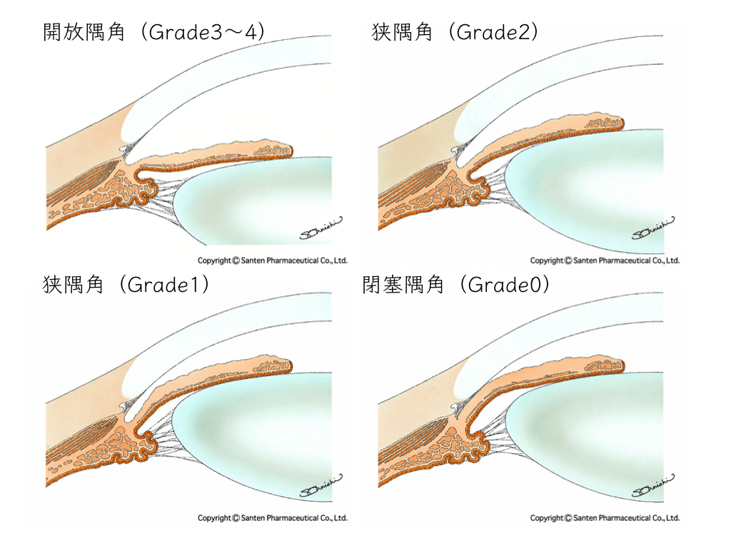
An enlarged lens can narrow the angle, the drainage pathway for fluid in the eye:
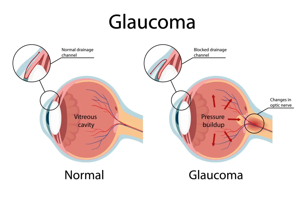
Eye pressure rises
→ damage progresses to the nerve tissue in the eye (the retina)
For this reason,
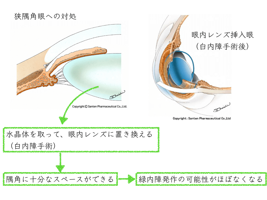
“Remove the lens to lower eye pressure”
= “perform cataract surgery early”
→ prevents “the transition from cataract to glaucoma”
is one line of thinking.
When a cataract progresses, it is often still at a stage that causes no trouble in daily life.
Careful judgment is required in deciding on surgery.
What does “if a cataract progresses, you develop glaucoma” mean?

Eye pressure: its relationship to how wide or narrow the angle, the drainage pathway for fluid in the eye, is
Most people have an open angle.
Aqueous humor, which also nourishes the front of the eye, is produced by the ciliary epithelium.
It flows out through the mesh structure of the trabecular meshwork in the angle and returns to the veins.
When this balance is maintained, eye pressure is normal.
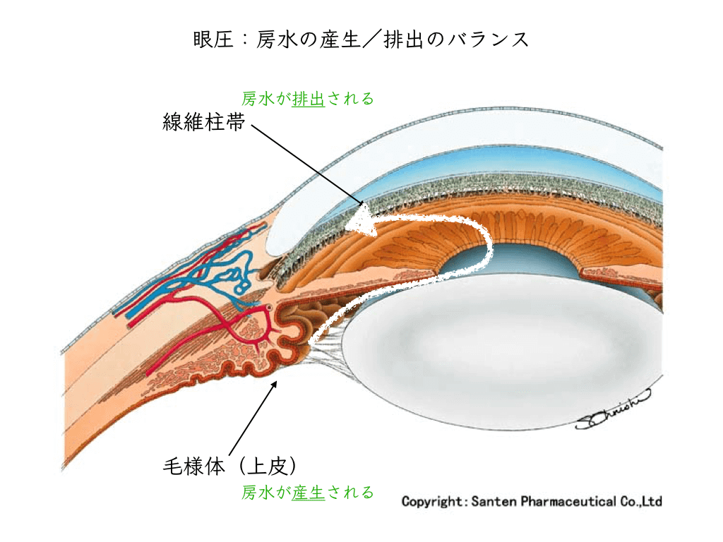
The normal range of eye pressure is about 10–20 mmHg (millimeters of mercury).
Pigment clogging the angle may impede the flow of aqueous humor and raise eye pressure (there are other causes too):
“Open-angle glaucoma”
On the other hand,
when the angle is narrow, aqueous humor also flows less easily and eye pressure may rise:
“Angle-closure glaucoma”

After aqueous humor has been flowing poorly for some time, the angle can suddenly close all the way around:
“Acute glaucoma / acute angle-closure glaucoma”
Eye pressure rises sharply, and severe symptoms such as headache and vomiting appear.
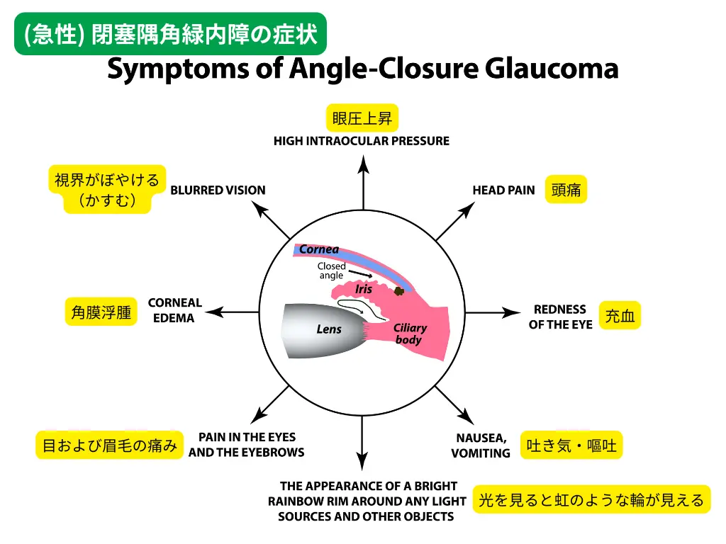
The “constitution” that makes glaucoma more likely as a cataract progresses
In a hyperopic (farsighted) eye, which is short from front to back, the lens takes up relatively more space.
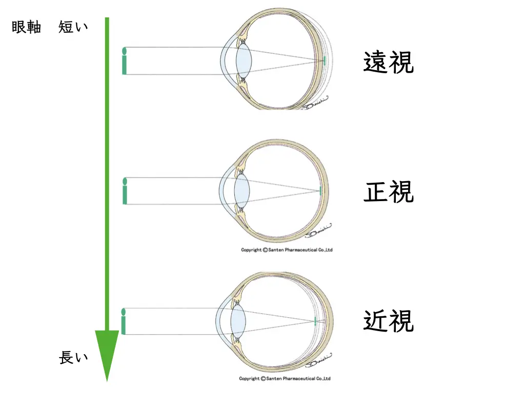
Because the lens occupies the angle, the angle, the drainage pathway for fluid, is often narrow.
This is called a “narrow angle.”
Eye pressure may also be slightly raised.
Whether a cataract is likely to lead to glaucoma: how to tell a “narrow angle”
What you can find out for yourself is
whether your eyes are farsighted
.
“I have good eyesight”
“I needed reading glasses early”
and the like suggest a tendency toward farsighted eyes,
and you may have a narrow angle.
As the cataract progresses, eye pressure is expected to rise.
At the eye examination,
we assess for a narrow angle in all patients and record it in the chart.
This is judged with a slit-lamp microscope (the Van Herick method).
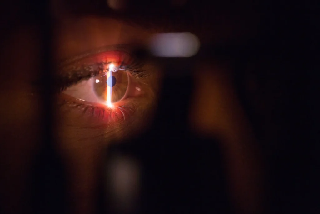
If needed, we also evaluate with gonioscopy and anterior segment OCT.
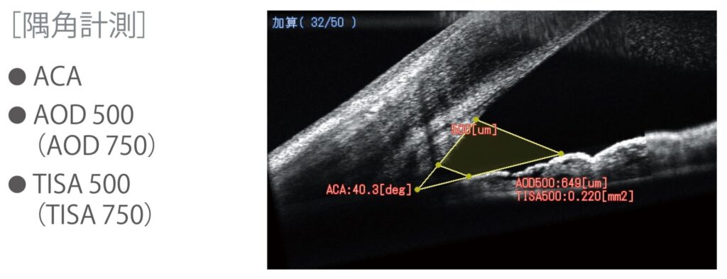
Narrow-angle eyes: what to watch for in daily life
There are medicines you cannot use.
We give people judged at the eye clinic to have a narrow anglea “glaucoma card”.
But “narrow angle” does not mean “glaucoma.”
We take the time to explain so that you do not have to feel excessiveworry.
Reading in the dark, using a smartphone, etc.
• In the dark, the pupil opens a little
• Looking down lets the lens move down
Together, these two effects increase the risk of developingacute angle-closure glaucoma.
Preventing a glaucoma attack: two approaches
Once it is known that the eye has a narrow angle, besides careful follow-up,
preventive measures may be considered.
There are mainly two.
“Laser iridotomy” and “cataract surgery”
Laser iridotomy

Cutting the iris with a laser requires a large amount of energy.
The major problem with laser iridotomy is that it can cause corneal endothelial damage decades later.1,2。
Corneal endothelial cells have a pump function that keeps aqueous humor from entering the cornea.
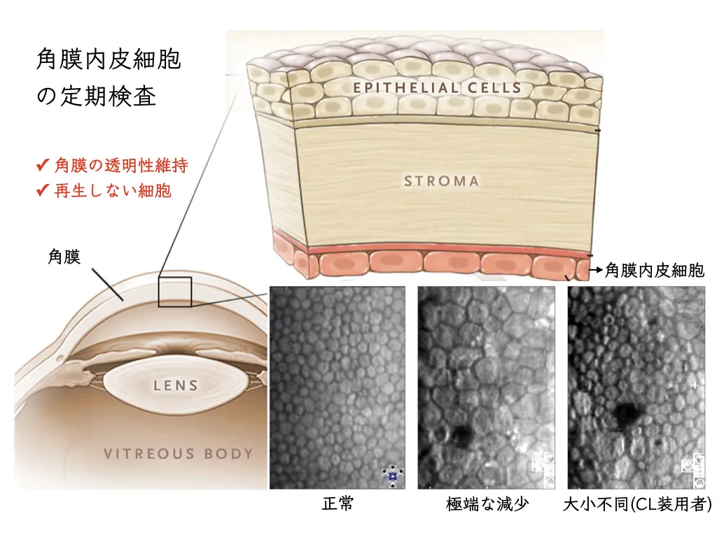
If this pump function declines, the cornea becomes swollen (= bullous keratopathy).
A swollen cornea loses transparency, and a cloudy cornea causes marked loss of vision.
This is a condition that requires a corneal transplant, and it places a heavy burden on the patient.
As described above,
laser iridotomy carries a risk of corneal endothelial damage and bullous keratopathy decades later.
Cataract surgery
Instead, removing the lens can lower that risk.
That is early cataract surgery.
Currently, early phacoemulsification appears to be superior to LI not only in terms of IOP control, but also in protecting the corneal endothelium.
Early phacoemulsification is thought to be better than laser iridotomy not only for eye pressure control but also for protecting the corneal endothelium3。
Wang, P.X., et al., 2014. Laser iridotomy and the corneal endothelium: a systemic review. Acta Ophthalmologica 92, 604–616. https://doi.org/10.1111/aos.12367
Eye pressure falls after cataract surgery
How much does cataract surgery lower eye pressure?
The normal range of eye pressure is about 10–20 mmHg (millimeters of mercury).

In eyes that have had cataract surgery, a reduction in eye pressure of about 2–4 mmHg is expected4。
Is laser iridotomy useful for narrow angles? Two important clinical studies
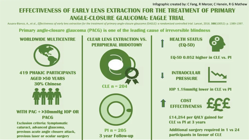
EAGLE trial5
ZAP trial6
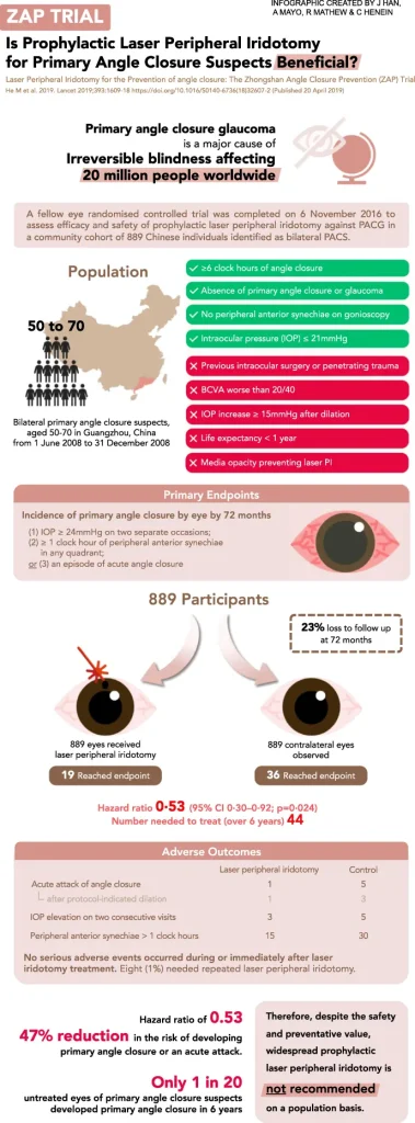
Based on these results,
preventive “laser iridotomy” is not currently recommended.
Disadvantages of cataract surgery in an “eye prone to glaucoma”
Eyes prone to acute glaucoma are “farsighted eyes”:
the so-called “good eyesight” state.
Even so,
“narrow angle”
= “eye pressure rises easily”
and these are judged to be high-risk eyes.
Even so,
Why is surgery needed on an eye that can see?
= What does “cataract surgery on an eye that sees well / cataract surgery for glaucoma” mean?
The disadvantages are
- that vision may actually be slightly worse after surgery
Pre-operative tests are performed to determine the power of the intraocular lens.
The intraocular lens calculation formulas use numbers based on statistics.
It is somewhat difficult to choose the right lens for a hyperopic eye (= short axial length).
Data on hyperopic eyes are limited, which suggests the result may deviate slightly from the prediction.
- In an eye that has already had a glaucoma attack, the intraocular lens may become unstable
The zonule, which supports the lens, may have become weak.
(Risk of intraocular lens dislocation or falling after a long period)
Even if an eye has had a glaucoma attack in the past, there may have been few symptoms and it may have gone unnoticed.
The decision on surgery must take the above into account as well.
References
- Ang, L.P.K., Higashihara, H., Sotozono, C., Shanmuganathan, V.A., Dua, H., Tan, D.T.H., Kinoshita, S., 2007. Argon laser iridotomy‐induced bullous keratopathy—a growing problem in Japan. Br J Ophthalmol 91, 1613–1615. https://doi.org/10.1136/bjo.2007.120261
- Wang, P.X., Koh, V.T.C., Loon, S.C., 2014. Laser iridotomy and the corneal endothelium: a systemic review. Acta Ophthalmologica 92, 604–616. https://doi.org/10.1111/aos.12367
- Wang, P.X., Koh, V.T.C., Loon, S.C., 2014. Laser iridotomy and the corneal endothelium: a systemic review. Acta Ophthalmologica 92, 604–616. https://doi.org/10.1111/aos.12367
- Shrivastava, A., Singh, K., 2014. The impact of cataract surgery on glaucoma care. Curr Opin Ophthalmol 25, 19–25. https://doi.org/10.1097/ICU.0000000000000010
- Azuara-Blanco, A., Burr, J., Ramsay, C., Cooper, D., Foster, P.J., Friedman, D.S., Scotland, G., Javanbakht, M., Cochrane, C., Norrie, J., 2016. Effectiveness of early lens extraction for the treatment of primary angle-closure glaucoma (EAGLE): a randomised controlled trial. The Lancet 388, 1389–1397. https://doi.org/10.1016/S0140-6736(16)30956-4
- He, M., Jiang, Y., Huang, S., Chang, D.S., Munoz, B., Aung, T., Foster, P.J., Friedman, D.S., 2019. Laser peripheral iridotomy for the prevention of angle closure: a single-centre, randomised controlled trial. The Lancet 393, 1609–1618. https://doi.org/10.1016/S0140-6736(18)32607-2