![Takeru Eye Clinic | Takatori Shopping Street, Sawara-ku, Fukuoka City [Nishijin Station / Fujisaki Station]](https://takeru-eye.com/wp-content/uploads/2022/10/takeru_logo_for-WP-header.png)

If a health checkup has flagged an enlarged optic disc cup, it can come as a shock.
It is often found in people with high myopia (very poor eyesight without correction).
This article is about evaluating the shape of the optic nerve inside the eye.
Imaging tests show whether there is any cause for concern.
“Enlarged Optic Disc Cup”: How Large Is the Cup in the Optic Nerve Inside Your Eye?

The optic nerve is a bundle of nerve fibers from the retina, and its shape tells us a great deal.
For almost every patient, regardless of the condition they come in with:
Using a slit lamp and a handheld lens (Superfield NC, Volk), I look at the optic nerve inside the eye.
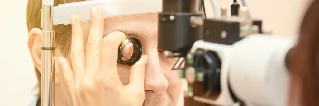
I assess the optic nerve instantly, without dilating the pupil with eye drops.
* Screening: deciding whether it looks normal or whether disease is suspected

- Size
- Condition of the rim (margin)
- Depth and shape of the cup
- Shape of the blood vessels
- Presence of hemorrhage
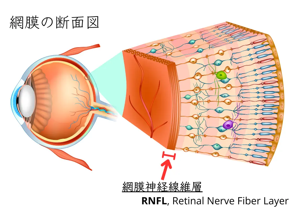
- Condition of the surrounding retina (retinal nerve fiber layer defect)
All of these are on the ophthalmologist’s mental checklist.
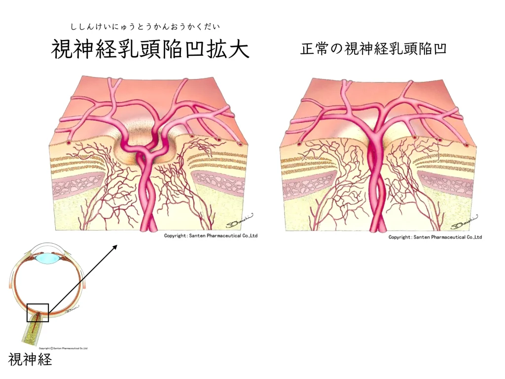
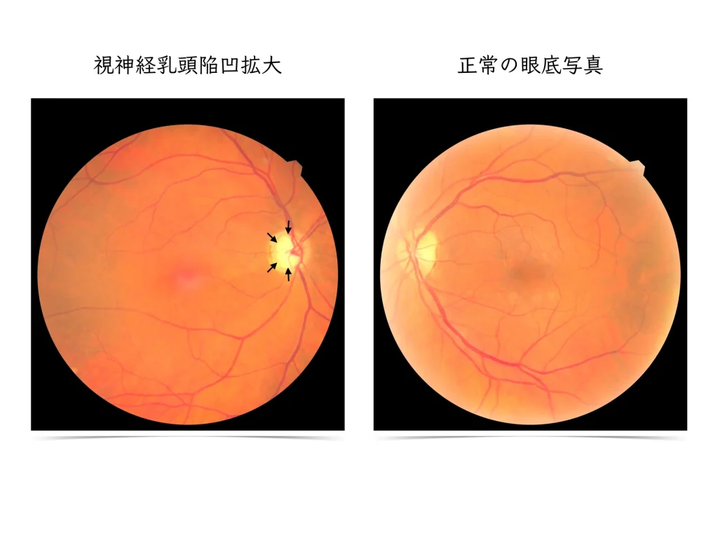
○ Enlarged optic disc cup(Shishinkei nyuto kan’o kakudai in Japanese)
It is very common in people with poor eyesight (high myopia).
People who have worn contact lenses or glasses for many years often do not notice that the shape of their optic nerve is abnormal.
It may also be pointed out from a fundus photograph at a company checkup or health checkup (a follow-up exam is advised).
Most cases of ocular hypertension and glaucoma have no symptoms.

Other eye diseases accompanied by optic nerve abnormalities
○ Optic disc hemorrhage(DH, disc hemorrhage)
○ Uveitis and optic neuroretinitis
○ Diabetic retinopathy and diabetic optic neuropathy
○ Optic neuritis
○ Hereditary optic neuropathy
Leber hereditary optic neuropathy
○ Toxic optic neuropathy
Caused by anti-tuberculosis drugs (ethambutol), thinners and organic solvents, antiarrhythmic drugs, and others
Insufficient nutrition (nutritional optic neuropathy) or insufficient blood flow (ischemic optic neuropathy), among others.
When needed, I also analyze the nerve with OCT (optical coherence tomography).
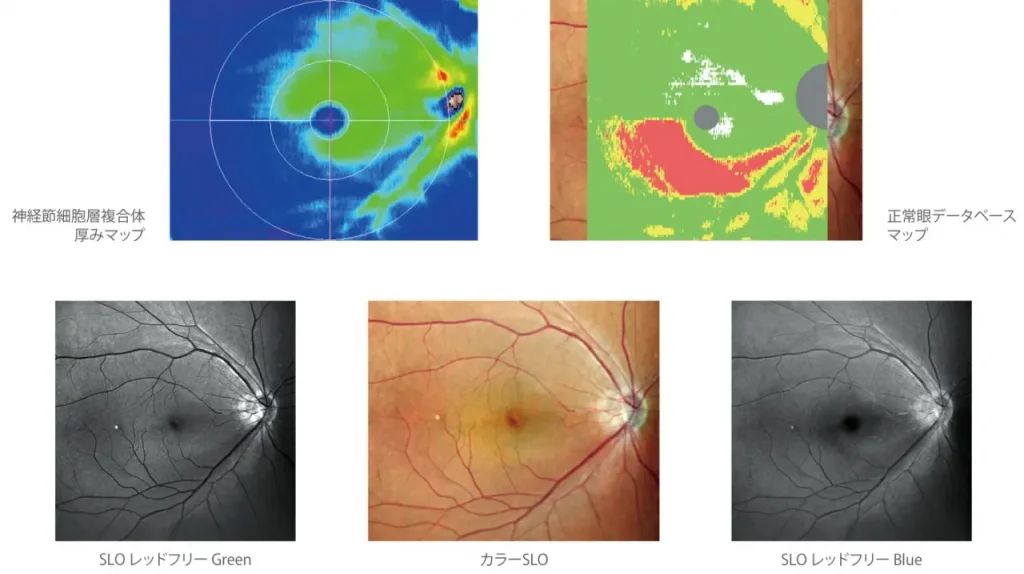
If a visual field test seems necessary, I first show you the fundus photograph and OCT results.
All results are shown and explained on the monitor in front of you.
If you would like your family to hear the explanation, they are welcome to come in at any time and I will show them the images.
Please do not hesitate to ask.