![Takeru Eye Clinic | Takatori Shopping Street, Sawara-ku, Fukuoka City [Nishijin Station / Fujisaki Station]](https://takeru-eye.com/wp-content/uploads/2022/10/takeru_logo_for-WP-header.png)

Age-related macular degeneration (AMD) care entered a major turning point with the 2024 revision of the clinical practice guidelines by the Japanese Ophthalmological Society.
The revision stresses that diagnosis should not be limited by age, and it establishes pachychoroid as a new disease concept.
As a result, care now has to take into account cases that begin at a relatively young age, even in the 40s.
Clinical Practice Guidelines for Neovascular Age-Related Macular Degeneration (Journal of the Japanese Ophthalmological Society, Vol. 128, No. 9)1 https://www.nichigan.or.jp/Portals/0/resources/member/guideline/nvAMD.pdf
Audio explanation (Google NotebookLM)
Age-Related Macular Degeneration (AMD): Revised Guidelines and How We Manage It in the Clinic
A New Understanding of AMD and the Importance of Early Detection

AMD was traditionally regarded as a disease of older people.
It is now recognized as a disease that can develop regardless of age. In particular, it has become clear that a condition called pachychoroid, which is common in Asian people, plays an important role in the development of AMD.
Pachychoroid refers to a state in which the blood vessels of the choroid (the vascular layer behind the retina) are dilated and no longer function normally.
Recent research recognizes pachychoroid neovasculopathy (PNV) as a new subtype of AMD, and it suggests that about 26% of Japanese patients may fall into this category.2
Are you worried about a retinal or macular abnormality?
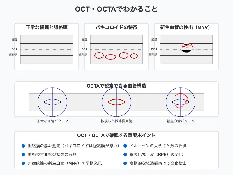
Using both OCT and OCTA imaging devices, we can detect these changes early.
Diagnosis and Management in a Solo-Doctor Eye Clinic

By using OCT and OCTA (OCT angiography), we can detect AMD early and manage it over time.
Three points are especially important.
The first is detailed observation through regular imaging tests.
OCT lets us observe the layered structure of the retina and evaluate in detail deposits called drusen and changes in the retinal pigment epithelium.OCTA lets us observe the state of the blood vessels without contrast dye, and in particular confirm the dilation of choroidal vessels that is characteristic of pachychoroid.
The second is talking with you about improving your lifestyle.
Smoking and diet are known to be strongly linked to the onset and progression of age-related macular degeneration. Smoking in particular is the most important environmental factor, and guidance on quitting smoking is the core of preventive management.
A diet centered on green and yellow vegetables also plays an important role in preventing age-related macular degeneration.
The third is continued follow-up.
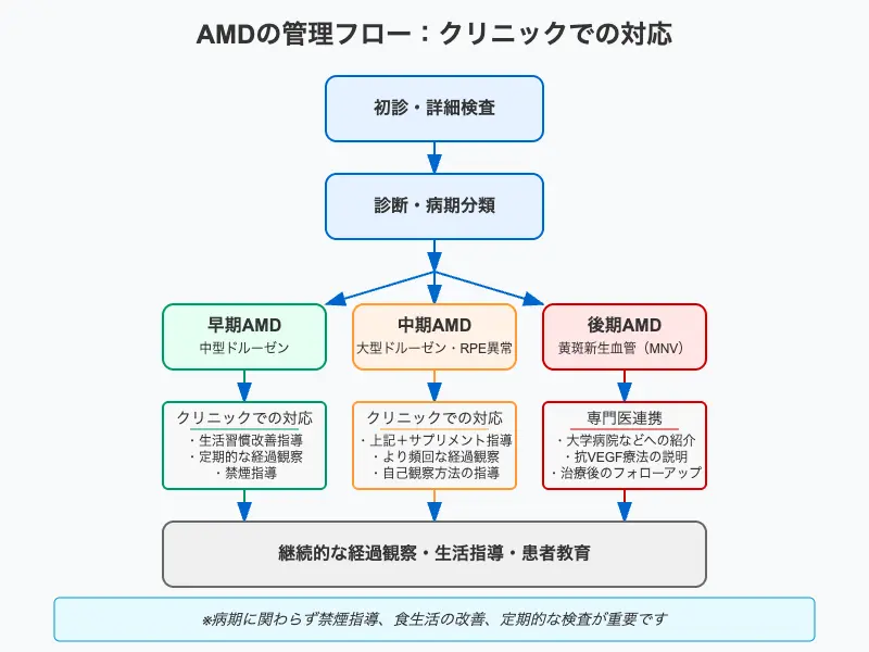
AMD is a slowly progressing disease, and it is extremely important to track changes through regular examinations. In particular, by detecting the appearance of macular neovascularization (MNV) early, we can refer you to a specialist medical institution at the right time.

Observation and Prevention You Can Do Yourself

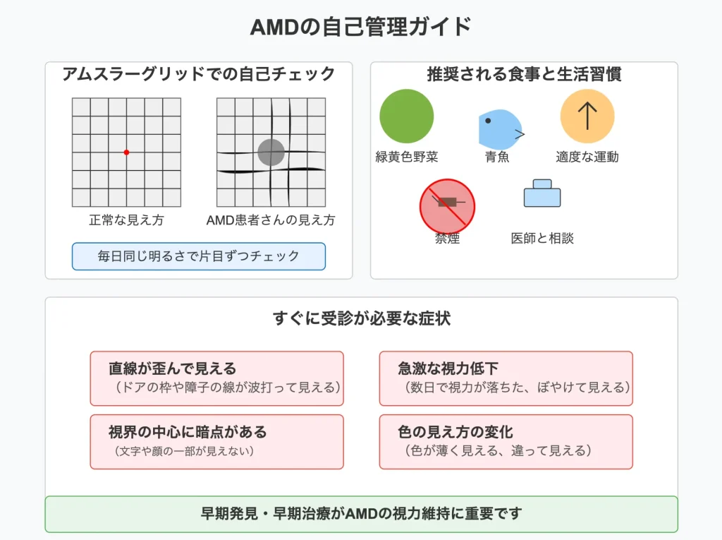
In managing AMD, regular observation by patients themselves also plays an important role.
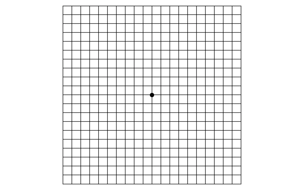
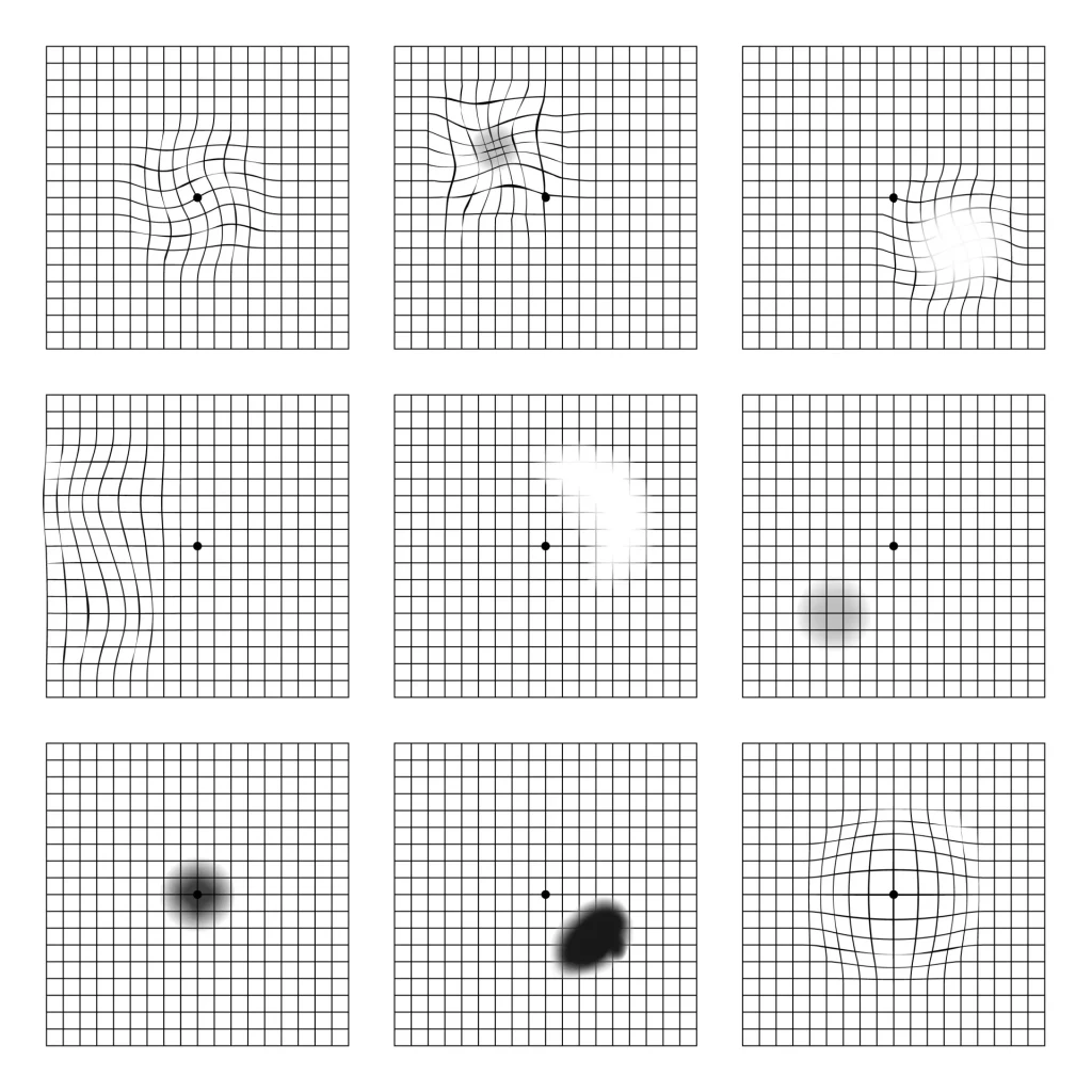

Please do a self-check with a grid-patterned chart called the Amsler chart, at the same time every day.
It is important to cover one eye while you check.
Distortion of the grid lines or the appearance of areas you cannot see is an important sign that your condition has changed.
From a preventive standpoint, we also recommend the following lifestyle improvements:
Eat plenty of green and yellow vegetables, which are rich in antioxidants.
Dark green vegetables such as spinach and broccoli contain nutrients that are important for keeping the retina healthy.
Building an exercise habit is also important. Moderate exercise improves blood flow and helps maintain retinal health.
However, avoid strenuous exercise, and we recommend continuing light exercise such as walking.

Coordination with Specialist Medical Institutions
In managing AMD, close coordination between the clinic and specialist medical institutions is essential.
In particular, when neovascular AMD is diagnosed, treatment with anti-VEGF drugs may be needed.
In such cases, we have a system in place with Kyushu University Hospital and other core hospitals so that appropriate treatment can be started promptly.
After treatment starts, we continue regular follow-up and evaluation with OCT to confirm the treatment effect and to detect any recurrence early.
So that things go in the best direction for you:
We will explain your current eye condition and what lies ahead, using the materials on this website as well.
References
- Japanese Ophthalmological Society (2024). Clinical Practice Guidelines for Neovascular Age-Related Macular Degeneration. Journal of the Japanese Ophthalmological Society, Vol. 128, No. 9.
- Kuranami, A., Maruko, I., Maruko, R., Nishihara, S., Iida, T., 2022. Age-related macular degeneration and new entity of pachychoroid neovasculopathy in Japanese patients. Investigative Ophthalmology & Visual Science 63, 335-F0166.
* Appropriate care differs from case to case.
Please feel free to consult us at any time if you have any symptoms that concern you.