![Takeru Eye Clinic | Takatori Shopping Street, Sawara-ku, Fukuoka City [Nishijin Station / Fujisaki Station]](https://takeru-eye.com/wp-content/uploads/2022/10/takeru_logo_for-WP-header.png)

Introduction
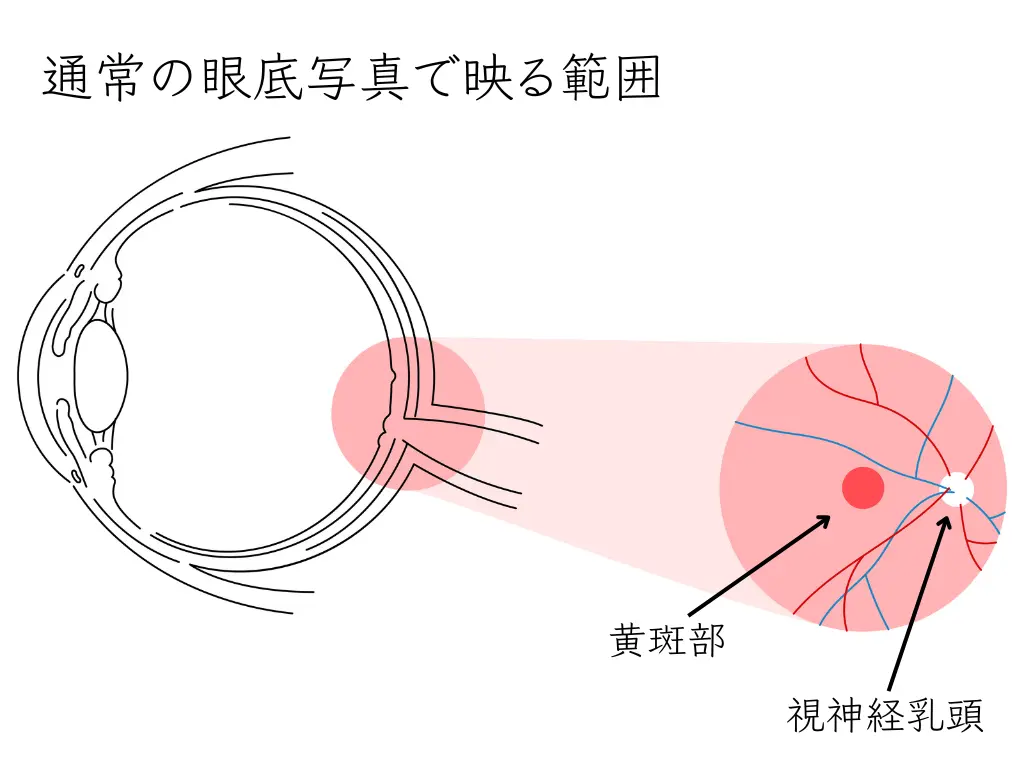
Central serous chorioretinopathy (CSC) is an eye disease that affects the macula, the most important part of the retina for seeing. This article explains its symptoms, causes, diagnosis and treatment in detail.
What is central serous chorioretinopathy (CSC)?
CSC is a disease in which circulatory disturbance and congestion of the choroid, which lies deeper than the retina at the back of the eye, lead to fluid collecting beneath the center of the retina (the macula). It is now considered part of the disease concept called “pachychoroid” (thick choroid).1
Main symptoms
- Objects look distorted (metamorphopsia)
- Sudden difficulty seeing
- Reduced central vision
Age and sex most often affected
It is seen most often in men in their 30s to 50s.2
Triggers
The following factors have long been known:3:
- Stress
- Smoking
- Oral steroids
- In women, it can also begin with pregnancy
Pachychoroid spectrum diseases
CSC is part of the broader pachychoroid spectrum of diseases. This spectrum includes the following:4:
Central serous chorioretinopathy (CSC)
- A disease in which fluid collects beneath the center of the retina (the macula), mainly because of a choroidal circulatory disturbance.
- In many cases it improves with just quitting smoking and observation, or with oral medication.
Pachychoroid pigment epitheliopathy
- A condition similar to CSC, but without fluid collecting beneath the retina (subretinal fluid).
Pachychoroid neovasculopathy
- A long-term complication that can develop after CSC or pachychoroid pigment epitheliopathy.
- New blood vessels form in the choroid, with a risk of reduced vision in the future.
Central serous chorioretinopathy (CSC): long-term complications
When CSC becomes chronic, the risk of the following complications may rise:
- Atrophy of the retinal pigment epithelium (RPE)
- Choroidal neovascularization (CNV)
- Fibrosis of the macula
- Permanent loss of vision
The risk of these complications may be related to how long CSC lasts and how many times it recurs. Regular examinations and appropriate management are important.
Diagnosis
A variety of imaging techniques are used to diagnose CSC and pachychoroid spectrum diseases.
Optical coherence tomography (OCT) and OCT angiography
It is now possible to use a device that combines OCT, OCT angiography and a fundus camera in one unit.
EDI-OCT (Enhanced Depth Imaging OCT)
EDI-OCT (enhanced depth imaging OCT) is a technique that images deeper structures more clearly than ordinary OCT. It is particularly useful for observing the pachychoroid.
Features of EDI-OCT:
- The detailed structure of the choroid can be observed
- The thickness of the choroid can be measured accurately
- It plays an important role in diagnosing pachychoroid
Mirante is equipped with an EDI-OCT function.
OCT angiography
It lets us observe the state of the retinal and choroidal blood vessels without using a contrast dye.
Fundus examination
A fundus examination lets us observe the inside of the eyeball: the retina and vitreous.
The ophthalmologist’s own eyes give a three-dimensional examination that detects swelling of the macula.
Retinal imaging with SLO: mainly images covering 89° from the center, used to evaluate the detailed state of the macula.
The importance of self-checking by patients
For early detection and appropriate treatment of eye disease, regular self-checks by patients are extremely important. In particular, checking the vision of each eye separately helps in the early detection of many eye diseases, not only central serous chorioretinopathy (CSC) and pachychoroid spectrum diseases.
Checking each eye separately: an important method common to all eye diseases
- Checking your vision with one eye covered is extremely important for the early detection of any eye disease.
- Specific steps:
- Lightly cover one eye with one hand.
- With the other eye, look carefully at objects and text around you.
- Then switch hands and look the same way with the other eye.
- Compare how things look with the two eyes and check whether there is any difference.
Please watch for the following:
- Objects look distorted (metamorphopsia)
- The center looks blurred
- There is a dark spot (a part you cannot see)
- Colors look different
- Objects look smaller (micropsia)
- Overall reduction in vision
This method is useful for the early detection of many eye diseases, not only CSC and pachychoroid spectrum diseases but also glaucoma, age-related macular degeneration and retinal detachment. It is a very important check so that you do not miss a disease that affects only one eye, or one that has progressed to different degrees in the two eyes.
Amsler grid test (Amsler chart)
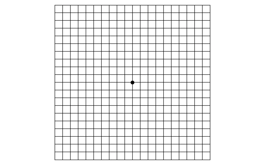
A self-check using an Amsler grid is also effective. An Amsler grid is a grid-like figure like the one below, with a dot in the center.
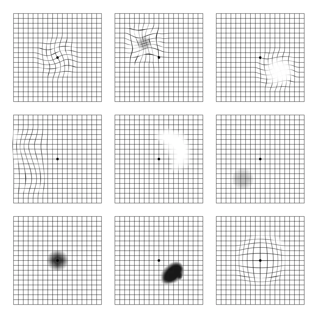
How to use the Amsler grid:
- Look at the grid from a normal reading distance (about 30 cm).
- Cover one eye and stare at the dot in the center with the other eye.
- Check the following:
- Straight lines look distorted
- Part of the grid disappears
- There is a blurred area or dark spot near the center
4. Check the other eye in the same way.
In a normal eye, all the lines look straight and no part of the grid is missing.
This self-check helps in the early detection of various eye diseases that affect the macula, such as central serous chorioretinopathy (CSC), pachychoroid spectrum diseases and age-related macular degeneration.
However, these checks are only a supplement and are no substitute for regular eye examinations.
If you notice even a slight abnormality, we strongly recommend seeing an ophthalmologist.
Central serous chorioretinopathy (CSC): treatment
Many cases of CSC improve with the following:
- Quitting smoking
- Observation
It is also common for the condition to “get better without doing anything.”
If symptoms persist, the following treatments may be considered:
- Photodynamic therapy (PDT)
- Laser photocoagulation
- Intravitreal injection of anti-VEGF drugs
We provide coordinated care, mainly with the Department of Ophthalmology at Kyushu University Hospital.
A possible new treatment: selective retinal therapy (SRT)
Research on a new treatment called selective retinal therapy (SRT) is under way, centered on the University of Lübeck in Germany. SRT may treat only the retinal pigment epithelium (RPE) cells while keeping damage to the surrounding tissue to a minimum.5
Features of SRT:
- It may promote regeneration of RPE cells
- It may cause less heat damage to the retina than conventional treatments
- It may make treatment near the fovea possible
In addition, SRT may have the following potential advantages over anti-VEGF therapy:6:
- Lower treatment cost: anti-VEGF drugs are expensive and often need to be given several times, whereas SRT may be effective with a single treatment.
- Fewer treatments: anti-VEGF therapy needs multiple injections, whereas SRT may achieve its effect with fewer treatments.
- Long-term cost-effectiveness: if the regenerative effect of SRT on RPE cells lasts, the need for retreatment would fall, which could reduce long-term medical costs.
However, SRT is still at the research stage, and further research is needed on its long-term effect, safety and actual cost-effectiveness. The best treatment also differs with each patient’s condition and the stage of the disease, so the choice of treatment should be made after thorough discussion with your doctor.
Central serous chorioretinopathy (CSC): summary
Central serous chorioretinopathy (CSC) is a disease that affects the macula, which is important for seeing. It is seen mainly in men in their 30s to 50s, and stress and smoking may trigger it.
The main symptoms of CSC are distorted vision (metamorphopsia), sudden difficulty seeing, and reduced central vision. If you notice these symptoms, it is important to see an ophthalmologist early.
Diagnosis uses the latest imaging technology, such as OCT and EDI-OCT, which allow the state of the choroid to be observed in detail. Regular self-checks by patients (checking each eye separately and the Amsler grid test) are also an important way to reach early detection and early treatment.
Treatment: in many cases, quitting smoking and observation lead to improvement. If symptoms persist, photodynamic therapy (PDT), laser photocoagulation and similar treatments are considered. Research on selective retinal therapy (SRT) as a new treatment is also under way.
With appropriate management and treatment, CSC often follows a good course, but if it becomes chronic the risk of complications such as reduced vision may rise. We therefore recommend paying attention to the following:
- Have regular eye examinations
- Make self-checks a habit
- Work on lifestyle improvements such as stress management and quitting smoking
- If you notice a change in your symptoms, see an ophthalmologist promptly
- Choose your treatment after thorough discussion with your doctor
The health of your eyes has a great effect on your quality of life.
If you have even a slight concern about your symptoms, please see an ophthalmologist early.
Early detection and early treatment help maintain good vision.
References
- Cheung, C.M.G., Lee, W.K., Koizumi, H., Dansingani, K., Lai, T.Y. and Freund, K.B., 2019. Pachychoroid disease. Eye, 33(1), pp.14-33. Available at: https://www.nature.com/articles/s41433-018-0158-4
- Daruich, A., Matet, A., Dirani, A., Bousquet, E., Zhao, M., Farman, N., Jaisser, F. and Behar-Cohen, F., 2015. Central serous chorioretinopathy: Recent findings and new physiopathology hypothesis. Progress in retinal and eye research, 48, pp.82-118.
- Nicholson, B., Noble, J., Forooghian, F. and Meyerle, C., 2013. Central serous chorioretinopathy: update on pathophysiology and treatment. Survey of ophthalmology, 58(2), pp.103-126.
- Dansingani, K.K., Balaratnasingam, C., Naysan, J. and Freund, K.B., 2016. En face imaging of pachychoroid spectrum disorders with swept-source optical coherence tomography. Retina, 36(3), pp.499-516.
- Neumann, J. and Brinkmann, R., 2006. Cell disintegration by laser-induced transient microbubbles and its simultaneous monitoring by interferometry. Journal of Biomedical Optics, 11(4), p.041112. Available at: https://doi.org/10.1117/1.2339815
- Klatt, C., Saeger, M., Oppermann, T., Pörksen, E., Treumer, F., Hillenkamp, J., Fritzer, E., Brinkmann, R., Birngruber, R. and Roider, J., 2011. Selective retina therapy for acute central serous chorioretinopathy. British Journal of Ophthalmology, 95(1), pp.83-88.