![Takeru Eye Clinic | Takatori Shopping Street, Sawara-ku, Fukuoka City [Nishijin Station / Fujisaki Station]](https://takeru-eye.com/wp-content/uploads/2022/10/takeru_logo_for-WP-header.png)

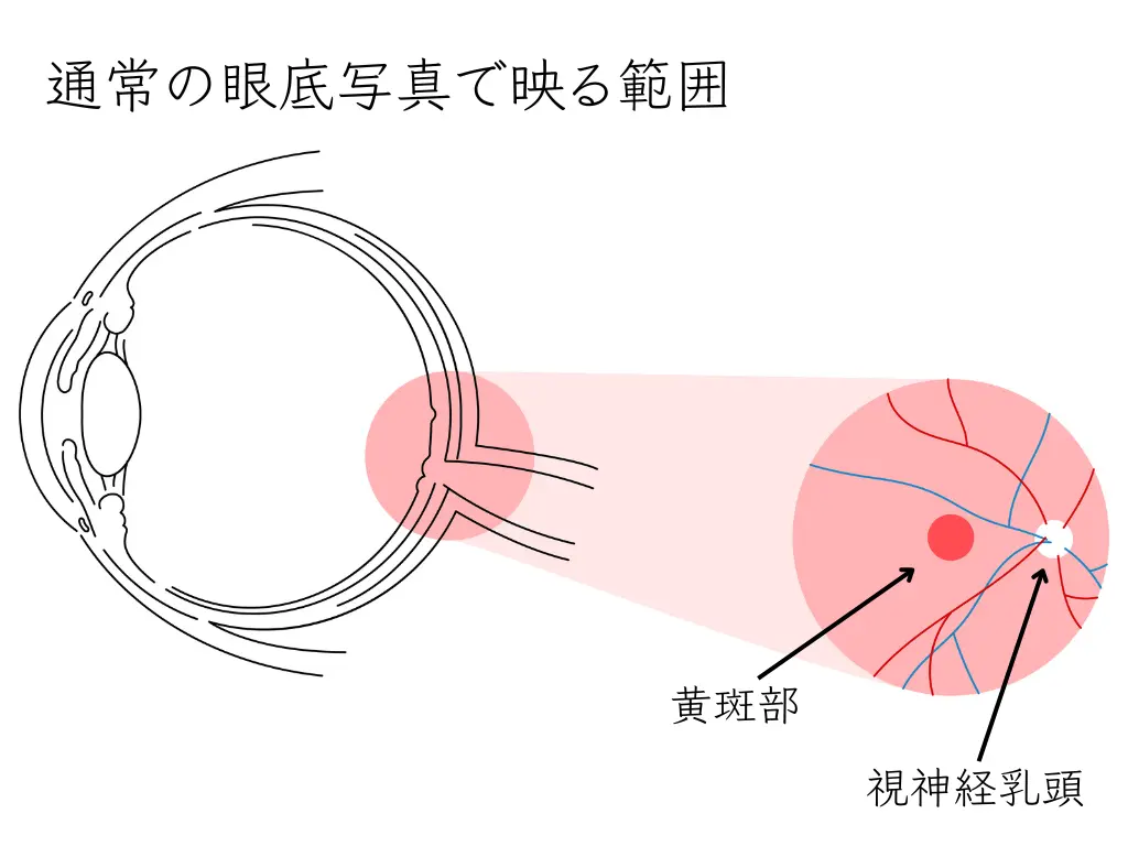
In a general medical checkup (ningen dock), fundus photographs are taken.
These evaluate only a narrow part of the broad retina, the macula, which is the most important area, and the part containing the optic nerve.
Even when they have symptoms such as “text on my smartphone has become hard to read lately” or “my eyes get tired in the evening,” many people tend to assume, “it must be my age.”
Not a few people also say, “I have no particular eye symptoms, but I would like to have my eyes examined thoroughly just in case.”
In daily practice, I sometimes receive requests such as “please examine everything about my eyes.”
However, under health insurance, comprehensive testing is limited when there are no symptoms.
For example, a car goes through a periodic inspection (shaken) even if the engine is running well.
The same applies to the eyes, and checking them before symptoms appear is considered important.
This is because many eye diseases progress without being noticed.
It is well known that glaucoma is the leading cause of blindness in Japan, developing in about 1 in 20 people aged 40 and over.
Morizane, Y., Morimoto, N., Fujiwara, A. et al. Incidence and causes of visual impairment in Japan: the first nation-wide complete enumeration survey of newly certified visually impaired individuals. Jpn J Ophthalmol 63, 26–33 (2019). https://doi.org/10.1007/s10384-018-0623-4
Take glaucoma as an example. This disease is known to progress without symptoms.
This is because the optic nerve is damaged gradually, and the visual field is lost without any subjective symptoms.
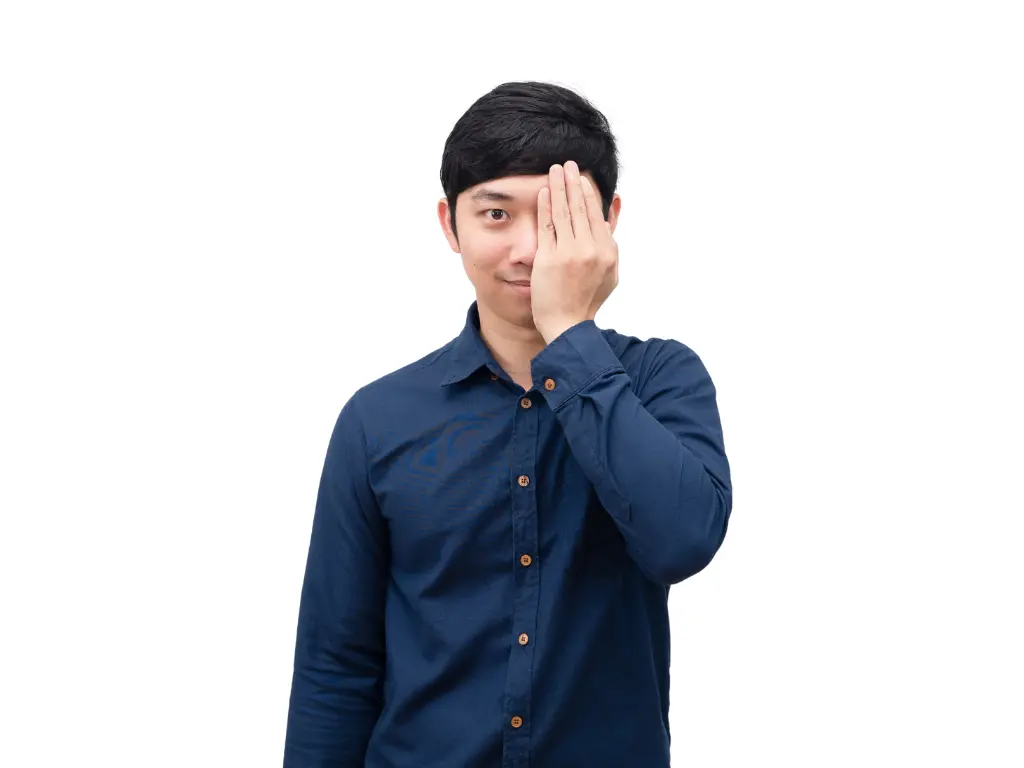
The human brain is remarkable: even if the visual field of one eye is lost, the other eye covers for it. For that reason, people often do not notice until the visual fields of both eyes have become quite narrow.
With current medicine, visual field that has been lost once cannot be restored.
However, if the disease is found early, progression can be slowed with treatment such as eye drops.
In light of this situation, I decided to set up a new Comprehensive Eye Exam (Ganka Dock).
Comprehensive Eye Examination (Eye Check-up): Examination Courses
| Test |
(1)
Comprehensive Eye Screening ¥22,000
|
(2)
Basic Eye Screening ¥11,000
|
(3)
Glaucoma Early Detection Screening ¥16,500
|
(4)
Presbyopia and Age-related Eye Disease Screening ¥16,500
|
(5)
Children and Students Eye Screening ¥11,000
|
(6)
Eye Strain and Dry Eye Screening ¥11,000
|
Additional
Eye Screening
(please ask us)
|
What it tells us |
|---|---|---|---|---|---|---|---|---|
| Eye examinations | ||||||||
| Objective refraction | The approximate power of your eyes, measured by an instrument | |||||||
| Corrected visual acuity (distance) | How well you can see at a distance with glasses or contact lenses | |||||||
| Corrected visual acuity (near) | How well you can see up close with glasses or contact lenses | |||||||
| Visual acuity with your current glasses | How well you see with the glasses you use now | |||||||
| Power of your current glasses | The power of the glasses you use now | |||||||
| Intraocular pressure | The firmness of the eye (a check for lifestyle-related eye disease) | |||||||
| Axial length | The front-to-back length of the eye (related to the progression of myopia) | |||||||
| Eye position (alignment) | How the eyes are aligned (whether there is strabismus or signs of it) | |||||||
| Eye movements | How the eyes move (whether they move smoothly) | |||||||
| Convergence | How the eyes turn inward when looking at near objects | |||||||
| Binocular function | How well the two eyes work together to see | |||||||
| Accommodation analysis | AccommoRef: the focusing power of the eye (a check for presbyopia and eye strain) | |||||||
| Wavefront aberrometry and corneal topography | The degree of distortion of the cornea and the lens | |||||||
| Corneal endothelial cells | The number and shape of the cells on the back of the cornea (especially important for contact lens wearers) | |||||||
| Anterior segment 3D imaging | A detailed three-dimensional examination of the front of the eye (whether there is anything to watch for regarding glaucoma) | |||||||
| Posterior segment 3D imaging (OCT) | A detailed three-dimensional examination of the back of the eye (signs of glaucoma, retinal disease and so on) | |||||||
| Widefield fundus photography | Imaging of a wide area inside the eye | |||||||
| OCT angiography | An examination of the blood vessels inside the eye | |||||||
| Additional tests | ||||||||
| Visual field | The extent of the area you can see | |||||||
| Pupil dilation | An examination using drops to widen the pupil (to look at the inside of the eye in detail) | |||||||
| Tear secretion | Assessment of the amount of tears (Schirmer test) | |||||||
| Consultation (board-certified ophthalmologist) | ||||||||
| Slit-lamp biomicroscopy | A magnified examination from the surface to the inside of the eye | |||||||
| Tear film breakup time (BUT) | Assessment of the quality and quantity of tears |
* The marks show which tests are included in each course. Additional eye screening can be selected in addition to any course.
* All eye examinations are performed by nationally certified orthoptists.
* Prices include tax.
* All of the Comprehensive Eye Examination courses above are by appointment only.
Since opening the clinic, I have been thinking about the importance of preventive medicine.
In 2025 we entered our seventh year, and thanks to everyone, we have come to see a great many patients.
What I keep realizing again through this is the importance of preventive testing before symptoms appear.
Why a Comprehensive Eye Exam Is Needed

Eye diseases that progress even without symptoms
Just as a crack in a building’s foundation does not immediately affect the whole building, eye diseases cause almost no subjective symptoms in their early stages.
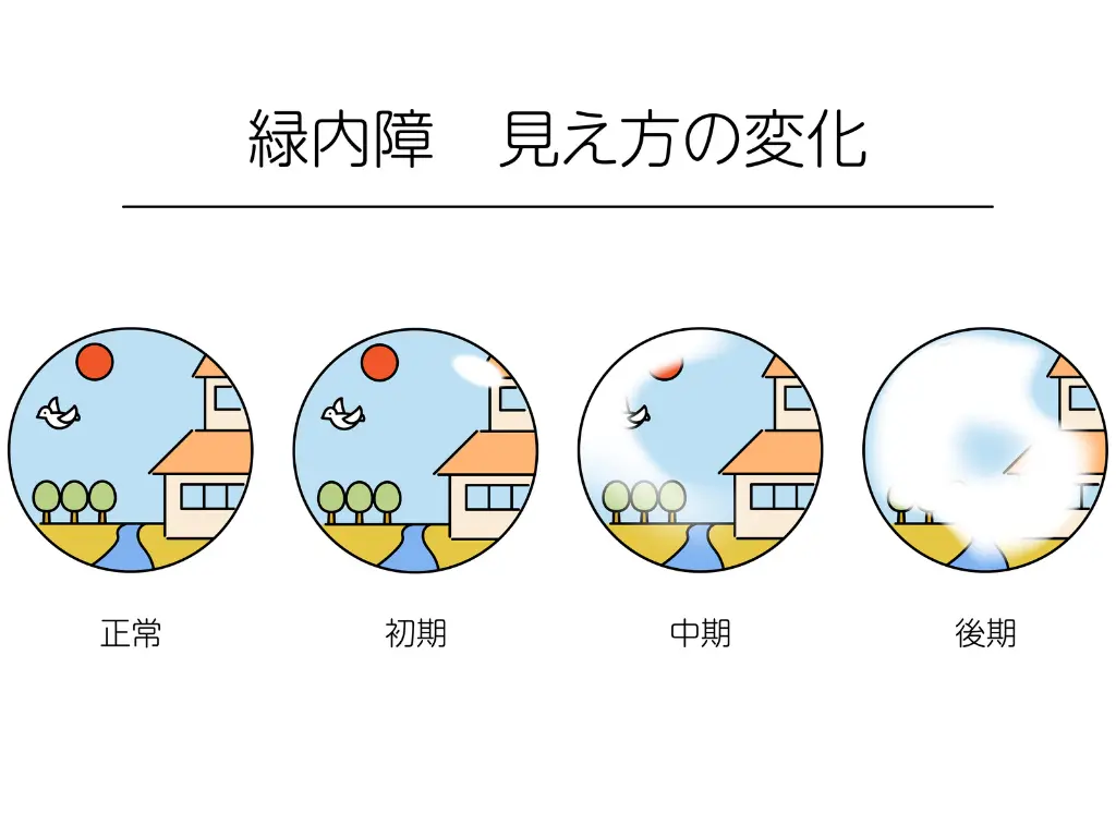
Glaucomais known to progress without symptoms.
It develops in about 1 in 20 Japanese people aged 40 and over, and about 1 in 8 aged 70 and over. Damage to the optic nerve is irreversible, and visual field that has been lost does not come back.
For example, even if some characters become invisible while reading a newspaper, the brain fills them in automatically, so it can go unnoticed for a long time.
Diabetic retinopathyis one of the three major complications of diabetes.
Imagine the walls of blood vessels being damaged by high blood sugar.
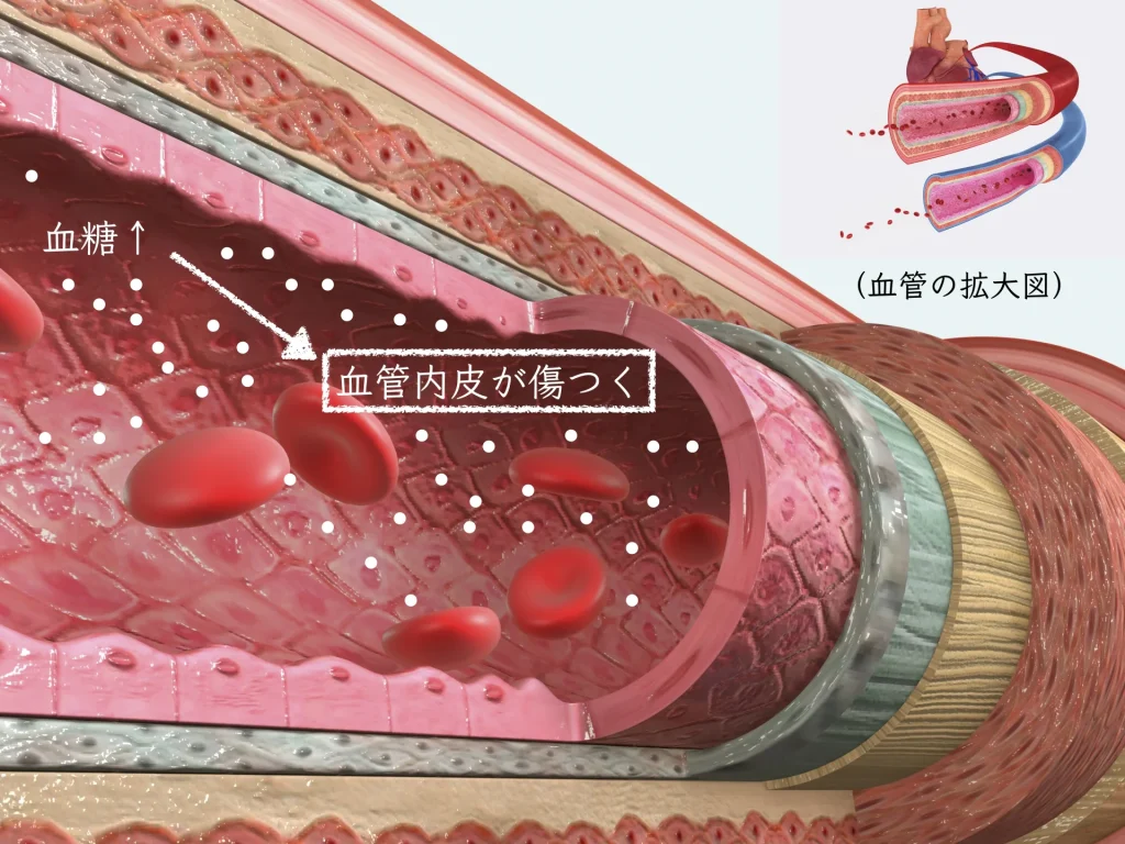
Diabetic retinopathy: the fine blood vessels of the retina become blocked, bleed, or leak fluid.
As it advances, it can lead to blindness.
Importantly, retinopathy may already be progressing even at the stage when you have only been told you are prediabetic.
-1024x768.jpg)
Age-related macular degenerationis a disease that damages the center of the retina (the macula), which corresponds to the film in a camera.
It is seen in about 1 in 80 people aged 50 and over.
Because central vision declines, it becomes hard to recognize people’s faces, and text may look distorted.
A characteristic symptom is that, when looking at a clock face, the hour hand at the center cannot be seen.

What these diseases have in common is that they are unlikely to cause subjective symptoms, and may have already progressed by the time they are noticed.
However, regular examinations make early detection possible, and appropriate treatment can slow progression.
Eye Diseases to Watch for by Age
The eye problems to watch for change with each stage of life. This is similar to how maintenance points change with the age of a house.
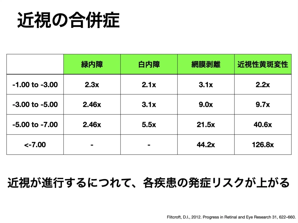
Childhood and the student yearsIn this period, the rise in myopia is a global problem. Compared with the past, the proportion of children who become myopic has increased rapidly.
Myopia is not simply a “condition that needs glasses.”
It has been shown to raise the risk of serious eye diseases such as glaucoma and retinal detachment in the future.
For example, there is research showing that elementary school children who spend two or more hours outdoors every day can slow the progression of myopia.
It is also important to keep an appropriate distance (30 cm or more) when reading or studying.
With the spread of smartphones and tablets, a new problem called “smartphone esotropia” has also appeared.
Looking at a screen for a long time upsets the balance of the muscles that move the eyeball, and one eye turns inward. As a result, diplopia, in which things look double, may also appear.
It may be something like how muscles become stiff when the same posture is held for a long time.
Young adulthoodIn this period, eye strain becomes the main problem. Long hours of work on computers and smartphones tire the eye’s focusing function. This is the same mechanism as eyes getting tired during long-distance driving.
However, eye strain (asthenopia) differs from simple tired eyes and refers to a condition that does not recover even with sufficient rest. Systemic symptoms such as headache, stiff shoulders, and reduced concentration may also appear.

Age 40 and overVarious age-related changes begin to appear.
Cataractis a disease in which the lens inside the eye becomes cloudy. It is like fogging on a camera lens, making vision hazy and light feel glaring. The incidence rises steadily with age: about 50% in the 50s, about 70% in the 60s, about 90% in the 70s, and almost 100% in the 80s.
Presbyopiais a phenomenon in which the lens loses elasticity, making it harder to focus on near objects. If you find yourself naturally holding a newspaper at arm’s length, it may be the beginning of presbyopia. Symptoms often appear after about age 40, and this is a natural aging phenomenon.
What matters is to understand these age-related changes and to receive appropriate examinations at the appropriate time.
Limits of Insurance Coverage and Self-Pay Options

When tests cannot be done under insurance
“Please examine everything about my eyes, just in case” is a natural wish for a patient.
However, health insurance has certain rules.
The insurance system is intended for “the treatment of disease.”
For that reason, tests can be performed only when the physician judges them medically necessary.
This rule exists to use limited medical resources efficiently and to provide appropriate care to those who truly need treatment.
The physician is obliged to record in detail in the medical chart why a test is necessary and how its results are interpreted.
(Of course, as before, insurance still applies when something was flagged in a medical checkup or screening, or when you have symptoms.)
Under the regulations for insured medical institutions and physicians (Ryoyo Tanto Kisoku),
tests done at the patient’s request, such as “I want to know whether I am healthy” or “please check just in case,” when there are no symptoms, are regarded as health checkups and are not covered by insurance.
For example, if you have symptoms such as headache or nausea and the physician suspects glaucoma, the test can be done under insurance.
But if you have no symptoms and say, “I am worried about glaucoma, so please test me,” insurance does not apply.
Physicians receive guidance from the Ministry of Health, Labour and Welfare (the regional bureaus of health and welfare) regarding the performance of “tests at the patient’s request.”
For Understanding Insured Medical Care – Ministry of Health, Labour and Welfare https://www.mhlw.go.jp/seisakunitsuite/bunya/kenkou_iryou/iryouhoken/shidou_kansa.html
This could be called a challenge arising between the patient’s wish to “feel reassured” and the rules of the insurance system.
Comprehensive Testing on a Self-Pay Basis
Based on the importance of preventive medicine
Partly to respond to patients’ wish to “have everything examined,”
I have newly set up a self-pay Comprehensive Eye Exam.
With self-pay care, comprehensive testing can be performed at the patient’s request, regardless of medical necessity.
This is the same idea as having tests such as a gastroscopy or electrocardiogram in a medical checkup (ningen dock).
With the Comprehensive Eye Exam, the health of your eyes can be examined in detail whether or not you have symptoms.
After the examination, rather than simply a result of “no abnormality,” we have also been able to prepare a detailed report that includes the examination images.
The report describes not only an explanation of the results but also points to watch in the future and a guide to when the next examination should be.
Eye Health Management in the Era of 100-Year Lives

Today is called the “era of 100-year lives.”
If you really live to 100, your eyes will also need to keep working for 100 years.
Just as regular maintenance is indispensable to keep using a car for a long time,
the eyes also need appropriate care to maintain good function over a long period.
Vision accounts for about 80% of the information we receive and is an important function that greatly affects the quality of daily life.
For example, many of life’s pleasures depend on vision: reading, watching movies, seeing family members’ faces, cooking, and going out to enjoy the scenery.
In today’s society, more people keep working into older age, and good vision is also indispensable in working life.
As the risk of various eye diseases rises with age,
the shift from the conventional idea of “treating after symptoms appear”
to the idea of “preventing before symptoms appear” is considered more important.
In recent years, the concept of “eye frailty” (eye frail) has attracted attention. Eye frailty refers to a state in which age-related decline in eye function begins to interfere with daily life. For example, becoming more likely to stumble on stairs or feeling anxious about driving at night may be related to a decline in visual function.
Eye frailty often appears earlier than physical frailty, and if left alone, it carries the risk of leading to cognitive decline and a need for nursing care. However, progression can be slowed by finding it early and responding appropriately.
Testing before symptoms appear can be thought of as an important “advance investment” in protecting future visual function. With early detection, progression of many eye diseases can be slowed, and as a result the chance of maintaining good vision throughout life is likely to rise.
Features of the Comprehensive Eye Exam
Completely by appointment
Only the Comprehensive Eye Exam is offered on a completely appointment-only basis.
Internet reservation slots are set on weekday mornings and from 2 p.m., so that examinations can be carried out with sufficient time.
Outpatient eye care is unpredictable in how long it takes,but this checkup finishes on schedule.
Specialized examinations by certified orthoptists
Certified orthoptists (shino kunrenshi) perform all of the eye examinations.
Orthoptists are national-licensed specialists in eye examination and can perform tests with high accuracy.
With staff who keep learning every day, we carry out high-standard examinations.
Comprehensive examination items
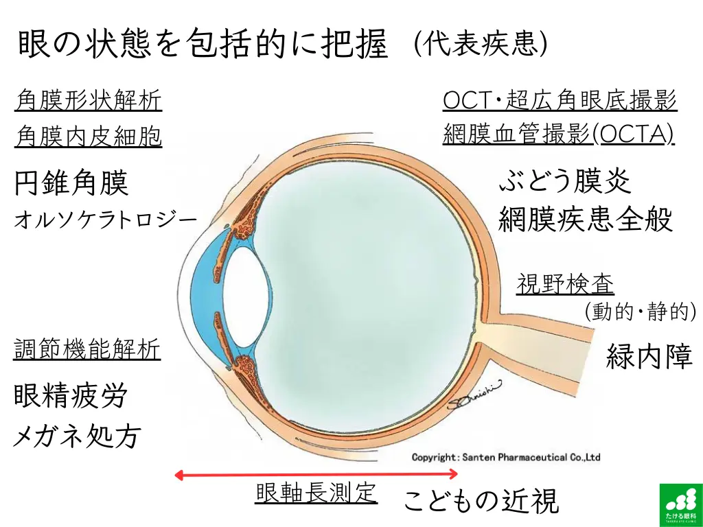
The Comprehensive Eye Exam offers comprehensive examination items that cover all eye diseases.
By combining visual acuity, intraocular pressure, visual field, and fundus examinations, optical coherence tomography (OCT), and more, we evaluate the condition of the eyes comprehensively.
For example, detailed testing of eye alignment is important for diagnosing smartphone esotropia.
For the fundus examination, we use Mirante®, an ultra-widefield fundus imaging device that does not require dilating the pupils.
Compared with a conventional fundus camera, it can photograph a wider area of the retina at once, and lesions in the periphery can be detected without being missed.
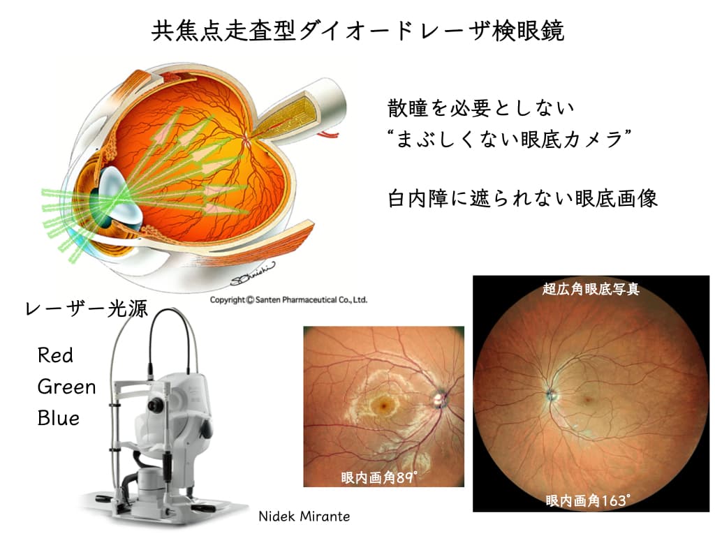
With the examinations performed by orthoptists, it is possible to find early signs.
Continued follow-up when an abnormality is found
If an abnormality is found in the examination, it can lead to early treatment under health insurance at a later date.
We have a cooperation system with core hospitals such as Kyushu University Hospital, so even when further workup or specialized treatment is needed, we can refer you to an appropriate medical institution.
Measures for Diseases with a High Risk of Blindness
According to research investigating the causes of blindness in Japan, the diseases with a high risk of blindness are as follows.
Again, this is cited from the paper by Dr. Morizane of the Department of Ophthalmology at Okayama University.
No. 1: Glaucoma (about 28.6%)
It is progressive, the visual field narrows gradually, and it is characterized by being hard to notice.
No. 2: Retinitis pigmentosa (about 14.0%)
A hereditary disease that causes night blindness, constriction of the visual field, and reduced visual acuity, and can lead to blindness as it progresses.
No. 3: Diabetic retinopathy (about 12.8%)
One of the three major complications of diabetes. As it progresses it can cause retinal hemorrhage, new blood vessels, and retinal detachment, raising the risk of blindness.
No. 4: Age-related macular degeneration (about 8.0%)
Common in older people, it impairs central vision. As it progresses it causes severe loss of vision or blindness.
Morizane, Y., Morimoto, N., Fujiwara, A. et al. Incidence and causes of visual impairment in Japan: the first nation-wide complete enumeration survey of newly certified visually impaired individuals. Jpn J Ophthalmol 63, 26–33 (2019). https://doi.org/10.1007/s10384-018-0623-4
For all of these diseases, early detection and early treatment are extremely important for preventing blindness.
Corneal Diseases and Dry Eye
For the cornea (the black part of the eye), it is important that the surface is smooth and transparent, that the cells on the inner side are sufficient in number, and that the shape is normal.
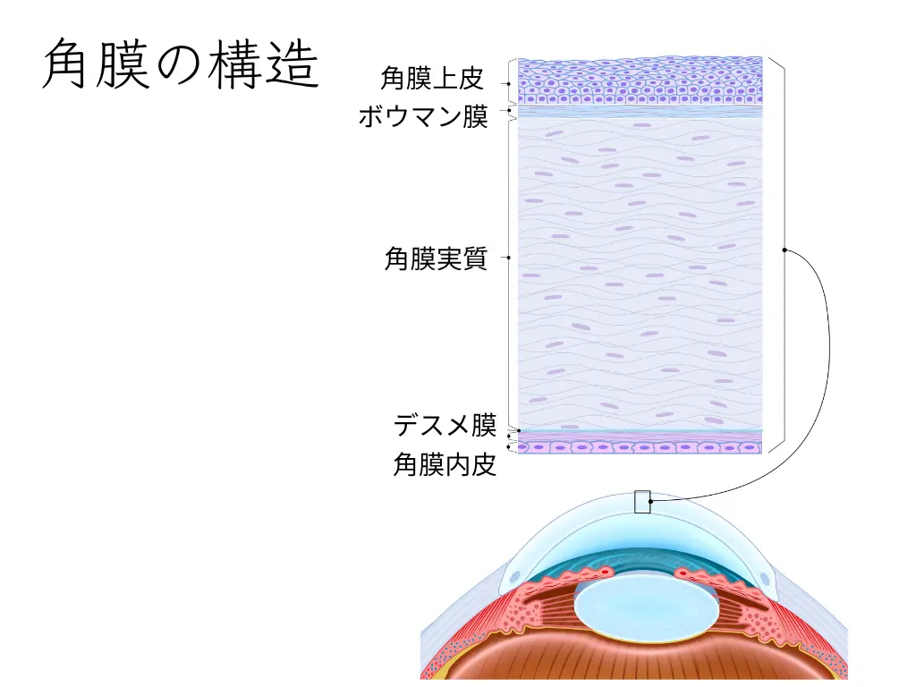
People who wear contact lenses, people with a history of refractive surgery, people who work on computers or smartphones for long hours, people in dry indoor environments, and people with habits such as smoking or insufficient blinking need regular checks.
Corneal diseases such as dry eye can be expected to improve with an appropriate diagnosis and treatment.
Appropriate Evaluation of Eye Strain

Causes of eye strain include working under inappropriate conditions, such as using glasses with the wrong prescription or not addressing presbyopia. In the Comprehensive Eye Exam, we investigate the causes of eye strain in detail and suggest appropriate ways to deal with it.
Summary

Eye diseases often progress even before symptoms appear, and by the time they are noticed, treatment may already be difficult.
In particular, glaucoma is the leading cause of blindness in Japan, and although about 1 in 20 people aged 40 and over develop it, it causes almost no subjective symptoms in the early stage.
With regular Comprehensive Eye Exams, diseases can be found before symptoms appear and appropriate treatment can be started, which can help protect future visual function.
In an era of 100-year lives, preventive testing plays an important role in maintaining good vision for as long as possible.
In the completely appointment-only Comprehensive Eye Exam, certified orthoptists carry out specialized examinations to assess the overall health of your eyes comprehensively.