![Takeru Eye Clinic | Takatori Shopping Street, Sawara-ku, Fukuoka City [Nishijin Station / Fujisaki Station]](https://takeru-eye.com/wp-content/uploads/2022/10/takeru_logo_for-WP-header.png)

“How long will treatment last?” “Can it be cured?”
These are the questions we are asked most often by patients who have been diagnosed with uveitis.
When the eye drops are reduced, the eye turns red again. When oral medicine is started, you worry about its effects on your body.
An unfamiliar term, “biologics,” comes up, and your anxiety grows.
In the middle of treatment, it often becomes hard to see which stage you are in now and what comes next.
This article lays out the whole picture of uveitis treatment, stage by stage.
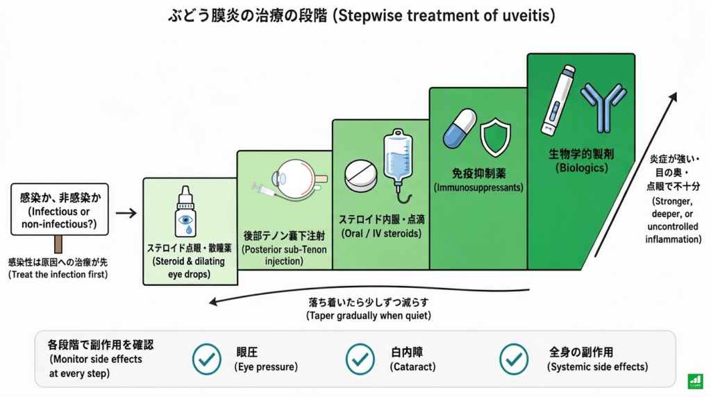
Uveitis treatment proceeds in the flow of “suppress the inflammation,” “prevent recurrence,” and “reduce the medicine”
Uveitis treatment starts by determining whether the cause is infection.
In non-infectious uveitis (uveitis not caused by infection), when the inflammation is in the front of the eye, steroid eye drops are the basis of treatment. When the inflammation is in the back of the eye, or cannot be controlled with eye drops, treatment is stepped up to injections around the eye, oral or intravenous steroids, immunosuppressants, and biologics.
The goals of treatment are to suppress inflammation and protect vision, and to keep the eye free of recurrence while gradually tapering the steroid.
The length of treatment varies greatly with the underlying disease: some people finish in a few weeks, while others continue for years.
What is uveitis?
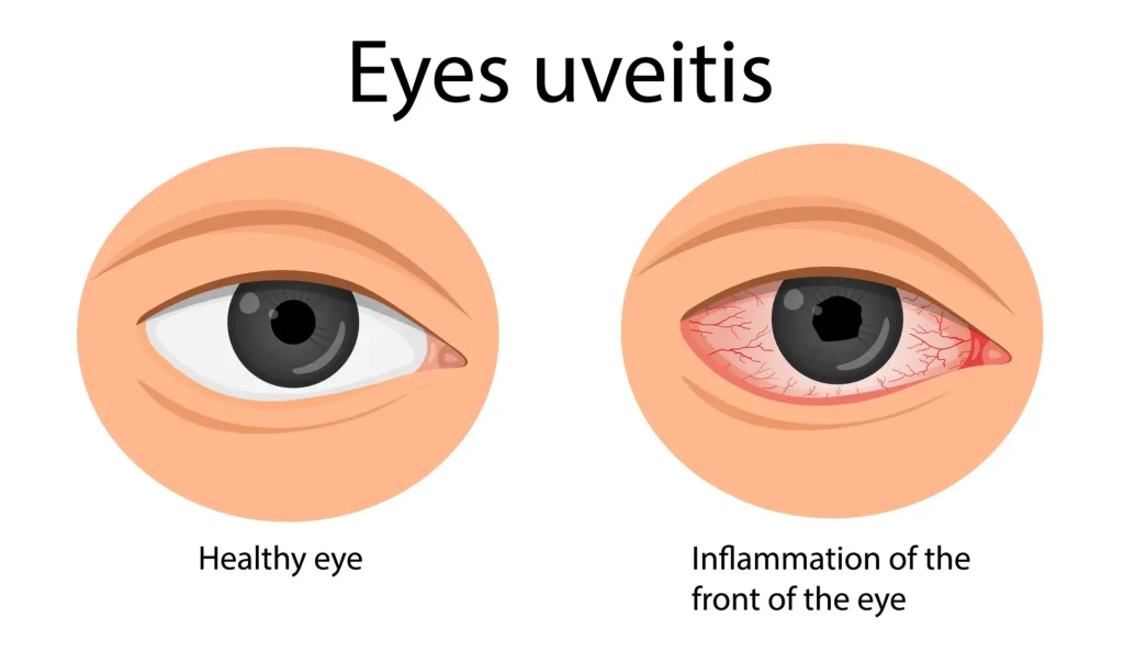
Uveitis is a disease in which inflammation occurs mainly in the “uvea” (iris, ciliary body, and choroid) inside the eye.
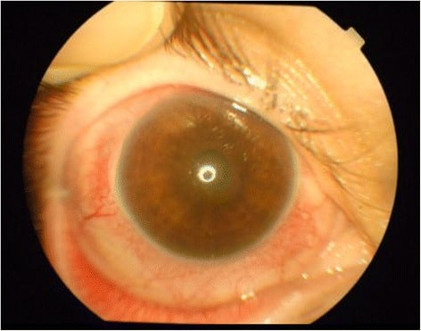
Source: Duman R, et al. Burns Trauma. 2015;3:19. Fig. 1(CC BY 4.0)
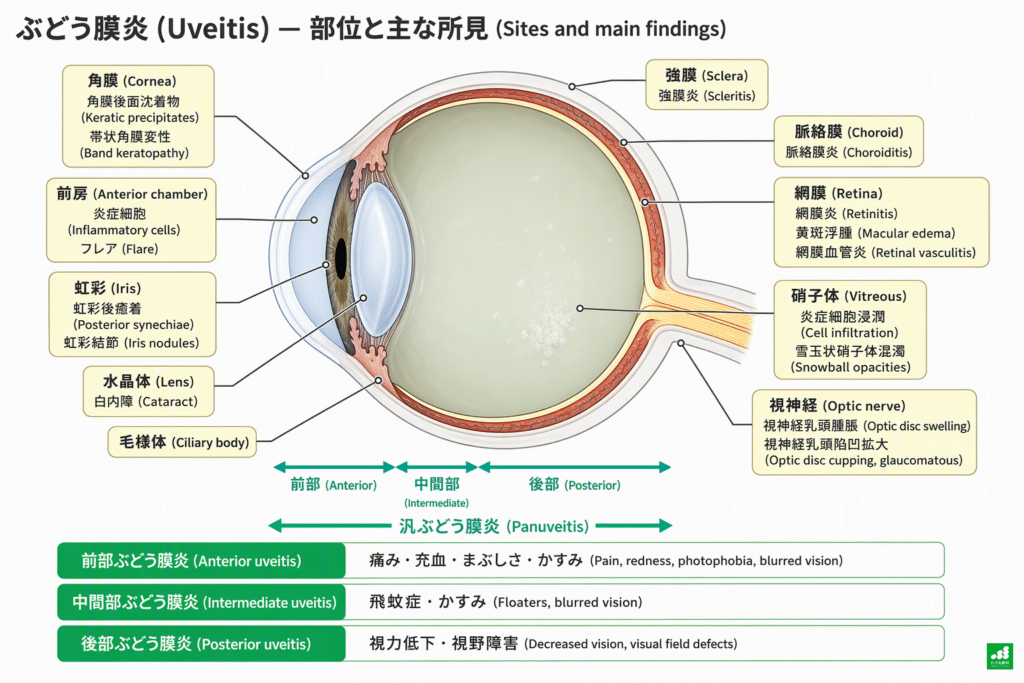
It is divided by where the inflammation mainly occurs into anterior (mainly the anterior chamber), intermediate (mainly the vitreous), and posterior (mainly the retina and choroid).
When inflammation is widespread across the anterior chamber, vitreous, retina and choroid and the main site cannot be narrowed to one, it is called panuveitis. The difference in location bears directly on which treatment is chosen.
A 2016 nationwide survey in Japan found that the causes, in order of frequency, were sarcoidosis, Vogt-Koyanagi-Harada disease (VKH; hereafter “Harada disease”), herpetic iritis, and acute anterior uveitis.
Behçet disease accounted for 4.2%, about the same as in the previous survey in 2009. In this survey, more than one in three people also had no identifiable cause1
The first thing to decide: is it caused by infection or not?
The first fork in uveitis treatment is whether the cause is infection (bacteria, viruses, fungi, parasites and so on).
For non-infectious uveitis, steroids that suppress inflammation are the mainstay. However, if steroids are used first in infectious uveitis, they weaken even the body’s ability to hold the pathogen in check, and the inflammation may suddenly worsen.
The guidelines of the Japanese Ocular Inflammation Society also state that a posterior sub-Tenon injection (described later) in infectious uveitis may cause rapid worsening. They therefore indicate treating first with antibacterial or antiviral drugs and the like, and adding a steroid after checking the response2
For this reason, at the first visit, in addition to the findings in the eye, we check blood tests and other examinations for infections such as syphilis and tuberculosis.
When an infection inside the eye caused by a herpes virus or similar is suspected, we take a very small amount of aqueous humor (the fluid behind the cornea) under topical anesthesia and examine it for viral genes by PCR2. Blood tests alone may not tell us whether a herpes virus is the cause of inflammation inside the eye.
A test developed mainly by Dr. Satoko Nakano and colleagues at Oita University can examine nine pathogens at once from a very small amount of aqueous humor or vitreous: members of the herpes virus family (herpes simplex, varicella-zoster, cytomegalovirus and others), HTLV-1, Treponema pallidum (syphilis), and Toxoplasma3. In a study using 475 specimens from 29 facilities across Japan, the aqueous humor results agreed 98.5% with conventional PCR testing3
The test kit that examines these nine pathogens received national approval in October 2025 and was launched in July 2026. According to an announcement by Oita University on July 28, 2026, efforts toward insurance coverage are under way.4
In Kyushu, including Fukuoka, uveitis related to the virus HTLV-1 is also known to be more common than in the rest of Japan.
Treatment by stage
From here, we explain the stages of treatment in order, focusing on non-infectious uveitis.
Not every patient moves through steps 1 to 5 in order. Mild anterior inflammation often settles with eye drops alone, and some diseases, such as Harada disease, need oral medicine or intravenous therapy from the start.
| Step | Target (main examples) | Purpose | Main side effects | Approximate duration |
|---|---|---|---|---|
| 1. Steroid eye drops and mydriatics | Anterior uveitis | Suppress inflammation in the anterior chamber and prevent adhesions of the iris | Raised eye pressure, cataract | Tapered over several weeks to months. Restarted if inflammation recurs |
| 2. Subconjunctival and posterior sub-Tenon injections | Strong anterior inflammation; intermediate or posterior inflammation; macular edema | Deliver the drug to places eye drops do not reach easily | Raised eye pressure, cataract, bleeding at the injection site | Effect lasts several weeks to months. Repeated as needed |
| 3. Oral or intravenous steroids | Strong bilateral or posterior inflammation (such as Harada disease) | Suppress inflammation in the back of the eye through the whole body | Osteoporosis and fractures, infections, stomach ulcer, worsening of diabetes and blood pressure, weight gain and moon face, mood changes | Tapered over several months |
| 4. Immunosuppressants | Non-infectious uveitis in which the steroid cannot be reduced or cannot be continued because of side effects | Reduce the steroid dose | Kidney and liver dysfunction, infections, long-term risk of malignancy | Often years |
| 5. Biologics | Non-infectious uveitis not controlled enough by existing treatment (such as Behçet disease) | Prevent attacks of inflammation and reduce the steroid | Infections such as tuberculosis and pneumonia; reactions to the injection or infusion | Years. Stopping is decided carefully |
* Durations are general guides and vary by individual depending on the underlying disease and the degree of inflammation. Targets and side effects are based on the Japanese Ocular Inflammation Society Uveitis Practice Guidelines (2019)2 and the package inserts of each drug.
1. Steroid eye drops and mydriatics
The basis of treatment for anterior uveitis is steroid eye drops.
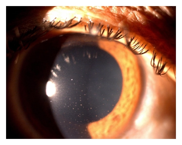
Source: Gogri PY, et al. Int Sch Res Notices. 2014;2014:347638. Figure(CC BY)
The guidelines start with 0.1% betamethasone (Rinderon®) eye drops. As the reduction of inflammatory cells is confirmed, the frequency of instillation is reduced and treatment is switched to a weaker steroid such as 0.1% fluorometholone (Flumetholon®)2
Mydriatics (drugs that dilate the pupil; Mydrin®-P and others) are often used at the same time.
When there is inflammation, the iris tends to stick to the lens behind it, causing “posterior synechiae.” Mydriatics are used to prevent these adhesions and to relax the ciliary muscle and ease pain.
In a study of Japanese insurance claims data, more than 60% of patients with non-infectious uveitis were treated at clinics rather than hospitals. Most prescriptions were eye drops5
Much uveitis treatment starts with eye drops at an ophthalmologist close to home.
The side effects of eye drops to watch for are a rise in eye pressure and the resulting glaucoma (steroid-induced glaucoma), and cataract.
In a study of children with uveitis associated with juvenile idiopathic arthritis (JIA), it was reported that when steroid eye drops were used more than three times a day, cataract occurred more often than when they were used three times a day or less6. It is important to suppress the inflammation while reducing the frequency to as low as possible.
2. Injections around the eye (subconjunctival and posterior sub-Tenon injections)
Eye drops reach the front of the eye, such as the cornea and anterior chamber, but they hardly reach the vitreous, retina, or choroid.
For strong anterior inflammation with fibrin deposition or hypopyon (pus in the anterior chamber), a “subconjunctival injection,” given under the membrane on the surface of the white of the eye, is used2. Our clinic offers subconjunctival injection of dexamethasone (Decadron®).
For intermediate or posterior inflammation that affects vision, such as vitreous haze or “macular edema,” swelling at the center of the retina, we perform a “posterior sub-Tenon injection”2. It is also called “STTA,” from the initials of the English “posterior sub-Tenon triamcinolone acetonide injection.”
The eyeball is wrapped by the conjunctiva on the surface of the white of the eye and, beneath it, a membrane called Tenon’s capsule. For a posterior sub-Tenon injection, after numbing the eye with drops, a small opening is made in the surface of the white of the eye, a thin tube with a rounded tip (a blunt cannula) is advanced along the wall of the eyeball toward the back, and the drug is placed behind the eyeball7
It is not an injection that puts a needle into the eye. The drug stays outside the eyeball, close to the retina and choroid, and is thought to act from there on inflammation deep in the eye.
In Japan, sub-Tenon administration of triamcinolone (MaQaid®) is approved to reduce macular edema associated with non-infectious uveitis. The label specifies an interval of at least 3 months before it is given again8
In a Japanese study of 51 eyes with uveitis of various causes, administration with a blunt cannula improved vitreous haze and macular edema, while cataract progression was seen in 31% of eyes and raised eye pressure in 27%7
In a randomized controlled trial of macular edema in uveitis (the POINT trial), macular thickness (swelling) at 8 weeks was reported to fall by 23% with periocular triamcinolone injection, 39% with intravitreal triamcinolone injection, and 46% with a dexamethasone sustained-release implant9
Antibiotic eye drops are used before and after administration. Bleeding, redness, or swelling of the white of the eye and eye pain may occur after administration8
The side effects that need attention are raised eye pressure and cataract. Cataract can appear more than six months after administration, and becomes more likely the more often injections are repeated2,8
At our clinic as well, we perform posterior sub-Tenon injections by making a small incision in the white of the eye and inserting a blunt cannula.
3. Oral or intravenous steroids
For strong inflammation in both eyes, or inflammation that reaches the retina, choroid, or optic nerve, steroids are given to the whole body by mouth or by infusion.
However, starting systemic steroids first can make it harder to interpret the tests that look for the cause. For this reason, before starting oral or intravenous treatment, our clinic places importance on having the tests for the cause done at the Uveitis Clinic of Kyushu University Hospital.
In the guidelines, the starting dose is 0.5 mg or more per kg of body weight per day, converted to oral prednisolone (Predonine®).
The drug is reduced only after confirming that inflammation has settled sufficiently. In severe cases, there is also a method of giving a large dose of methylprednisolone (Solu-Medrol®) by infusion2
If inflammation flares up during the taper, the dose is raised a little and then reduced more slowly than before2. “Not reducing it all at once” is important for preventing recurrence.
The systemic side effects that call for particular attention are osteoporosis and fractures.
Infections, stomach ulcer, worsening of diabetes and high blood pressure, weight gain, and a rounder face (moon face) can also occur.
Attention is also needed for mood changes such as insomnia, elation, or low mood, effects on children’s growth, and decreased adrenal function after long use2. When used for a long time, we discuss bone density measurement and the use of bone-protecting drugs with the internal medicine physician.
Stopping oral steroids suddenly on your own can lead to a flare of inflammation or a worse physical condition. It is important to decide how to reduce them in consultation with your doctor.
4. Immunosuppressants
When inflammation recurs if the steroid is reduced below a certain dose, or when the steroid cannot be continued because of side effects, immunosuppressants are considered. The purpose is to reduce the steroid dose (to “take over” part of the steroid’s role)2
In Japan, the drug covered by insurance for uveitis is cyclosporine (Neoral®). It is indicated for Behçet disease (with eye symptoms) and for active intermediate or posterior non-infectious uveitis in which existing treatment is not effective enough and there is a risk of vision loss10
Methotrexate and mycophenolate mofetil are also widely used overseas, but they are not covered by insurance for uveitis in Japan (as of the 2019 edition of the guidelines)2
In a randomized controlled trial comparing the two drugs (the FAST trial), the proportion with controlled inflammation after reducing the steroid to a low dose (treatment success) at 6 months was 66.7% with methotrexate and 57.1% with mycophenolate mofetil, with no statistically significant difference. When limited to posterior uveitis and panuveitis, the proportion was higher with methotrexate11
Immunosuppressants carry a risk of kidney and liver dysfunction, infections, and, over the long term, malignancy, and need regular checks such as blood tests2
5. Biologics (biologic drugs)
Biologics are drugs that target a specific molecule that causes inflammation and block its action. In uveitis, two drugs that suppress the molecule TNF-α (tumor necrosis factor) are used in Japan.
Adalimumab (Humira®) is a subcutaneous injection used for non-infectious intermediate, posterior, and panuveitis that is not controlled enough by existing treatment.
In the VISUAL I trial, which enrolled patients with active inflammation, the median time to treatment failure, such as from worsening inflammation, was 24 weeks in the adalimumab group and 13 weeks in the placebo group12. In the VISUAL II trial, which enrolled patients whose inflammation was controlled with steroids, the proportion of treatment failure, such as from flare-up, was 39% in the adalimumab group and 55% in the placebo group13
The domestic dosing is a subcutaneous injection of 80 mg for the first dose, 40 mg one week later, and then 40 mg every two weeks14. It was approved in Japan in September 2016 and became the first biologic usable for non-infectious uveitis regardless of the underlying disease.15
Infliximab (Remicade®) is an intravenous infusion used for refractory retinouveitis due to Behçet disease. It is given at the first dose, 2 weeks, and 6 weeks later, and then every 8 weeks16
In Japan, it became covered by insurance in 2007, ahead of the rest of the world17
In a Japanese multicenter study, the frequency of ocular attacks decreased after starting treatment, while about 60% of patients had a relapse. However, it was reported that most relapses settled with an increased dose of eye drops or a shorter interval between infusions18
| Trial | Target | Comparison | Main results |
|---|---|---|---|
| VISUAL I (NEJM 2016)12 | Active non-infectious intermediate, posterior, and panuveitis, 217 patients | Adalimumab vs. placebo | Median time to treatment failure 24 weeks vs. 13 weeks (HR 0.50) |
| VISUAL II (Lancet 2016)13 | Non-infectious uveitis controlled with steroids, 226 patients | Adalimumab vs. placebo | Treatment failure 39% vs. 55% (HR 0.57) |
| FAST (JAMA 2019)11 | Non-infectious uveitis, 216 patients | Methotrexate vs. mycophenolate mofetil | Treatment success at 6 months (including steroid reduction) 66.7% vs. 57.1% (difference not significant) |
| POINT (Ophthalmology 2019)9 | Macular edema in uveitis | Periocular triamcinolone vs. intravitreal triamcinolone and dexamethasone sustained-release implant | Reduction in retinal thickness at 8 weeks 23% vs. 39% and 46% |
* Because the participants and conditions differ between trials, the treatments cannot simply be compared with one another. HR: hazard ratio (the smaller it is below 1, the less likely treatment failure is).
What to check before starting a biologic
Drugs that suppress TNF-α also weaken the body’s ability to hold infections such as tuberculosis in check.
For this reason, before starting, we check for tuberculosis (history, chest X-ray, blood test [IGRA], and chest CT if needed) and for infection with hepatitis B and C viruses. If a past tuberculosis infection is suspected, depending on the test results, drugs for tuberculosis (preventive treatment) are started first, and the timing of starting the biologic is decided2,14
The package insert of adalimumab clearly states that, when it is used for uveitis, it should be used in close cooperation with physicians in internal medicine and other specialties14. Treatment with a biologic is not something that can be completed by the ophthalmologist alone.
A biologic is not a drug that eliminates the disease itself, but a drug that keeps the eye in a state where inflammation is less likely to occur.
Treatments that target cytokines: what research has shown and what is still unknown
I myself previously did basic research on cytokines involved in uveitis at Kyushu University. Cytokines are small proteins that act as messengers between immune cells.
In our basic research, we reported that IL-6 increases Th17 cells, the cells that cause inflammation, and that blocking the IL-6 receptor with an antibody reduces inflammation19. We also reported that without IL-17 the onset of inflammation is unchanged and only the later phase is milder.20
However, the results of basic research cannot be applied to people as they are.
In human uveitis, the drugs whose effect has been confirmed in randomized controlled trials and that are approved in Japan are the drugs that suppress TNF-α.
On the other hand, drugs that suppress IL-17A did not produce the expected results in uveitis trials.
For IL-6, the anti-IL-6 antibody vamikibart was submitted for approval in Japan in August 2026 for macular edema associated with non-infectious uveitis21. Vamikibart is a drug injected directly into the eye.
The application is based on the results of phase III trials21. As of September 2026, it has not yet been approved in Japan.
Side effects of each, and what we check at our clinic
Steroids and drugs that suppress the immune system have side effects on both the eyes and the whole body. If you are worried about side effects, please do not hesitate to talk to your doctor; finding side effects early and adjusting treatment is important.
| What we check | Why | Main method |
|---|---|---|
| Eye pressure | It can rise with steroid eye drops or injections | Measured at every visit |
| Lens (cataract) | It can progress with both steroids and inflammation | Slit-lamp microscopy |
| Macular swelling | The main cause of reduced vision | OCT (optical coherence tomography) |
| Inflammation of the retina and choroid, vasculitis | To look at findings in the back of the eye and changes in blood flow | Fundus examination, ultra-widefield fundus imaging, OCTA |
| Systemic side effects (blood sugar, blood pressure, bone, infection) | Can occur with oral drugs, immunosuppressants, and biologics | Coordination with internal medicine and your family doctor |
Some people are prone to a rise in eye pressure with steroids (steroid responders). Even when eye pressure rises, there are almost no symptoms, so it is a change that is hard to notice without measuring.
How long treatment lasts, how the dose is reduced, and recurrence
The answer to “How long will uveitis treatment continue?” depends on the underlying disease and the type of inflammation.
By an international standard, inflammation is classified as “chronic” when it persists and worsens again less than 3 months after stopping treatment, and as “recurrent” when attacks repeat with periods of 3 months or more of quiescence without treatment2. Chronic cases tend to need long-term treatment, while in recurrent cases, treatment is given at each attack and the drugs are reduced again once things settle.
The dose is reduced gradually while confirming that there is no inflammation. A common step is to reduce the eye drops to once a day before stopping.
There is not yet an answer common to all patients on when and how to stop a biologic22
In a trial of children with uveitis associated with juvenile idiopathic arthritis (JIA) who had been quiet on adalimumab for at least one year (the ADJUST trial), uveitis or arthritis flared in 14% of the group that continued adalimumab, compared with 68% of the group that stopped it23
Results from studies in children do not necessarily apply to adults as they are. However, the ADJUST results show that “being quiet does not necessarily mean the drug is no longer needed.”
When a recurrence occurs, what matters is to notice it early and step up treatment early. “Redness,” “glare,” “blurring,” or “more floaters” can be signs of a recurrence.
What we do at Takeru Eye Clinic (Takatori Shopping Street, Sawara-ku, Fukuoka City)
Takeru Eye Clinic has continued to care for uveitis and ocular inflammation since opening in 2018.
At every visit, we check visual acuity, eye pressure, and inflammation in the anterior chamber and vitreous, and, as needed, evaluate inflammation in the back of the eye and macular swelling with OCT, ultra-widefield fundus imaging, and OCTA.
As a clinic with a single physician, shared care with Kyushu University Hospital is at the center of our practice.
We handle adjustment of eye drop frequency, subconjunctival dexamethasone injection, posterior sub-Tenon injection, eye pressure management, and early response to recurrence at our clinic.
Detailed testing for the cause, systemic steroid treatment, immunosuppressants, and the start and continuation of biologics are done at the Uveitis Clinic of Kyushu University Hospital. We ask patients to have the tests for the cause done at Kyushu University Hospital before systemic steroids are started because, if the drug is started first, it can become harder to interpret the test results.
Our clinic and Kyushu University Hospital are connected by a single subway line, so it is easy to attend both.
As a guide, visits are every 1 to 2 weeks while inflammation is strong, and every 1 to 3 months once it has settled.
What we want to tell patients
In uveitis treatment, when inflammation is strong, treatment is stepped up as if climbing stairs, and when it settles, it is stepped down gradually as if coming down the stairs.
Which step you are on now, and what the next goal is. What to do if inflammation returns during the taper.
We believe that sharing the outlook for treatment as we go is a support for continuing a long course of treatment.
Please do not stop your eye drops or oral medicines on your own, and if you have symptoms or concerns about side effects, please talk to us early.
If you have any concerns, please speak up at your visit.
Revision history
First published: September 23, 2026
References
- Sonoda KH, Hasegawa E, Namba K, Okada AA, Ohguro N, Goto H; JOIS (Japanese Ocular Inflammation Society) Uveitis Survey Working Group. Epidemiology of uveitis in Japan: a 2016 retrospective nationwide survey. Jpn J Ophthalmol. 2021;65(2):184-190. https://pubmed.ncbi.nlm.nih.gov/33694024/
- Japanese Ocular Inflammation Society, Uveitis Practice Guidelines Committee. Uveitis Practice Guidelines. Journal of the Japanese Ophthalmological Society. 2019;123(6):635-696. (In Japanese) https://www.nichigan.or.jp/Portals/0/resources/member/guideline/uveitis_guideline.pdf
- Nakano S, Sugita S, Takase H, et al. Clinical performance of the multiplex solid-phase “Direct Strip PCR” for infectious uveitis: a multicenter diagnostic accuracy study. Jpn J Ophthalmol. 2026;70(4):966-982. https://pubmed.ncbi.nlm.nih.gov/41926010/
- Oita University. Testing for nine of the main pathogens of infectious uveitis at once by PCR: launch of the in vitro diagnostic “Ampdirect Infectious Uveitis Pathogen Detection Kit” (press release). July 28, 2026. (In Japanese) Accessed September 23, 2026. https://www.oita-u.ac.jp/000071601.pdf
- Umazume A, Ohguro N, Okada AA, et al. Prevalence and incidence rates and treatment patterns of non-infectious uveitis in Japan: real-world data using a claims database. Jpn J Ophthalmol. 2021;65(5):657-665. https://pubmed.ncbi.nlm.nih.gov/34181111/
- Thorne JE, Woreta FA, Dunn JP, Jabs DA. Risk of cataract development among children with juvenile idiopathic arthritis-related uveitis treated with topical corticosteroids. Ophthalmology. 2010;117(7):1436-1441. https://pubmed.ncbi.nlm.nih.gov/20363502/
- Okada AA, Wakabayashi T, Morimura Y, et al. Trans-Tenon’s retrobulbar triamcinolone infusion for the treatment of uveitis. Br J Ophthalmol. 2003;87(8):968-971. https://pubmed.ncbi.nlm.nih.gov/12881336/
- Wakamoto Pharmaceutical. MaQaid Intraocular Injection 40 mg, package insert. KEGG MEDICUS, Prescription Drug Information. (In Japanese) https://www.kegg.jp/medicus-bin/japic_med?japic_code=00066924
- Thorne JE, Sugar EA, Holbrook JT, et al. Periocular triamcinolone vs. intravitreal triamcinolone vs. intravitreal dexamethasone implant for the treatment of uveitic macular edema: the PeriOcular vs. INTravitreal corticosteroids for uveitic macular edema (POINT) trial. Ophthalmology. 2019;126(2):283-295. https://pubmed.ncbi.nlm.nih.gov/30269924/
- Novartis Pharma. Neoral, package insert. KEGG MEDICUS, Prescription Drug Information. (In Japanese) https://www.kegg.jp/medicus-bin/japic_med?japic_code=00057440
- Rathinam SR, Gonzales JA, Thundikandy R, et al. Effect of corticosteroid-sparing treatment with mycophenolate mofetil vs methotrexate on inflammation in patients with uveitis: a randomized clinical trial. JAMA. 2019;322(10):936-945. https://pubmed.ncbi.nlm.nih.gov/31503307/
- Jaffe GJ, Dick AD, Brézin AP, et al. Adalimumab in patients with active noninfectious uveitis. N Engl J Med. 2016;375(10):932-943. https://pubmed.ncbi.nlm.nih.gov/27602665/
- Nguyen QD, Merrill PT, Jaffe GJ, et al. Adalimumab for prevention of uveitic flare in patients with inactive non-infectious uveitis controlled by corticosteroids (VISUAL II): a multicentre, double-masked, randomised, placebo-controlled phase 3 trial. Lancet. 2016;388(10050):1183-1192. https://pubmed.ncbi.nlm.nih.gov/27542302/
- AbbVie. Humira Subcutaneous Injection, package insert. KEGG MEDICUS, Prescription Drug Information. (In Japanese) https://www.kegg.jp/medicus-bin/japic_med?japic_code=00068024
- AbbVie GK, Eisai Co., Ltd. Approval obtained for the indication of non-infectious uveitis for the fully human anti-human TNFα monoclonal antibody Humira®. September 28, 2016. (In Japanese) https://www.eisai.co.jp/news/news201667.html
- Tanabe Pharma. Remicade for Intravenous Infusion 100, package insert. KEGG MEDICUS, Prescription Drug Information. (In Japanese) https://www.kegg.jp/medicus-bin/japic_med?japic_code=00048579
- Research Group on Behçet Disease. New treatments (information for patients). (In Japanese) https://www.nms-behcet.jp/patient/behcet/newremedy.html
- Takeuchi M, Kezuka T, Sugita S, et al. Evaluation of the long-term efficacy and safety of infliximab treatment for uveitis in Behçet’s disease: a multicenter study. Ophthalmology. 2014;121(10):1877-1884. https://pubmed.ncbi.nlm.nih.gov/24950593/
- Yoshimura T, Sonoda KH, Ohguro N, et al. Involvement of Th17 cells and the effect of anti-IL-6 therapy in autoimmune uveitis. Rheumatology (Oxford). 2009;48(4):347-354. https://pubmed.ncbi.nlm.nih.gov/19164426/
- Yoshimura T, Sonoda KH, Miyazaki Y, et al. Differential roles for IFN-gamma and IL-17 in experimental autoimmune uveoretinitis. Int Immunol. 2008;20(2):209-214. https://pubmed.ncbi.nlm.nih.gov/18156624/
- Chugai Pharmaceutical. Application for manufacturing and marketing approval in Japan of vamikibart for macular edema associated with non-infectious uveitis. August 25, 2026. (In Japanese) https://www.chugai-pharm.co.jp/news/detail/20260825153000_1610.html
- Leclercq M, Desbois AC, Domont F, et al. Biotherapies in uveitis. J Clin Med. 2020;9(11):3599. https://pubmed.ncbi.nlm.nih.gov/33171664/
- Acharya NR, Ramanan AV, Coyne AB, et al. Stopping of adalimumab in juvenile idiopathic arthritis-associated uveitis (ADJUST): a multicentre, double-masked, randomised controlled trial. Lancet. 2025;405(10475):303-313. https://pubmed.ncbi.nlm.nih.gov/39863370/