![Takeru Eye Clinic | Takatori Shopping Street, Sawara-ku, Fukuoka City [Nishijin Station / Fujisaki Station]](https://takeru-eye.com/wp-content/uploads/2022/10/takeru_logo_for-WP-header.png)

In my practice treating uveitis, I have noticed that certain groups of patients are increasing.
More and more people come to the eye clinic with complaints such as “my eyes are red,” “my vision is blurry,” or “things look distorted” while being treated for rheumatoid arthritis, psoriasis, or ankylosing spondylitis, or while receiving cancer immunotherapy.
When I check their medications, I often find the name of a biologic or a molecular targeted drug.
I once did basic research on cytokines at a Kyushu University immunology laboratory (the Medical Institute of Bioregulation, Kyushu University).
IL-6, TNF-α, IL-17A: for researchers, it was a perfectly natural idea that controlling these inflammatory molecules should make it possible to treat disease.
I felt that we had entered an era in which the very molecules we handled in the laboratory were appearing, one after another, as real drugs.
📌 What are cytokines?
Cytokines are small proteins secreted by immune cells that act as messengers between cells.
They act like a command center that regulates the body’s immune response, triggering or suppressing inflammation and calling immune cells to where they are needed.
IL-6, TNF-α, and IL-17A are typical examples, and when they act excessively, autoimmune diseases such as rheumatoid arthritis and psoriasis develop.
Biologics target these cytokines precisely.
In daily practice, however, I have encountered some unexpected realities.
Inflammation develops in the eye even though TNF-α has been blocked. Inflammation persists even though IL-17A has been suppressed. When the brakes on the immune system are released, it begins to attack the patient’s own eye!
What happens in the eyes of these patients cannot always be explained by the simple word “side effect.”
This is because the eye has a special immune system of its own.
How do biologics and immunotherapy drugs rewrite that system?
And why does infectious uveitis arise not as a “chance occurrence” but as a “breakdown of containment”?
In this article, I will lay out the thinking behind these questions.

The Eye Is an “Immune Privileged Site”
From an immunological standpoint, the eye is an extremely special organ.
In experiments conducted by Sir Peter Medawar in the 1940s, tissue transplanted into the eye (the anterior chamber) was observed to survive longer than tissue transplanted elsewhere in the body¹.
This was the starting point of the concept of “ocular immune privilege“.
The reason the eye is special is to protect vision.
Inflammation in the eye is a special case: it is important that it heals while preserving transparency as much as possible.
The retina does not regenerate once it is damaged.
If eye tissue heals with scarring, vision is affected for life.
For this reason, the eye has a special suppression system so that inflammation does not damage the eye itself.
A physical barrier called the blood-retinal barrier (BRB) prevents immune cells and pathogens in the blood from entering the eye.
In addition, the fluid inside the eye (aqueous humor and vitreous) contains substances that suppress inflammation, and even if immune cells do enter, these substances work to dampen their activity².
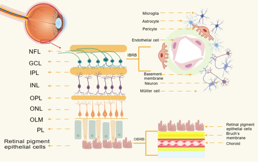
The blood-retinal barrier consists of two layers, an inner barrier (iBRB) and an outer barrier (oBRB). The iBRB is formed by the neurovascular unit, made up of retinal vascular endothelial cells, pericytes, microglia, astrocytes, and Müller cells, while the outer barrier (oBRB) consists of retinal pigment epithelial cells, Bruch’s membrane, and the choriocapillaris. These two barriers work together to maintain retinal homeostasis, and the breakdown of either one contributes to many retinal diseases, including diabetic retinopathy.
Schematic diagram of the blood-retinal barrier (BRB) structure. The inner BRB (iBRB) comprises retinal vascular endothelial cells, pericytes, microglia, astrocytes, and Müller cells forming the neurovascular unit. The outer BRB (oBRB) consists of retinal pigment epithelial cells, Bruch’s membrane, and the choriocapillaris. Disruption of either barrier underlies the pathogenesis of diabetic retinopathy and other retinal diseases.
Li M et al. Front Endocrinol. 2025;16:1617797. Adapted under CC BY 4.0. https://www.frontiersin.org/journals/endocrinology/articles/10.3389/fendo.2025.1617797/full
In addition, when an antigen enters the eye, a systemic signal is generated via the spleen telling the body not to overreact to that antigen. This is called anterior chamber-associated immune deviation (ACAID)³.
In modern immunology, ocular immune privilege is understood not as mere “isolation” but as an “active immune regulatory system”⁴.
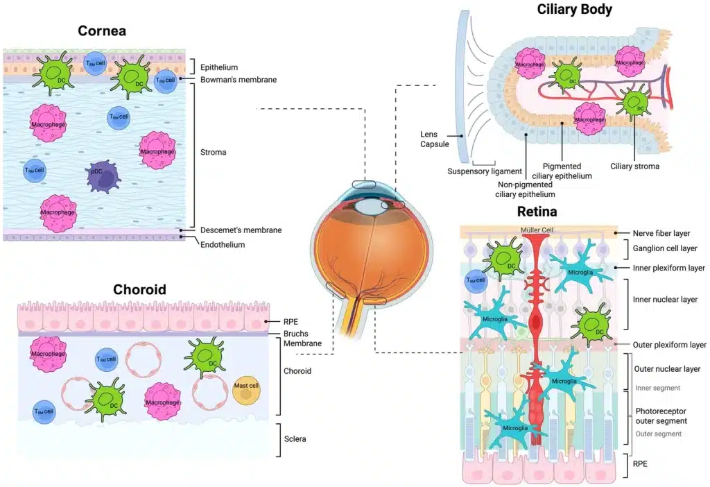
The eye is an immune-privileged organ, yet tissue-resident immune cells (such as tissue-resident memory T cells, dendritic cells, and plasmacytoid dendritic cells) are present in the cornea, ciliary body, choroid, and retina, where they maintain homeostasis and regulate inflammatory responses. A disturbance in the balance of these resident immune cells is thought to contribute to the onset of uveitis.
Figure showing tissue-resident immune populations present in healthy normal eyes across distinct ocular microenvironments, including tissue-resident memory T cells (T_RM), dendritic cells (DCs), and plasmacytoid dendritic cells (pDCs).
Han et al. Front Med. 2026;13:1717056. Adapted under CC BY 4.0. https://www.frontiersin.org/journals/medicine/articles/10.3389/fmed.2026.1717056/full
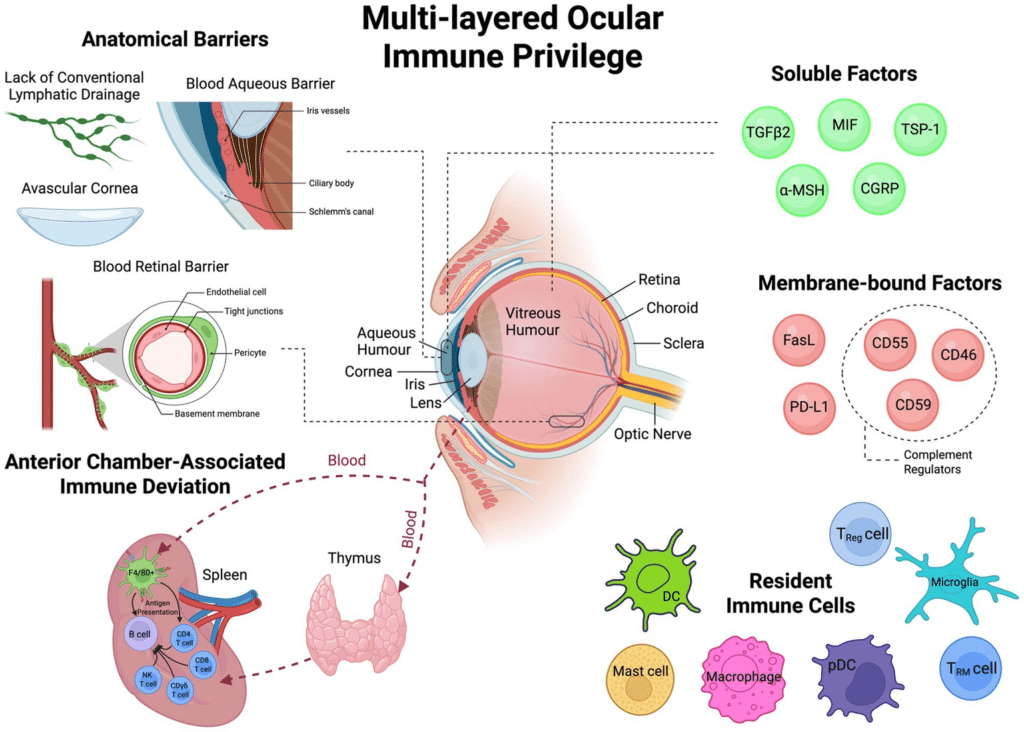
Several defense mechanisms overlap to make the eye an “immune privileged site.” Intraocular inflammation is tightly controlled by a combination of anatomical barriers (the avascular cornea, the blood-aqueous barrier, the blood-retinal barrier, and the absence of lymphatic vessels), immunosuppressive soluble factors (TGFβ2, MIF, TSP-1, α-MSH, CGRP, and others), membrane-bound factors (FasL, PD-L1, and complement regulatory proteins), and the induction of systemic immune tolerance through anterior chamber-associated immune deviation (ACAID). Resident immune cells (dendritic cells, macrophages, microglia, regulatory T cells, and others) also play an important role in maintaining this homeostasis.
Schematic diagram illustrating the multi-layered mechanisms of ocular immune privilege, including anatomical barriers, soluble immunosuppressive factors, membrane-bound regulators, anterior chamber-associated immune deviation (ACAID), and resident immune cells.
Han et al. Front Med. 2026;13:1717056. Adapted under CC BY 4.0. https://www.frontiersin.org/journals/medicine/articles/10.3389/fmed.2026.1717056/full
What are biologics?
Biologics are drugs that target specific molecules directly involved in immune responses, such as cytokines and cell-surface receptors.
They are used for a wide range of conditions, including rheumatoid arthritis, psoriasis, ankylosing spondylitis, Crohn’s disease, and cancer treatment.
The main classes of biologics are summarized below.
Anti-TNF-α agents (TNF inhibitors) have the longest history; typical examples are infliximab, adalimumab, and etanercept.
IL-17 inhibitors (secukinumab, ixekizumab, and others) are used especially for psoriasis and ankylosing spondylitis.
JAK inhibitors (upadacitinib, tofacitinib, and others) are oral drugs that block the signaling of multiple cytokines at once.
And immune checkpoint inhibitors (pembrolizumab, nivolumab, ipilimumab, and others) are used as cancer immunotherapy.
These drugs control systemic disease by suppressing “excessive reactions” of the immune system. At the same time, however, they can upset the immune balance that the eye maintains on its own.
At which stage biologics are used in the treatment of uveitis is explained in an article that summarizes the whole picture of treatment.

Why do biologics cause uveitis?
The mechanism by which each class of drug affects the eye is different. Let us look at them in turn.
Anti-TNF-α agents: paradoxical inflammation can occur
Anti-TNF agents suppress inflammation in the joints and skin, but paradoxical side effects in which they instead trigger inflammation in the eye have been reported.
This is called a “paradoxical reaction”⁵.
Etanercept has attracted particular attention.
Several studies have shown that, even among anti-TNF agents, etanercept carries a higher risk of new-onset or recurrent uveitis than the monoclonal antibody types (infliximab and adalimumab)⁶.
A network meta-analysis of patients with ankylosing spondylitis reported that the risk of uveitis with etanercept was more than twice that with golimumab or infliximab⁷.
Why does this difference arise?
TNF-α is an inflammatory cytokine, but it also has the role of suppressing the production of an inflammatory signal (interferon-α) by a type of immune cell called plasmacytoid dendritic cells (pDCs).
Etanercept binds only soluble TNF and has a weak effect on inducing T-cell apoptosis, so TNF blockade is thought to increase interferon production instead and to make granulomatous inflammation more likely.
Monoclonal antibody types suppress this shift more strongly, so the risk of uveitis is considered relatively lower⁸.
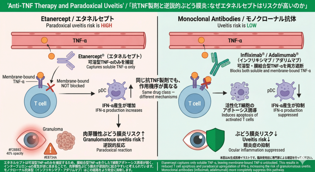
Even among anti-TNF agents, the effect on the eye differs depending on the mechanism of action. (Left) Etanercept captures only soluble TNF-α and is weak at inducing T-cell apoptosis via membrane-bound TNF-α, so IFN-α production by plasmacytoid dendritic cells (pDCs) increases instead, and granulomatous uveitis is thought to occur paradoxically. (Right) Monoclonal antibodies such as infliximab and adalimumab block both soluble and membrane-bound TNF-α and suppress this pathway more completely.
Schematic diagram illustrating the differential mechanisms of etanercept versus anti-TNF monoclonal antibodies in paradoxical uveitis. Etanercept captures only soluble TNF-α, leaving membrane-bound TNF-α unblocked, resulting in reduced T cell apoptosis and paradoxical IFN-α upregulation that increases granulomatous uveitis risk. Monoclonal antibodies (infliximab, adalimumab) block both soluble and membrane-bound TNF-α, more completely suppressing this pathway.
AI-generated medical illustration / Takeru Eye Clinic, 2026.
IL-17 inhibitors: blocking a substance that protected the barrier
IL-17 inhibitors such as secukinumab and ixekizumab are highly effective for psoriasis and ankylosing spondylitis, but their relationship with uveitis is complex.
IL-17A is an inflammatory cytokine, but it also helps protect the barrier function of the eye (tight junctions).
Secukinumab was expected to be a treatment for non-infectious uveitis, and three randomized controlled trials (SHIELD, INSURE, and ENDURE) were conducted, but they did not meet their primary endpoints⁹.
On the other hand, there are also reports that, when used in patients with ankylosing spondylitis, it can trigger new-onset uveitis or cause it to recur.
A large network meta-analysis in 2024 showed that inhibiting IL-17A alone (secukinumab, ixekizumab) may be less favorable for the eye than placebo, whereas bimekizumab, which inhibits both IL-17A and IL-17F, significantly reduced the risk of uveitis⁷.
This is thought to be because blocking IL-17F as well as IL-17A suppresses the pathogenic inflammatory pathway more completely while preserving barrier protection.
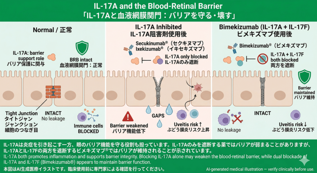
With agents that block only IL-17A (secukinumab, ixekizumab), barrier function declines and the risk of uveitis rises, whereas with bimekizumab, which blocks both IL-17A and IL-17F, the barrier is maintained and the risk falls.
Schematic diagram illustrating the effect of IL-17 inhibitors on the blood-retinal barrier. Blocking IL-17A alone (secukinumab, ixekizumab) may weaken barrier function and increase uveitis risk, while dual blockade of IL-17A and IL-17F (bimekizumab) appears to maintain barrier integrity.
AI-generated medical illustration / Takeru Eye Clinic, 2026.
(An aside) I once published a paper reporting that when I tried to induce experimental uveitis in mice lacking the IL-17A gene, there was little difference in the inflammation I had expected to be suppressed. I found it puzzling at the time, but now it makes sense.
JAK inhibitors: losing the ability to contain viruses
JAK inhibitors (JAK1/2/3 inhibitors) broadly block the signaling of multiple cytokines, including interleukin-2, interleukin-6, and interferons.
This broad blockade can undermine the interferon-dependent defenses that keep viruses in check within the eye.
Cytomegalovirus (CMV) retinitis and necrotizing retinitis caused by varicella-zoster virus (VZV), which rarely occur when the immune system is normal, have been reported during JAK inhibitor therapy.
Moreover, these infectious retinitis cases can occur with almost none of the inflammatory findings usually seen, such as vitreous opacity.
This means that when the dysfunction that allows viral replication and the blockade of signals that recruit inflammatory cells occur at the same time, the result can be a very dangerous clinical picture of “silently progressing necrosis”¹⁰.
When visual symptoms appear during JAK inhibitor therapy, infectious causes must be actively ruled out.
Immune checkpoint inhibitors: releasing the eye’s “self-tolerance”
PD-1/PD-L1 inhibitors (pembrolizumab, nivolumab, and others) and CTLA-4 inhibitors (ipilimumab) used in cancer treatment strengthen the attack on cancer cells by releasing the “brakes” on the immune system.
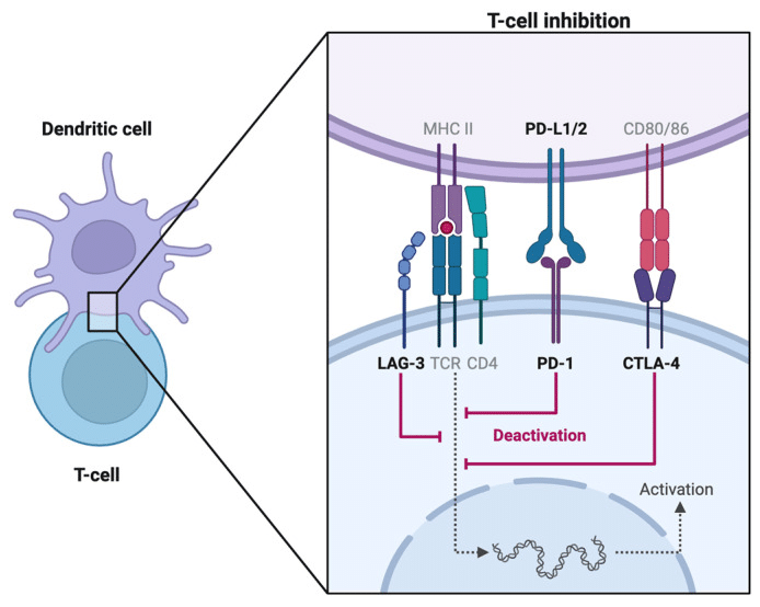
(Left) Normally, PD-1 on T cells binds to PD-L1 on cancer cells or antigen-presenting cells, which suppresses T-cell activity and allows the tumor to escape immune attack. Similarly, CTLA-4 competes with CD28 and suppresses T-cell activation at an early stage. (Right) Anti-PD-1/PD-L1 antibodies and anti-CTLA-4 antibodies block these inhibitory signals, releasing the “brakes” on T cells and restoring the attack on cancer cells.
Schematic diagram illustrating the mechanisms of immune checkpoint inhibition. (Left) Under normal conditions, PD-1 on T cells binds to PD-L1 on cancer cells or antigen-presenting cells, suppressing T cell activity and allowing tumor immune evasion. CTLA-4 similarly dampens T cell activation at an earlier stage. (Right) Anti-PD-1/PD-L1 and anti-CTLA-4 antibodies block these inhibitory interactions, restoring T cell activity against cancer cells.
Zhang H et al. Diagnostics. 2024;14(3):336. Adapted under CC BY 4.0. https://pmc.ncbi.nlm.nih.gov/articles/PMC10855398/
However, this action of “releasing the brakes” is also what originally suppresses attacks on healthy self-tissue.
In the eye, the PD-1/PD-L1 pathway plays a role in suppressing autoimmune reactions against melanocytes (pigment cells) in the retina and uveal tract.
When this pathway is blocked, an immune attack on melanocytes in the eye begins, and bilateral granulomatous panuveitis closely resembling Vogt-Koyanagi-Harada disease (VKH) can develop¹¹.
A 2024 systematic review reported 52 cases of VKH-like uveitis, 69% of which occurred in patients with malignant melanoma¹².
Because malignant melanoma is originally a malignant transformation of melanocytes, the tumor and the melanocytes in the eye are likely to become “the same target.”
In addition, the risk of developing uveitis with ipilimumab (a CTLA-4 inhibitor) is high, with a hazard ratio of 5.86 (95% confidence interval 1.99 to 17.24), which stands out compared with a PD-1 inhibitor alone (hazard ratio: 1.98)¹³.
Interestingly, it has also been suggested that such immune reactions in the eye may be a marker of a strong systemic immune response against cancer, and studies examining the correlation with tumor prognosis have begun to appear.
Infectious uveitis does not occur “by chance”
Here is the heart of this article.
In patients receiving biologics or immunotherapy drugs, infections such as tuberculosis, syphilis, herpesviruses, and CMV can develop.
It is not accurate to regard these as “bad luck” or infections acquired “by chance.”
The eye originally has the ability to “contain” pathogens. When a drug destroys that ability, what was being contained starts to move. That is the structure behind what happens.
Herpesviruses and CMV: latent viruses reactivate
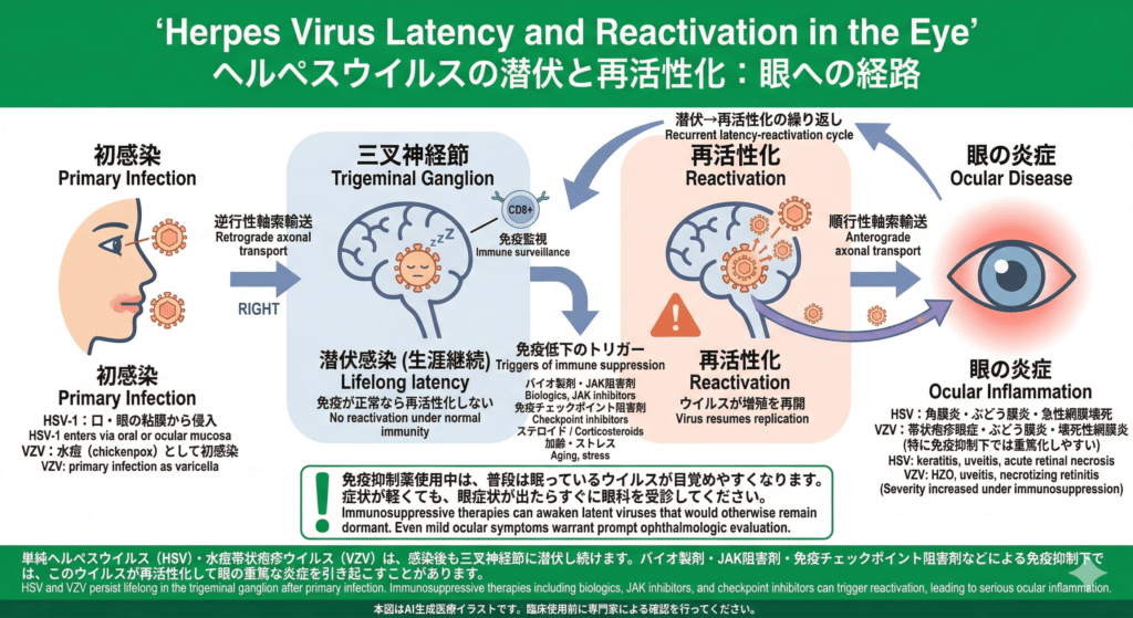
Herpes simplex virus (HSV) and varicella-zoster virus (VZV) remain latent in the trigeminal ganglion for life after primary infection. When immunity is normal they do not reactivate, but under immunosuppression from biologics, JAK inhibitors, immune checkpoint inhibitors, steroids, and similar drugs, the latent virus can reactivate and reach the eye by anterograde axonal transport (traveling along the nerve pathway to the eye), causing severe inflammation. With HSV, keratitis, uveitis, and acute retinal necrosis can occur; with VZV, herpes zoster ophthalmicus, uveitis, and necrotizing retinitis can occur.
Schematic diagram illustrating the latency-reactivation cycle of HSV and VZV in the eye. After primary infection, both viruses establish lifelong latency in the trigeminal ganglion. Immunosuppressive therapies including biologics, JAK inhibitors, and checkpoint inhibitors can trigger reactivation, with anterograde axonal transport leading to serious ocular inflammation including keratitis, uveitis, and retinal necrosis.
AI-generated medical illustration / Takeru Eye Clinic, 2026.
Herpes simplex virus (HSV) and varicella-zoster virus (VZV) lie dormant in sensory ganglia.
When immunity is normal, the virus stays latent.
However, when interferon-based defenses are weakened by steroids or immunosuppressants, especially JAK inhibitors, the virus reactivates and can cause keratitis, uveitis, and acute retinal necrosis¹⁰.

CMV can remain latent in the corneal endothelium and intraocular tissues.
When local immune surveillance is lost during treatment with JAK inhibitors or other strong immunosuppressants, CMV begins to multiply.
The defining feature of this type of infection is that the eye’s interferon-dependent defenses collapse “from the inside.”
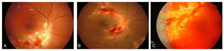
When CMV reactivates in the eye in an immunocompromised state, it causes retinitis.(A) Early stage: yellowish-white necrotic lesions around the optic disc and intraretinal hemorrhage.(B) Advanced, hemorrhagic type: full-thickness retinal necrosis and hemorrhage centered on the posterior pole, with satellite lesions at the active margin, the so-called “pizza pie” appearance.(C) Granular type: confluent white necrotic lesions spreading from the periphery toward the posterior pole.
Fundus photographs of cytomegalovirus (CMV) retinitis showing three distinct presentations. (A) Early-stage retinitis with yellowish-white necrotic lesions and intraretinal hemorrhage around the optic disc. (B) Advanced hemorrhagic variant with full-thickness retinal necrosis and “pizza pie” appearance. (C) Peripheral granular variant with confluent white necrotic lesions spreading toward the posterior pole.
Zhang J et al. Viruses. 2024;16(9):1427. Adapted under CC BY 4.0. https://pmc.ncbi.nlm.nih.gov/articles/PMC11437412/
Tuberculosis: an unnoticed coexistence that suddenly breaks down
Latent intraocular tuberculosis is relatively rare, but tubercle bacilli that have been lying dormant somewhere in the body can be reactivated by anti-TNF agents and reach the eye.
Ordinary intraocular tuberculosis presents with relatively recognizable findings such as choroidal nodules, but when it occurs under anti-TNF therapy it can take an atypical form with extensive tissue necrosis, such as serpiginous-like choroiditis.
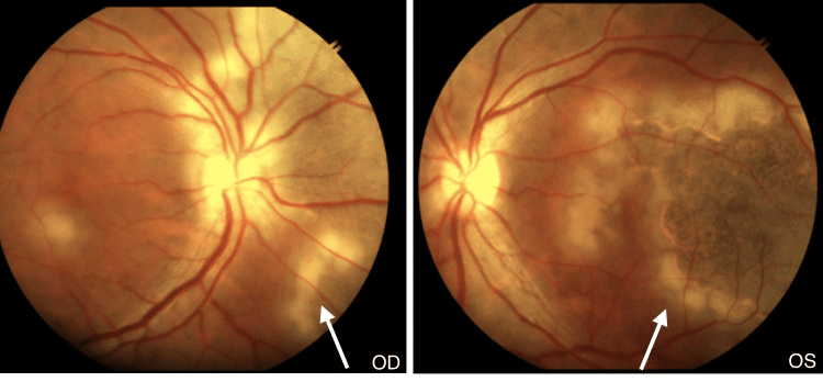
Uveitis associated with Mycobacterium tuberculosis infection can show not only typical findings such as choroidal nodules but also extensive tissue necrosis resembling serpiginous choroiditis. In both the right eye (OD) and the left eye (OS), grey-yellowish subretinal infiltrates can be seen spreading in a serpiginous pattern in the areas indicated by the arrows.
Fundus photographs of both eyes showing tubercular serpiginous-like choroiditis. Grey-yellowish subretinal infiltrates spreading in a serpiginous pattern are visible in the right eye (OD) and left eye (OS), with active scalloped edges at the advancing margin (arrows).
Magdum R et al. Cureus. 2024;16(3):e57093. Adapted under CC BY 4.0. https://pmc.ncbi.nlm.nih.gov/articles/PMC11055604/
This is thought to result not only from the growth of the tubercle bacilli themselves, but also from the collapse of the granuloma containment structure that had been maintained by TNF-α, leading to a “disordered hypersensitivity reaction”¹⁴.
This is why screening for latent tuberculosis with an interferon-γ release assay (such as the QuantiFERON® test) is required before starting anti-TNF therapy.
Syphilis: what had been quiet starts to move
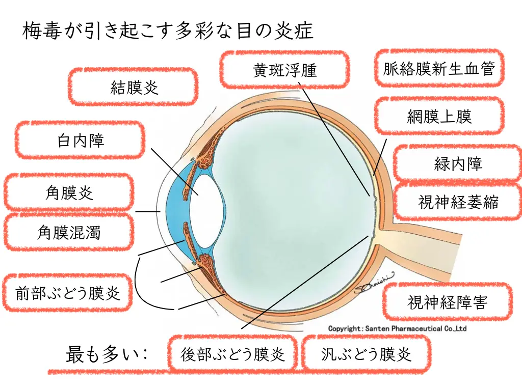
Syphilis has been on the rise in Japan in recent years. In an immunocompromised state, more severe ocular syphilis than usual can develop.
It appears in many forms in the eye, including uveitis, optic neuritis, and chorioretinitis.
When you see unexplained uveitis in a patient receiving biologics or immune checkpoint inhibitors, infection screening, including serologic testing for syphilis, is important¹⁰.
Thinking in terms of the eye’s “immune containment function”
Summarizing what we have seen so far, we can say the following.
The eye has a built-in ability to “contain” microorganisms and pro-inflammatory substances.
The blood-retinal barrier, suppressive cytokines, interferon-based defenses, and ACAID work together to protect the eye from inflammation.
Biologics and immunotherapy drugs are necessary for treating systemic disease.
But when they act on the eye’s immune balance, different types of uveitis can be triggered depending on which defense mechanism is affected.
| Drug class | Main effect on the eye | Typical condition |
|---|---|---|
| Anti-TNF agents (especially etanercept) | Paradoxical increase in interferon production | Granulomatous uveitis, paradoxical inflammation |
| IL-17A inhibitors | Weakening of barrier-protective factors | New-onset or recurrent anterior uveitis |
| JAK inhibitors | Loss of antiviral defense (IFN system) | CMV and VZV retinitis (atypical) |
| Immune checkpoint inhibitors | Release of self-tolerance checkpoints | VKH-like panuveitis |
| Infections (TB, syphilis, herpes, etc.) | Reactivation due to collapse of containment mechanisms | Granulomatous and necrotizing uveitis |
Infectious uveitis occurs where the “infection” row of this table intersects with the other drug rows. The sequence is that the drug weakens immune containment, and pathogens that had been lying dormant begin to move.
When these symptoms appear
If you are using biologics or immune checkpoint inhibitors and develop any of the following symptoms, an eye examination is recommended.
Persistent redness, eye pain, blurred vision, distorted vision, light sensitivity, or any change in vision: these are common signs of uveitis.
However, during treatment with JAK inhibitors or steroids, signs of inflammation may be less apparent, so caution is needed even when symptoms are mild.
Even a complaint as mild as “my eyes feel a little off” can lead to irreversible vision loss if left untreated.
It is important to undergo a detailed eye examination while coordinating with the physician who knows the drug names and doses (rheumatology, medical oncology, dermatology, and so on).

What lies ahead
Research on biologics and ocular inflammation is still advancing rapidly.
Bimekizumab, which inhibits both IL-17A and IL-17F, is increasingly shown to strongly improve joint and skin symptoms while lowering the risk of uveitis compared with conventional IL-17A-only inhibition⁷.
For ocular adverse events related to immune checkpoint inhibitors, data are accumulating on the timing of onset, severity, and optimization of treatment¹²¹³.
For JAK inhibitors, researchers are examining which pathway blockade contributes most to the risk of ocular infection.
It is also known that after anti-HIV therapy or the withdrawal of systemic immunosuppression, immunity against pathogens that had been contained can suddenly rise and cause inflammation called “immune recovery uveitis,” which can be understood in the same context of a “rebound of the containment mechanism”¹⁴.
As an ophthalmologist,
I believe that the importance of regularly examining the eyes of patients using these biologic drugs will only grow.
To protect vision while using biologics, information sharing between the ophthalmologist and the prescribing physician is essential.
Main drugs mentioned in this article
Anti-TNF-α agents (TNF inhibitors)
| Generic name | Brand name (Japan) | Generic / brand (international) | Route |
|---|---|---|---|
| Infliximab | Remicade® | infliximab / Remicade® | Intravenous infusion |
| Adalimumab | Humira® | adalimumab / Humira® | Subcutaneous injection |
| Etanercept | Enbrel® | etanercept / Enbrel® | Subcutaneous injection |
| Golimumab | Simponi® | golimumab / Simponi® | Subcutaneous injection |
IL-17 inhibitors
| Generic name | Brand name (Japan) | Generic / brand (international) | Route |
|---|---|---|---|
| Secukinumab | Cosentyx® | secukinumab / Cosentyx® | Subcutaneous injection |
| Ixekizumab | Taltz® | ixekizumab / Taltz® | Subcutaneous injection |
| Bimekizumab | Bimzelx® | bimekizumab / Bimzelx® | Subcutaneous injection |
JAK inhibitors
| Generic name | Brand name (Japan) | Generic / brand (international) | Route |
|---|---|---|---|
| Upadacitinib | Rinvoq® | upadacitinib / Rinvoq® | Oral |
| Tofacitinib | Xeljanz® | tofacitinib / Xeljanz® | Oral |
Immune checkpoint inhibitors
| Generic name | Brand name (Japan) | Generic / brand (international) | Route |
|---|---|---|---|
| Pembrolizumab | Keytruda® | pembrolizumab / Keytruda® | Intravenous infusion |
| Nivolumab | Opdivo® | nivolumab / Opdivo® | Intravenous infusion |
| Ipilimumab | Yervoy® | ipilimumab / Yervoy® | Intravenous infusion |
* Drug names in this article are given with generic names, representative Japanese brand names, and English names. For actual prescriptions and dosing, please check your medication notebook, the package insert, or your prescribing physician. For effects on the eyes, please consult an ophthalmologist.
References
- Medawar PB. Immunity to homologous grafted skin; the fate of skin homografts transplanted to the brain, to subcutaneous tissue, and to the anterior chamber of the eye. Br J Exp Pathol. 1948;29(1):58-69. https://www.ncbi.nlm.nih.gov/pmc/articles/PMC2073079/
- Streilein JW. Ocular immune privilege: therapeutic opportunities from an experiment of nature. Nat Rev Immunol. 2003;3(11):879-889. https://pubmed.ncbi.nlm.nih.gov/14668804/
- Taylor AW. Ocular immune privilege. Eye (Lond). 2009;23(10):1885-1889. https://doi.org/10.1038/eye.2008.382
- Redefining our vision: an updated guide to the ocular immune system. Nat Rev Immunol. 2024. https://www.nature.com/articles/s41577-024-01064-y
- Nicolela Susanna F, Pavesio C. A review of ocular adverse events of biological anti-TNF drugs. J Ophthalmic Inflamm Infect. 2020;10(1):11. https://link.springer.com/article/10.1186/s12348-020-00202-6
- Fabiani C, Vitale A, Lopalco G, et al. Different roles of TNF inhibitors in acute anterior uveitis associated with ankylosing spondylitis: state of the art. Clin Rheumatol. 2016;35(11):2631-2638. https://pubmed.ncbi.nlm.nih.gov/27686662/
- Zhao X, He X, et al. Risk of new-onset and recurrent uveitis with different biologics for ankylosing spondylitis: a network meta-analysis. Front Immunol. 2025. https://pmc.ncbi.nlm.nih.gov/articles/PMC12226306/
- Baughman RP, Lower EE, Bradley DA, Raymond LA, Kaufman A. Etanercept for refractory ocular sarcoidosis: results of a double-blind randomized trial. Chest. 2005;128(2):1062-1047. https://journal.chestnet.org/article/S0012-3692(15)50471-6/
- Dick AD, Tugal-Tutkun I, Foster S, et al. Secukinumab in the treatment of noninfectious uveitis: results of three randomized, controlled clinical trials. Ophthalmology. 2013;120(4):777-787. https://pubmed.ncbi.nlm.nih.gov/23290985/
- Teabagy S, Wood E, Bilsbury E, et al. Ocular immunosuppressive microenvironment and novel drug delivery for control of uveitis. Adv Drug Deliv Rev. 2023;198:114869. https://pubmed.ncbi.nlm.nih.gov/37172782/
- Sun MM, Levinson RD, Filipowicz A, et al. Uveitis in patients treated with CTLA-4 and PD-1 checkpoint blockade inhibition. Ocul Immunol Inflamm. 2020;28(2):217-227. https://pmc.ncbi.nlm.nih.gov/articles/PMC6832811/
- Zhang H, Houadj L, Wu KY, Tran SD. Immune checkpoint inhibitor-associated Vogt-Koyanagi-Harada-like syndrome: a descriptive systematic review. J Ophthal Inflamm Infect. 2025. https://link.springer.com/article/10.1186/s12348-025-00484-8
- Kuo HT, Chen CY, Hsu AY, et al. Association between immune checkpoint inhibitor medication and uveitis: a population-based cohort study utilizing TriNetX database. Front Immunol. 2023;14:1302293. https://pubmed.ncbi.nlm.nih.gov/38264654/
- Alves NR, Barão C, Mota C, Costa L, Pinto Proença R. Immune recovery uveitis: a focus review. Graefes Arch Clin Exp Ophthalmol. 2024;262(8):2703-2712. https://pubmed.ncbi.nlm.nih.gov/38381160/