![Takeru Eye Clinic | Takatori Shopping Street, Sawara-ku, Fukuoka City [Nishijin Station / Fujisaki Station]](https://takeru-eye.com/wp-content/uploads/2022/10/takeru_logo_for-WP-header.png)

Inflammation in which the body recognizes part of itself as foreign and attacks it: “autoimmune inflammation”
Rheumatoid arthritis:
Autoimmune inflammation gradually damages the joints over a long period1。
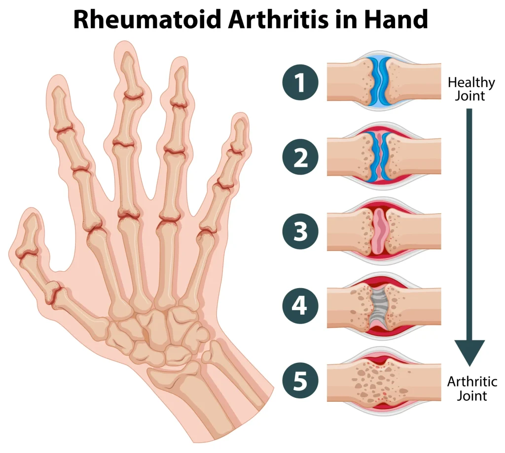
What it has in common with the eye:
The eye (cornea and sclera) and articular cartilage have in common that they contain a lot of collagen and proteoglycans (glycoproteins).
For this reason, patients with rheumatoid arthritis may develop inflammation of the cornea, conjunctiva, and sclera.
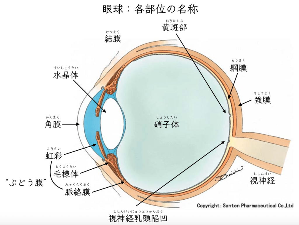
Uveitis may also occur.
- Dry eye (keratoconjunctivitis sicca)
- Peripheral ulcerative keratitis2
- Scleritis
- Uveitis3
Ocular inflammatory diseases can be brought to healing by intensifying local treatment such as eye drops and ointments.
In some cases, systemic treatment (oral medication or infusion) may be necessary.

Rheumatoid Arthritis and the Eyes: How to Think About Inflammatory Disease
For inflammatory disease, it is important to determine whether it is infectious or autoimmune (non-infectious).
Our practice is based on knowledge of immunology, thinking about what kind of reaction is occurring in the body.
If the inflammation is judged to be autoimmune, drug treatment to suppress inflammation is given.

Corticosteroids suppress inflammation powerfully, but they are a “double-edged sword” because they can also worsen infections.
Even with local treatment using eye drops and ointments, great care is needed in how they are used.
To determine whether inflammation is infectious, auxiliary tests are performed as needed.
- Gram staining (available at our clinic): classifies bacteria using dyes
- Bacterial culture (outsourced), etc.
Depending on the results, an appropriate antibiotic is selected.
For light to enter the eye and reach the retina, the tissues (cornea, lens, vitreous) must be transparent.
Even if an eye disease is cured, if it leaves a scar, it can lead to permanent loss of vision.
Prompt judgment and response are important, and our goal is“maintaining the transparency of the eye.”
The results are explained clearly using images.
We have a system in place to refer patients to Kyushu University Hospital or core hospitals without missing the right timing, as needed.
References:
1. Extra-articular manifestations of rheumatoid arthritis: an update
Autoimmun Rev. 2011 Dec;11(2):123-31.
https://www.ncbi.nlm.nih.gov/pubmed/21939785
2. Ocular inflammatory diseases associated with rheumatoid arthritis.
Nat Rev Rheumatol. 2014 Feb;10(2):108-16.
https://www.ncbi.nlm.nih.gov/pubmed/24323074
3. Arthritis and inflammatory eye disease
Rheumatology. 2005 Oct;44(10):1207-1209
https://doi.org/10.1093/rheumatology/kei027
In More Detail: A 2026 Perspective
(Added September 2026)
Seven years have passed since I wrote the article above.
The core of my thinking has not changed.
But I would like to add a little of what has become clear since.
From here on, the content is for those who want to know a little more.
If you do not need it, please feel free to skip it.
A Sign of “Vasculitis” in the Eye: Peripheral Ulcerative Keratitis (PUK)
This is a condition that occurs in roughly 1–2% of patients with rheumatoid arthritis4。
Not the center of the cornea (the black part of the eye), but only the “rim” at the border with the white of the eye erodes away like a crescent.
Why only the rim?
The cornea is originally a transparent tissue without blood vessels.
At the rim, however, fine blood vessels (the limbal vascular arcade) reach in from the white of the eye.
Immune complexes circulating in the blood (antibodies such as rheumatoid factor bound to antigen) are deposited in these vessels.
There, complement is activated, neutrophils are recruited, and enzymes that dissolve collagen (MMPs) are released4。
In other words, PUK is “vasculitis” appearing on the surface of the eye.
What matters here is that the same thing may be happening in other blood vessels in the body.
There is a classic report from 1984.
Of 34 patients with rheumatoid arthritis and necrotizing scleritis or PUK who were followed, 9 of the 17 managed with steroids and anti-inflammatory painkillers alone died of vascular complications.
Among the 17 who received systemic immunosuppressive treatment, it was 15。
This is data from 40 years ago, before biologics existed.
Treatment and patient backgrounds differ between then and now, so these numbers cannot be applied directly to today’s patients.
Even so, the position that “the eye is a sensor for hidden systemic vasculitis” has been carried over as it is into a 2026 review4。
When we see PUK or necrotizing scleritis, we ask the rheumatologist for a systemic evaluation at the same time as treating the eye.
I do not think this is a condition that can be managed with eye drops alone.
The Five Types of Scleritis
The classification by Watson and Hayreh in 1976 is still used for scleritis6。
| Type | Features | Severity |
|---|---|---|
| Diffuse anterior scleritis | The white of the eye is broadly red and painful. The most common | Relatively mild |
| Nodular anterior scleritis | A lump (nodule) forms within the redness | Moderate |
| Necrotizing scleritis (with inflammation) | Severe pain. The white of the eye melts and thins, and the purple tissue underneath (the uvea) may show through | Most severe |
| Scleromalacia (without inflammation) | The white of the eye thins with little pain or redness. Seen in people with long-standing rheumatoid arthritis | Severe (easily overlooked) |
| Posterior scleritis | Inflammation of the sclera at the back of the eye. Hard to see from the front; found with imaging tests | Moderate to severe |
Source: Watson & Hayreh 19766、Sainz de la Maza 20127
In a study of 500 patients with scleritis, 90% of the necrotizing type had ocular complications and 80% were found to have a systemic disease.
The odds ratio indicating the likelihood of vision loss was reported as 6.67。
It is known that scleritis in rheumatoid arthritis is often of the “diffuse” or “necrotizing” type8。
Note that “episcleritis,” in which only the surface of the white of the eye turns red, is a different disease.
The pain is mild, and it rarely reduces vision.
It is also said that episcleritis does not progress to scleritis6。
The rule of thumb for telling scleritis apart is “pain.”
If pain wakes you up at night, or you feel pain deep in the eye or in the temple, scleritis rather than episcleritis is suspected.
However, symptoms alone cannot reliably tell them apart. If you have strong pain, have your eyes examined at an eye clinic early.
What Has Changed in the Era of Biologics and JAK Inhibitors
Since the 2000s, the treatment of rheumatoid arthritis has changed greatly.
With TNF inhibitors, IL-6 inhibitors, and oral JAK inhibitors now available, joint destruction can be prevented considerably.
So have eye complications decreased?
Honestly, the answer at this point is that “they have decreased a little, but their severity when they occur has not changed.”
In rheumatoid arthritis overall, extra-articular complications (such as vasculitis) have been decreasing8。
There is a study of 1,623 patients with rheumatoid arthritis at a university hospital in the Netherlands from 2011 to 2021.
The incidence of scleritis was 0.25% over 10 years, slightly lower than the figures reported before biologics arrived (0.7–0.8%).
On the other hand, scleral necrosis was seen in more than 80% of the scleritis cases that developed.
Mortality within 9 years of onset was 30%, unchanged from older reports9。
Only 10 people developed scleritis, so the numbers carry a wide margin. Even so, the results cannot be called milder.
It cannot yet be said that “treatment has advanced, so eye complications are no longer a concern.”
Treatment options have increased.
However, the strength of the evidence for ocular inflammation differs considerably from drug to drug.
| Drug | Evidence for ocular inflammation | Role |
|---|---|---|
| Steroids and conventional immunosuppressants (methotrexate, etc.) | Long experience of use. Reduced mortality in the 1984 report5 | Basic treatment |
| TNF inhibitors (infliximab, adalimumab) | High response rates in case series of refractory scleritis. No randomized trials8 | Often recommended for refractory cases |
| Rituximab (anti-CD20 antibody) | The only randomized trial in scleritis (12 cases, dose comparison). 9 improved, of whom 7 relapsed and were retreated10 | An option when vasculitis is present |
| IL-6 inhibitor (tocilizumab) | Case-report level. There is also a report of 4 cases that developed PUK during treatment11 | Effect on the eye is inconsistent |
| JAK inhibitors (tofacitinib, etc.) | Effective in 9 of 10 cases of refractory scleritis. However, herpes reactivation within 1 month in 2 cases12 | A new option. Beware of infection |
Source: stated in each cell. Because the subjects and conditions differ from trial to trial, the drugs cannot simply be compared with one another.
The drugs themselves can also cause ocular inflammation.
Specifically, these include paradoxical uveitis with TNF inhibitors (especially etanercept), herpes zoster with JAK inhibitors, and retinopathy with hydroxychloroquine13。
I have written about these in detail in another article.

Cytokines: An Honest Summary as a Researcher
I previously studied cytokines using animal models of uveitis at the Medical Institute of Bioregulation, Kyushu University.
When IL-6 acts, Th17 cells increase and uveitis worsens, and blocking IL-6 with an antibody makes it milder14。
Even without the molecule IL-17, the onset of inflammation does not change, and only the later phase becomes milder15。
The molecule IL-27 has two faces, both promoting and suppressing inflammation16。
I have watched the era in which molecules like these became drugs for rheumatoid arthritis (IL-6 inhibitors, IL-17 inhibitors).
So, in the “eye” inflammation of rheumatoid arthritis, are the same molecules the main players?
Here I will separate what is clear from what is not yet clear.
What Is Clear
- In scleritis tissue, T cells, macrophages, and B cells gather, and immune complexes are deposited in the vessel walls (a study of scleral tissue from 30 patients)17。
- Enzymes that dissolve collagen (MMPs) are involved. In PUK, MMP-8 and MMP-9 are increased in the tears18。
- Because TNF inhibitors work in refractory cases, an involvement of TNF-α is indirectly supported8。
What Is Not Yet Clear
- I have not found a study that measured IL-17 directly in scleral tissue or in the tears of patients with PUK. All that is known is that Th17 cells increase in the blood during the active phase19。
- A 2024 study that comprehensively examined the serum and tears of 121 cases of scleritis found no clear signal for TNF, IL-6, or IL-1720。
- The drug that blocks IL-17A (secukinumab) failed to meet the primary endpoint in three randomized trials in uveitis21. There are not yet any trials in scleritis.
- IL-6 inhibitors work very well for the joints, but for the eye, reports of effectiveness and reports of ocular inflammation appearing during treatment are mixed11。
In other words, the current understanding is that joint inflammation and eye inflammation share the same molecular network to some extent, while the eye has stronger features of “vasculitis” and “enzymatic tissue dissolution.”
I cannot simply apply what I saw in uveitis in mice to human scleritis.
As a researcher, I would like to draw a line there.
Restating “Infectious or Autoimmune?” in the Language of Immunology
In the article above, I wrote that “it is important to determine whether it is infectious or autoimmune.”
Why is this distinction difficult?
Let me add a little from the perspective of immunology.
The immune system has “sensors” that switch on inflammation.
These sensors react to components of bacteria and viruses (called PAMPs).
But the same sensors also react to components leaking from damaged cells of the body itself (called DAMPs)22。
Because the entry point is the same, the redness, pain, and swelling that follow look very similar whether the cause is infection or autoimmunity.
This is why the two cannot be told apart by appearance alone.
One more thing.
The immune system has “innate immunity” (neutrophils and macrophages: fast but rough), which acts first, and “adaptive immunity” (T cells and B cells: slow but precisely targeted, and leaving memory), which acts later22。
The inflammation of rheumatoid arthritis is a state in which adaptive immunity has targeted the body’s own tissues.
In bacterial infection, on the other hand, inflammation arises as innate immunity tries to eliminate the bacteria.
Steroids weaken both.
With autoimmune inflammation, there is no opponent to eliminate, so suppressing it is itself the treatment.
If the same is done for infectious inflammation, it deprives the body only of its power to eliminate the bacteria.
This is what I meant by “double-edged sword.”
Patients with rheumatoid arthritis are often already taking drugs that suppress immunity.
They are prone not only to autoimmune scleritis and corneal ulcers but also to infectious scleritis and keratitis.
That is why we follow a set order at our clinic.
- First, check for any findings that suggest infection (Gram stain, culture)
- If infectious, choose an antibacterial drug
- Only after judging it to be autoimmune, move on to treatment that suppresses inflammation
- For PUK or necrotizing scleritis, contact rheumatology and request a systemic evaluation
For Patients with Rheumatoid Arthritis: When to See a Doctor
Please see an eye doctor early in the following situations.
- Redness of the white of the eye lasting several days or more, tender to the touch
- Waking at night from eye pain
- The edge of the black of the eye has become cloudy white, or seems to have thinned
- Sensitivity to light, or difficulty seeing
When you visit, please bring your medication notebook (okusuri techo).
Which condition we need to consider depends on the drugs you are using.
We examine the eyes while sharing information with your rheumatologist.
If you are concerned, please let us know at your visit.
References (Additional)
- Forestier MG, Calderón RM, Oliver AL. An overview of rheumatoid arthritis-associated dry eye disease, scleritis, and peripheral ulcerative keratitis. J Clin Med. 2026;15(9):3207. https://doi.org/10.3390/jcm15093207
- Foster CS, Forstot SL, Wilson LA. Mortality rate in rheumatoid arthritis patients developing necrotizing scleritis or peripheral ulcerative keratitis. Effects of systemic immunosuppression. Ophthalmology. 1984;91(10):1253-1263. https://pubmed.ncbi.nlm.nih.gov/6514289/
- Watson PG, Hayreh SS. Scleritis and episcleritis. Br J Ophthalmol. 1976;60(3):163-191. https://pubmed.ncbi.nlm.nih.gov/1268179/
- Sainz de la Maza M, Molina N, Gonzalez-Gonzalez LA, Doctor PP, Tauber J, Foster CS. Clinical characteristics of a large cohort of patients with scleritis and episcleritis. Ophthalmology. 2012;119(1):43-50. https://pubmed.ncbi.nlm.nih.gov/21963265/
- Promelle V, Goeb V, Gueudry J. Rheumatoid arthritis associated episcleritis and scleritis: an update on treatment perspectives. J Clin Med. 2021;10(10):2118. https://pubmed.ncbi.nlm.nih.gov/34068884/
- Vergouwen DPC, de Jong PHP, Schreurs MWJ, ten Berge JC, Rothova A. Scleritis in rheumatoid arthritis: before and during biologic era. Front Ophthalmol (Lausanne). 2023;3:1106419. https://pubmed.ncbi.nlm.nih.gov/38983001/
- Suhler EB, Lim LL, Beardsley RM, et al. Rituximab therapy for refractory scleritis: results of a phase I/II dose-ranging, randomized, clinical trial. Ophthalmology. 2014;121(10):1885-1891. https://pubmed.ncbi.nlm.nih.gov/24953794/
- Cohen F, Gabison EE, Stéphan S, et al. Peripheral ulcerative keratitis in rheumatoid arthritis patients taking tocilizumab: paradoxical manifestation or insufficient efficacy? Rheumatology (Oxford). 2021;60(11):5413-5418. https://pubmed.ncbi.nlm.nih.gov/33528012/
- Pyare R, Dutta Majumder P, Shah M, Kaushik V, Agarwal M, Biswas J. Tofacitinib in scleritis: a case series. Ocul Immunol Inflamm. 2024;32(6):884-890. https://pubmed.ncbi.nlm.nih.gov/36126052/
- Dammacco R, Guerriero S, Alessio G, Dammacco F. Natural and iatrogenic ocular manifestations of rheumatoid arthritis: a systematic review. Int Ophthalmol. 2022;42(2):689-711. https://pubmed.ncbi.nlm.nih.gov/34802085/
- Yoshimura T, Sonoda KH, Ohguro N, et al. Involvement of Th17 cells and the effect of anti-IL-6 therapy in autoimmune uveitis. Rheumatology (Oxford). 2009;48(4):347-354. https://pubmed.ncbi.nlm.nih.gov/19164426/
- Yoshimura T, Sonoda KH, Miyazaki Y, et al. Differential roles for IFN-gamma and IL-17 in experimental autoimmune uveoretinitis. Int Immunol. 2008;20(2):209-214. https://pubmed.ncbi.nlm.nih.gov/18156624/
- Yoshimura T, Takeda A, Hamano S, et al. Two-sided roles of IL-27: induction of Th1 differentiation on naive CD4+ T cells versus suppression of proinflammatory cytokine production including IL-23-induced IL-17 on activated CD4+ T cells partially through STAT3-dependent mechanism. J Immunol. 2006;177(8):5377-5385. https://pubmed.ncbi.nlm.nih.gov/17015723/
- Fong LP, Sainz de la Maza M, Rice BA, Kupferman AE, Foster CS. Immunopathology of scleritis. Ophthalmology. 1991;98(4):472-479. https://pubmed.ncbi.nlm.nih.gov/1828871/
- Sakimoto T, Ohnishi T, Ishimori A. Simultaneous study of matrix metalloproteinases, proinflammatory cytokines, and soluble cytokine receptors in the tears of noninfectious corneal ulcer patients. Graefes Arch Clin Exp Ophthalmol. 2014;252(9):1451-1456. https://pubmed.ncbi.nlm.nih.gov/25005620/
- Amadi-Obi A, Yu CR, Liu X, et al. TH17 cells contribute to uveitis and scleritis and are expanded by IL-2 and inhibited by IL-27/STAT1. Nat Med. 2007;13(6):711-718. https://pubmed.ncbi.nlm.nih.gov/17496900/
- Vergouwen DPC, Kolijn PM, de Hoog J, et al. Potential biomarkers for noninfectious scleritis identified by serum and tear fluid proteomics. Ophthalmol Sci. 2024;4(1):100407. https://pubmed.ncbi.nlm.nih.gov/38054106/
- Dick AD, Tugal-Tutkun I, Foster S, et al. Secukinumab in the treatment of noninfectious uveitis: results of three randomized, controlled clinical trials. Ophthalmology. 2013;120(4):777-787. https://pubmed.ncbi.nlm.nih.gov/23290985/
- Chen GY, Nuñez G. Sterile inflammation: sensing and reacting to damage. Nat Rev Immunol. 2010;10(12):826-837. https://pubmed.ncbi.nlm.nih.gov/21088683/