![Takeru Eye Clinic | Takatori Shopping Street, Sawara-ku, Fukuoka City [Nishijin Station / Fujisaki Station]](https://takeru-eye.com/wp-content/uploads/2022/10/takeru_logo_for-WP-header.png)

“I see specks floating in my vision.” “My eyesight feels like it has gotten worse.”
For the cause of floaters—clouding of the “vitreous”—
there are ways to treat it.
Breaking up the vitreous opacities into pieces so small you no longer notice them
:Laser vitreolysis
Removing the vitreous itself
:Vitrectomy
Currently there are mainly two.
Both carry a small chance of serious complications.
For physiologic (normal) floaters,
you need to make a decision based on how much risk you are willing to take.
Two Treatments for Floaters and Their Complications: Making a Decision That Weighs the Risks

Causes of Floaters
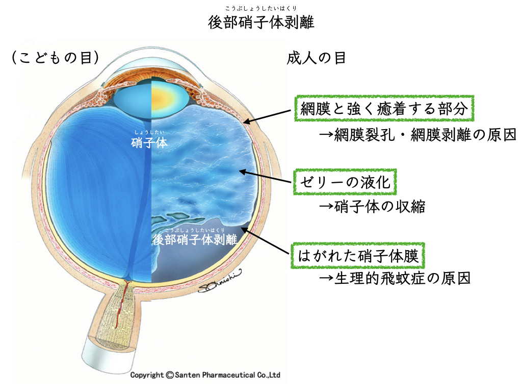
Most are caused by “posterior vitreous detachment.”
The gel inside the eye, the “vitreous,” is wrapped in a membrane.
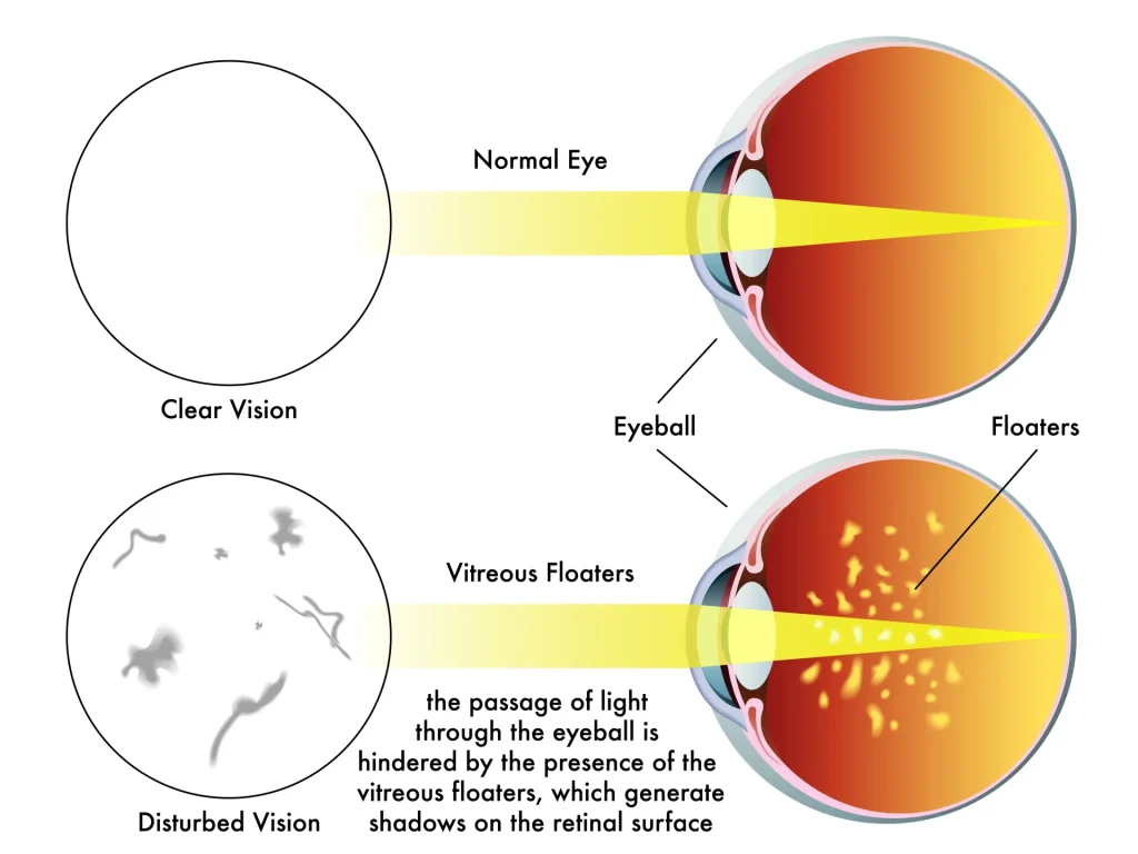
The detached vitreous membrane causes floaters.
Stress itself does not cause floaters.
Floaters arise from changes in the vitreous, regardless of changes in your state of mind.

The following diseases may also be the cause, in which case it can be very dangerous.
• Retinal tear, retinal detachment
• Inflammation inside the eye (uveitis)
• Advanced diabetic retinopathy: proliferative membranes, vitreous hemorrhage
It is very important to see an ophthalmologist and confirm that nothing is wrong.
Most cases are judged to be physiologic (normal) floaters.
However, studies show that floaters impair the quality of vision more than is generally thought.
(Physiologic) floaters can sometimes be very bothersome, and people may wish to get rid of them completely.
Research on How Bothersome Floaters Are

A study of 266 patients with floaters (aged 21 to 97; mean age 52.9).
The bother caused by floaters is directly tied to quality of life (QOL).
The younger the patient, the more they
want to get rid of their floaters even if it means taking risks
1.
Anything that affects quality of life is a major issue.
In ophthalmology too, floaters and QOL have become an important topic2.
Two Treatments for Floaters
Breaking up the vitreous opacities into pieces so small you no longer notice them
:Laser vitreolysis
Removing the vitreous itself
:Vitrectomy
Careful judgment is needed about whether you can accept iatrogenic complications (complications caused by the procedure itself).
Treating Physiologic Floaters with a YAG Laser: Laser Vitreolysis
Laser vitreolysis
Vitreo (vitreous) + lysis (to dissolve)
The YAG (yttrium-aluminum-garnet) laser is a treatment that uses shock waves.
In ophthalmology, the YAG laser is commonly used for:
• Posterior capsule opacification
• Laser treatment of glaucoma (SLT)
The same YAG laser is used to apply shock waves to the cloudy areas of the vitreous.
Solid opacities are broken into fine pieces and become harder to notice.
You may actually feel that “the floaters are gone”3.
But not all of the opacities are removed.
Complications of Laser Vitreolysis
• Cataract
• Retinal tear, retinal detachment
• Retinal hemorrhage

These are complications caused by
the YAG laser accidentally hitting the lens, the retina, or retinal blood vessels.
• Elevated intraocular pressure
• Glaucoma
• Cataract (localized clouding of the lens)
have also been reported4.
The long-term complications of laser vitreolysis have not yet been adequately studied5.
Laser vitreolysis is not covered by insurance.
Treatment for any complications that occur would also be self-pay (not covered by insurance).
This may lead to a heavy financial burden.
Treating Physiologic Floaters with Surgery: Vitrectomy
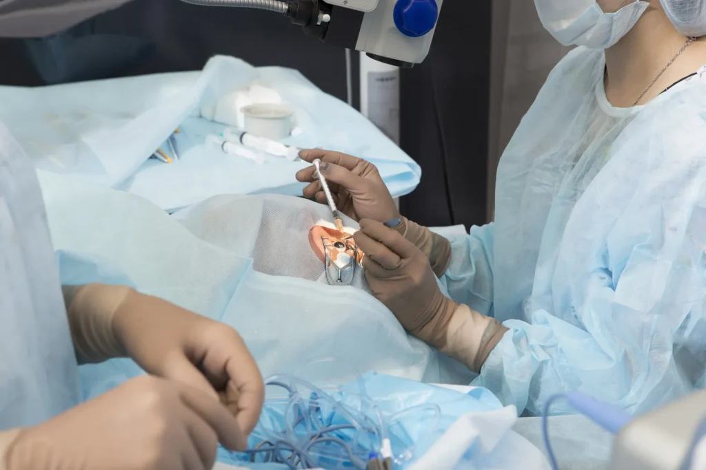
Conditions treated with vitrectomy:
• Diabetic retinopathy
• Retinal detachment
• Vitreous hemorrhage
• Uveitis
• Intraocular infection
• Eye injury
• Macular hole
• Epiretinal membrane
• Complications after cataract surgery, and others
Among eye surgeries, it is one of the larger procedures.
Vitrectomy is performed in exchange for various risks.

In vitrectomy, the aim is a complete removal of the “vitreous,” which acts as a scaffold for harmful changes inside the eye (removing all of it).
Harmful changes include:
• The remaining vitreous pulls on the retina and causes retinal detachment
• Proteins in the vitreous, “cytokines,” further worsen the environment inside the eye
Vitrectomy for Floaters
With the vitreous itself gone, improvement of floater symptoms can be expected.
The vitreous is a gel.
It cannot simply be suctioned out as it is.
That is because it would pull on the retina it is attached to.
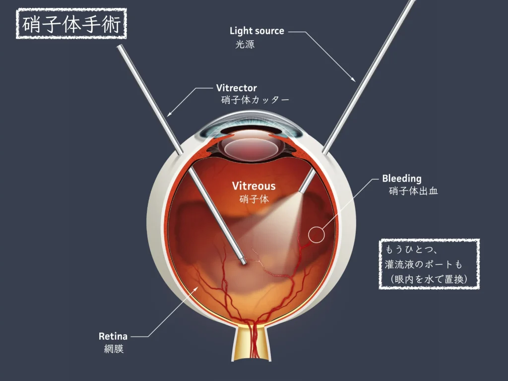
So a “vitreous cutter” is used6.
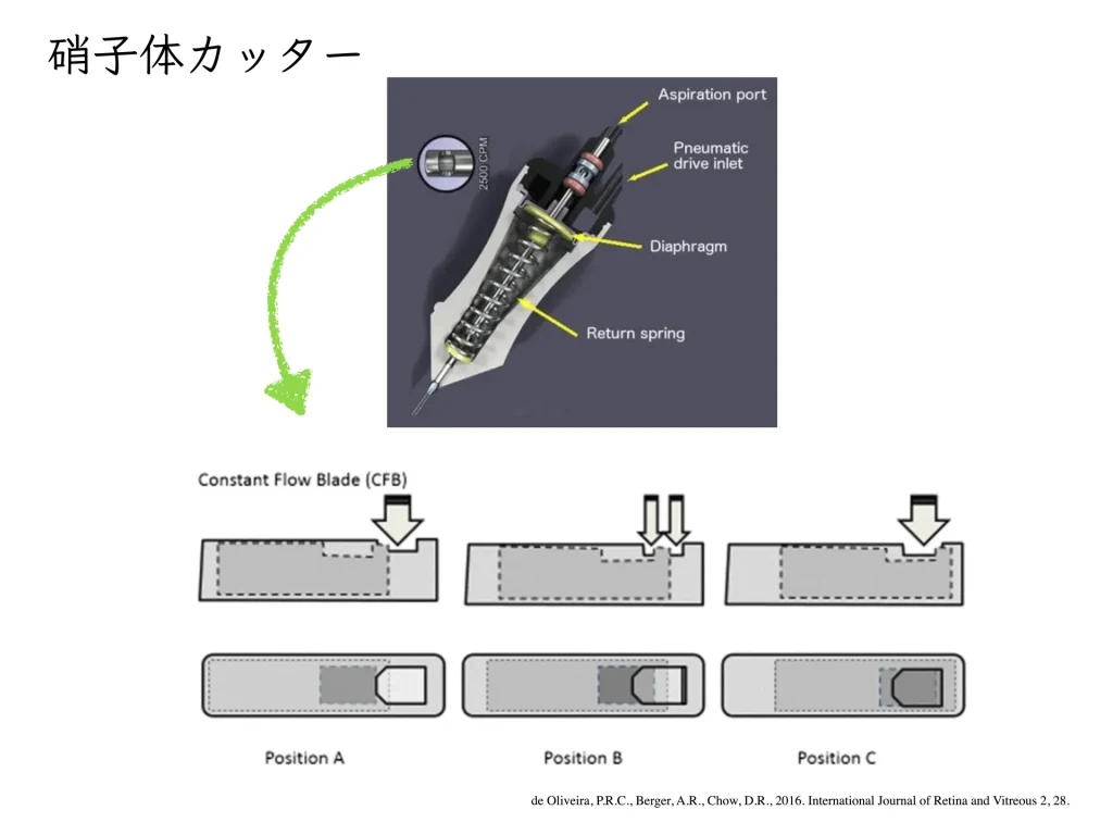
The vitreous cutter sits inside a thin tube.
The vitreous that enters is cut in a guillotine fashion and aspirated.
It is replaced with fluid (irrigating solution).
If complications such as a retinal tear or retinal detachment occur during surgery,
• Air (passed through a sterile filter)
• Gas
• Silicone oil
may be used as the replacement.
A video of about one minute:
To clear the vitreous opacities,
a suspended steroid is injected into the eye so that the vitreous can be removed completely,
membrane and all.
Vitreous left behind at the macula can also cause a macular hole.
For that reason, methods are used to remove the vitreous as thoroughly as possible.
A suspended steroid (triamcinolone acetonide) is a great help in this7.
Complications of Vitrectomy
• Cataract8
• Retinal tear, retinal detachment9
• Infection (endophthalmitis)
Wearing a mask after surgery increases the risk of endophthalmitis10
• Bleeding (vitreous hemorrhage)
• Macular hole11
Even when the eye is judged to be normal, (physiologic) floaters can greatly affect quality of life.
It is important to judge how much risk you are willing to accept in having vitrectomy for floaters.
References
- Wagle, A.M., Lim, W.-Y., Yap, T.-P., Neelam, K., Au Eong, K.-G., 2011. Utility Values Associated With Vitreous Floaters. American Journal of Ophthalmology 152, 60-65.e1. https://doi.org/10.1016/j.ajo.2011.01.026
- Sebag, J., 2011. Floaters and the Quality of Life. American Journal of Ophthalmology 152, 3-4.e1. https://doi.org/10.1016/j.ajo.2011.02.015
- Katsanos, A., Tsaldari, N., Gorgoli, K., Lalos, F., Stefaniotou, M., Asproudis, I., 2020. Safety and Efficacy of YAG Laser Vitreolysis for the Treatment of Vitreous Floaters: An Overview. Adv Ther 37, 1319–1327. https://doi.org/10.1007/s12325-020-01261-w
- Hahn, P., Schneider, E.W., Tabandeh, H., Wong, R.W., Emerson, G.G., American Society of Retina Specialists Research and Safety in Therapeutics (ASRS ReST) Committee, 2017. Reported Complications Following Laser Vitreolysis. JAMA Ophthalmol 135, 973–976. https://doi.org/10.1001/jamaophthalmol.2017.2477
- Su, D., Shah, C.P., Hsu, J., 2020. Laser vitreolysis for symptomatic floaters is not yet ready for widespread adoption. Survey of Ophthalmology 65, 589–591. https://doi.org/10.1016/j.survophthal.2020.02.007
- de Oliveira, P.R.C., Berger, A.R. & Chow, D.R. Vitreoretinal instruments: vitrectomy cutters, endoillumination and wide-angle viewing systems. Int J Retin Vitr 2, 28 (2016). https://doi.org/10.1186/s40942-016-0052-9
- Chen, T.Y., Yang, C.M., Liu, K.R., 2006. Intravitreal triamcinolone staining observation of residual undetached cortical vitreous after posterior vitreous detachment. Eye 20, 423–427. https://doi.org/10.1038/sj.eye.6701892
- Do, D.V., Gichuhi, S., Vedula, S.S., Hawkins, B.S., 2018. Surgery for postvitrectomy cataract. Cochrane Database Syst Rev 2018, CD006366. https://doi.org/10.1002/14651858.CD006366.pub4
- Wimpissinger, B., Binder, S., 2007. Entry-site-related retinal detachment after pars plana vitrectomy. Acta Ophthalmologica Scandinavica 85, 782–785. https://doi.org/10.1111/j.1600-0420.2007.00930.x
- Sakamoto, T., Terasaki, H., Yamashita, T., Shiihara, H., Funatsu, R., Uemura, A., 2022. Increased incidence of endophthalmitis after vitrectomy relative to face mask wearing during COVID-19 pandemic. British Journal of Ophthalmology. https://doi.org/10.1136/bjophthalmol-2022-321357
- Lee, S.H., Park, K.H., Kim, J.H., Heo, J.W., Yu, H.G., Yu, Y.S., Chung, H., 2010. SECONDARY MACULAR HOLE FORMATION AFTER VITRECTOMY. RETINA 30, 1072–1077. https://doi.org/10.1097/IAE.0b013e3181cd4819