![Takeru Eye Clinic | Takatori Shopping Street, Sawara-ku, Fukuoka City [Nishijin Station / Fujisaki Station]](https://takeru-eye.com/wp-content/uploads/2022/10/takeru_logo_for-WP-header.png)

It is not that having LASIK surgery makes you more likely to develop glaucoma.
Eyes that are candidates for LASIK (eyes that need LASIK)
= eyes that are prone to glaucoma (at higher risk).
In addition, because eye pressure tends to read lower, glaucoma can slip through screening tests (the eye pressure test in a health checkup).
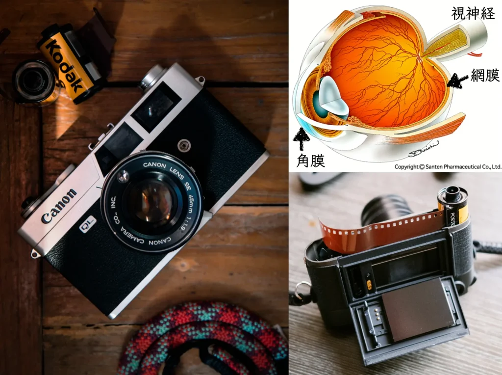
Glaucoma is a disease of the “retina” and “optic nerve” at the back of the eye,
while LASIK (laser in situ keratomileusis) treats only the “cornea” at the front of the eye.

The fact that you had LASIK, and
developing glaucoma,
are not directly related.
People with high myopia (“severe myopia”) have a relatively high risk of developing glaucoma after their 40s.

Myopic eyes, particularly highly myopic eyes, already carry an increased risk of ocular complications before LASIK.
During LASIK surgery, eye pressure is temporarily raised by suction with a device called a suction ring.
With LASIK using a microkeratome, it rises to 122–141 mmHg,
and with a femtosecond laser, it rises to 62–81 mmHg1.
I found no literature with scientific evidence that glaucoma develops because of this sudden rise in eye pressure.
LASIK operates on the cornea so that focus falls farther back, which is why vision becomes clear.
When myopia returns, it is because the thinned cornea has become thicker again.
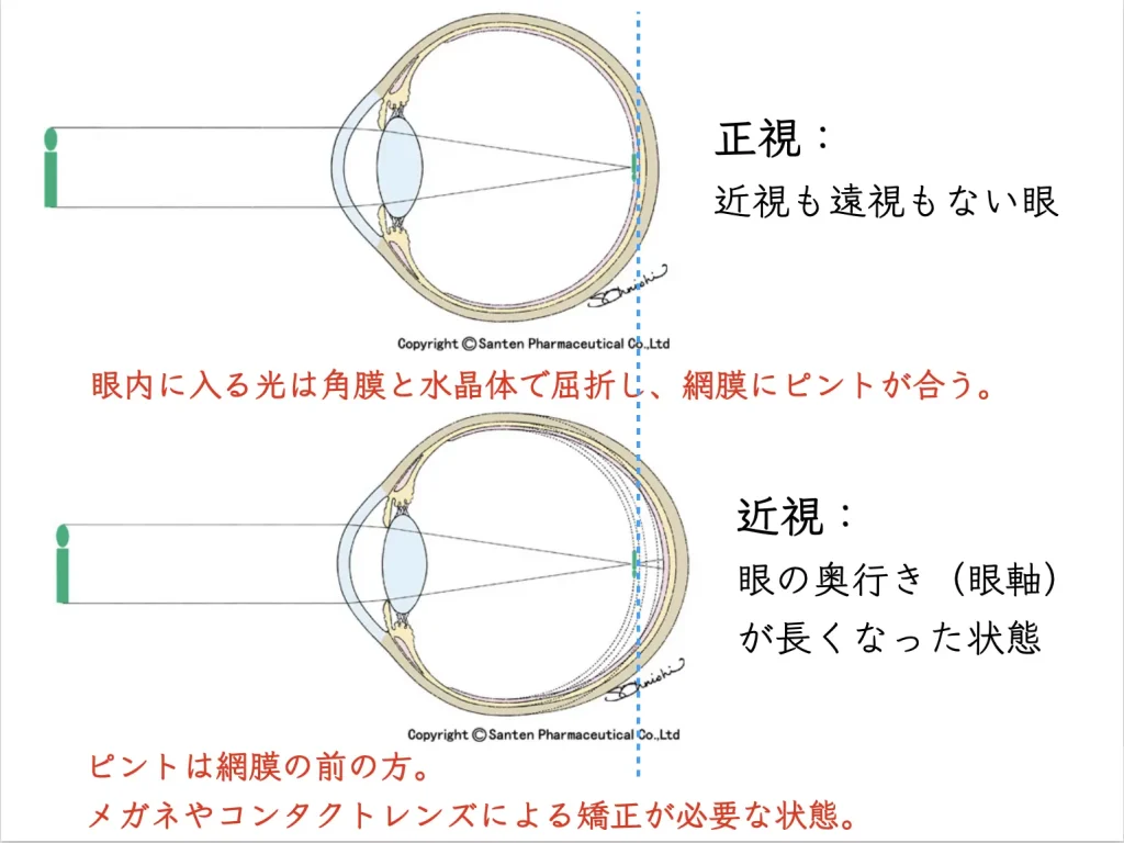
But the eye remains long
= the risks that come with high myopia remain
.
Eyes that need LASIK
= high myopia
= long axial length
= high risk of glaucoma
After LASIK, you reach your 40s
= the risk of glaucoma rises further.
Glaucoma After LASIK: The Problem of “Eye Pressure Reading Lower Than It Is”

In eyes after LASIK, the “accurate” eye pressure cannot be known
Because the cornea is thinner after LASIK, eye pressure cannot be measured accurately
The problem is that
after LASIK, glaucoma (or suspected glaucoma) may not be detected
.
In eyes after LASIK, the measured eye pressure value reads low.
This is because the cornea has become thinner.
In reality, a high eye pressure may show up as a low value2.
After LASIK, eye pressure is underestimated,
and glaucoma may slip through screening tests such as health checkups.
How Eye Pressure Is Measured
There are mainly two methods of measuring eye pressure.
Air puff method (non-contact tonometry, NCT)
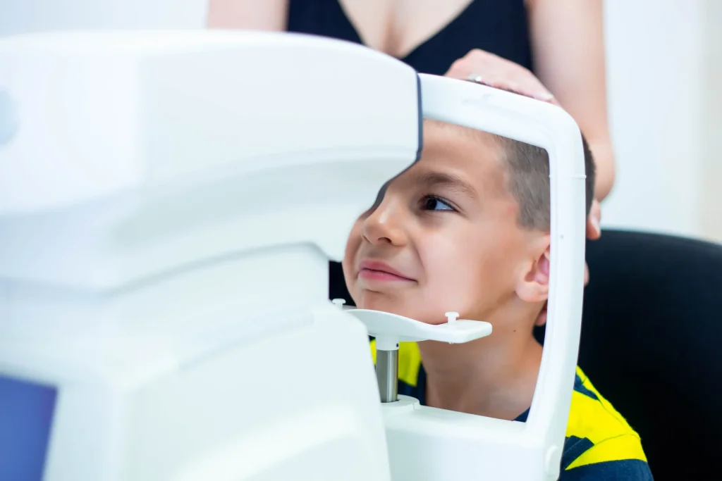
Performed in the testing room.
Goldmann applanation tonometry (GAT)

Performed in the consultation room.
It is known that the values measured by the two methods differ3.
There are also methods for continuous monitoring of eye pressure.
They are not yet at the practical stage.

Eye Pressure Values Depend on Corneal Thickness: How to Identify Glaucoma “Suspects”
Both methods of measuring eye pressure depend on corneal thickness.
Thick cornea: the eye pressure value reads higher.
Thin cornea: the eye pressure value reads lower.
There are mainly two glaucoma screening tests (health checkups):
- Eye pressure
- Fundus photograph
Of these, because eye pressure reads low,
glaucoma (or suspected glaucoma) may be missed.
The fundus photograph may show “enlargement of the optic disc cup.”
With advanced myopia, the length of the eye (axial length) has become longer.
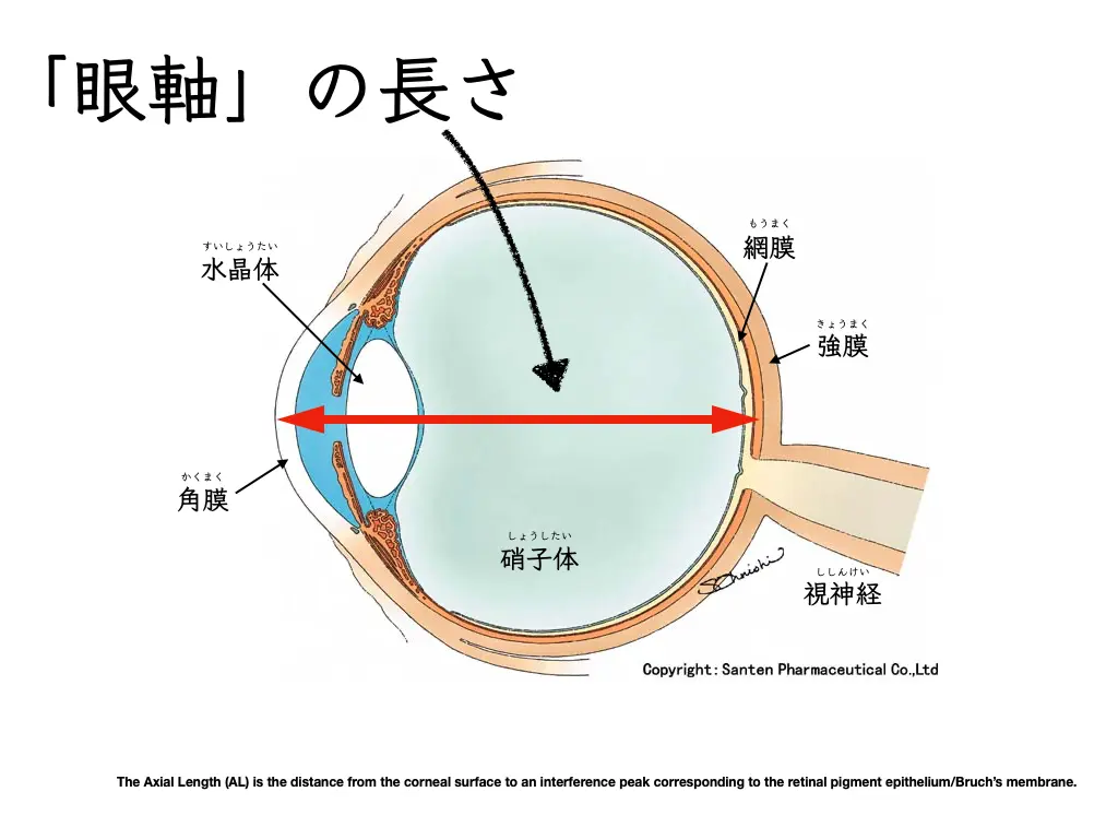
LASIK surgery, which works on the “cornea” at the front of the eye, improves uncorrected vision.
And it is wonderful that quality of life improves.
But the long axial length has not improved.
The cornea is operated on so that focus falls farther back, and vision is clear
The eye remains long, and the risk of high myopia remains
This is the situation.
(Among people who work in eye care, including ophthalmologists, most seem to wear glasses or contact lenses without having LASIK or ICL surgery.)
What to Watch For in Eyes After LASIK
- If you have had LASIK
- and are over 40,
it is important to determine
whether you have a risk of glaucoma.
In people who have had LASIK, eye pressure may read lower than the actual value.
Even if eye pressure is not high (even if you have not been told you have high eye pressure at a health checkup), glaucoma may be hidden.
Findings that make glaucoma suspected are determined with three-dimensional fundus imaging (optical coherence tomography, OCT).
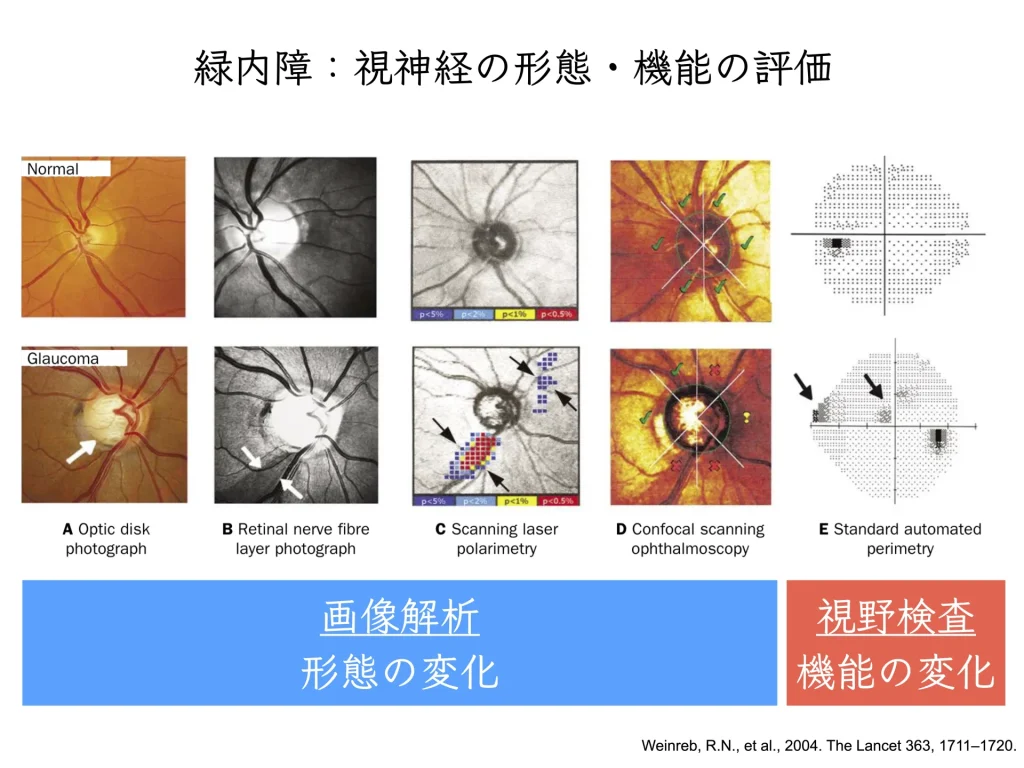
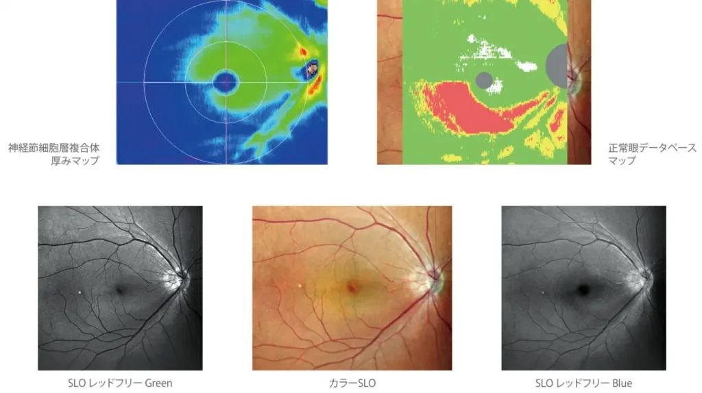
In modern ophthalmology, imaging analysis has advanced, and OCT is now a test that takes only a few seconds.
But please do not worry excessively.
References
- Chaurasia, SS, Luengo Gimeno, F, Tan, K, Yu, S, Tan, DT, Beuerman, RW & Mehta, JS 2010, ‘In Vivo Real-Time Intraocular Pressure Variations during LASIK Flap Creation’, Investigative Ophthalmology & Visual Science, vol. 51, no. 9, pp. 4641-4645.
- Fournier, A.V., Podtetenev, M., Lemire, J., Thompson, P., Duchesne, R., Perreault, C., Chehade, N., Blondeau, P., 1998. Intraocular pressure change measured by Goldmann tonometry after laser in situ keratomileusis. J Cataract Refract Surg 24, 905–910. https://doi.org/10.1016/s0886-3350(98)80041-2
- Wang, P., Song, Y., Lin, F., Wang, Z., Gao, X., Cheng, W., Chen, M., Peng, Y., Liu, Y., Zhang, X., Chen, S., 2022. Comparison of Non-contact Tonometry and Goldmann Applanation Tonometry Measurements in Non-pathologic High Myopia. Frontiers in Medicine 9.